[Preprint]. 2024 Aug 27:2024.03.29.587381.
doi: 10.1101/2024.03.29.587381.

Recharacterization of RSL3 reveals that the selenoproteome is a druggable target in colorectal cancer

Affiliations

Recharacterization of RSL3 reveals that the selenoproteome is a druggable target in colorectal cancer

Stephen L DeAngelo et al. bioRxiv. .

Update in

Abstract

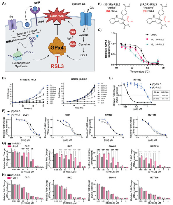
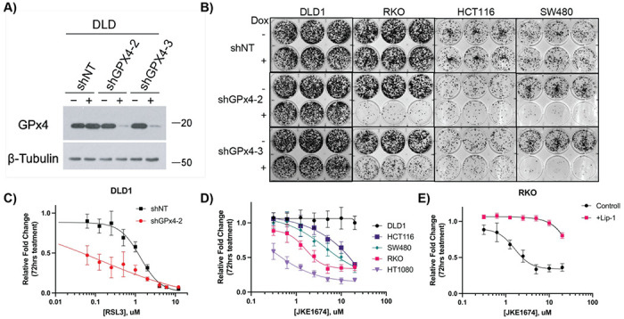
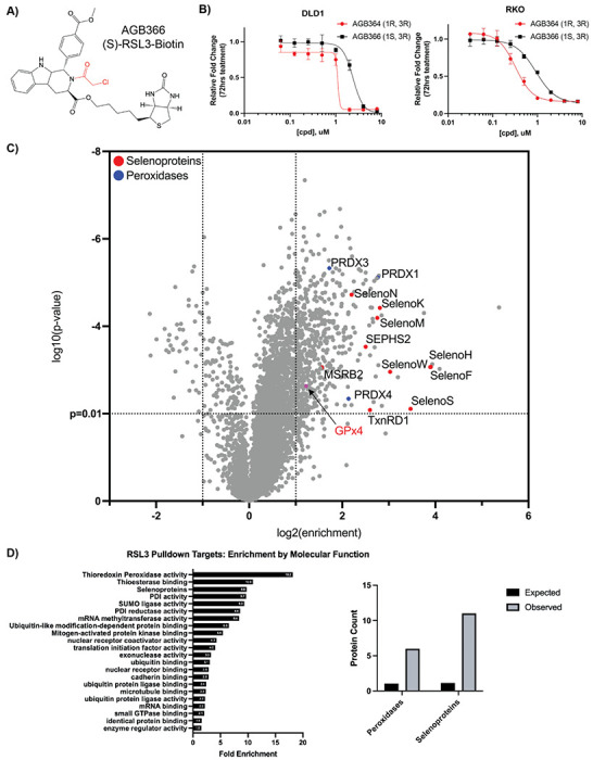
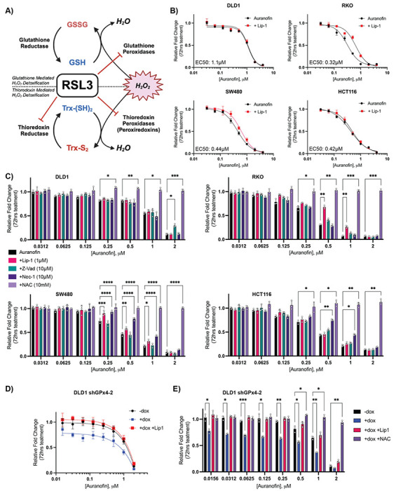
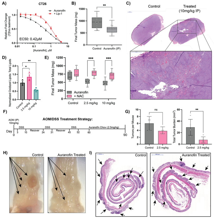
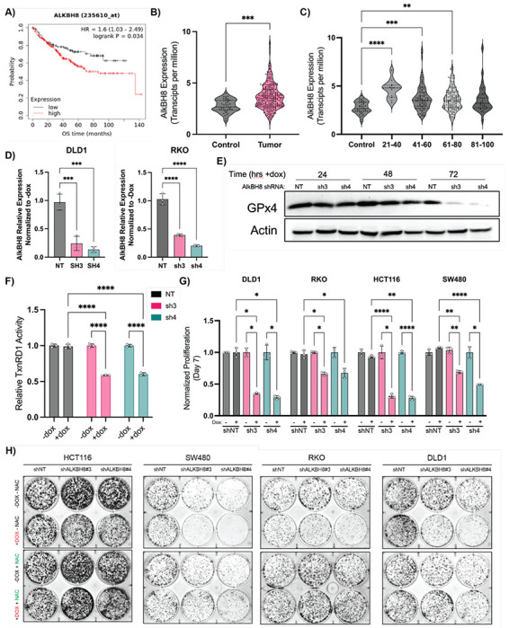
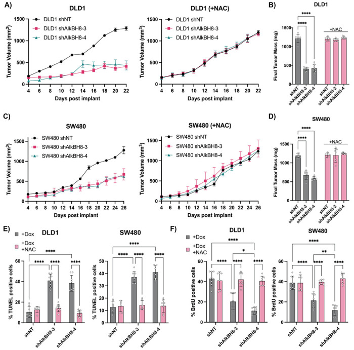
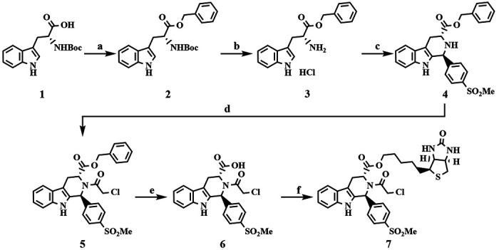
Ferroptosis is a non-apoptotic form of cell death resulting from the iron-dependent accumulation of lipid peroxides. Colorectal cancer (CRC) cells accumulate high levels of intracellular iron and reactive oxygen species (ROS) and are thus particularly sensitive to ferroptosis. The compound (S)-RSL3 ([1S,3R]-RSL3) is a commonly used ferroptosis inducing compound that is currently characterized as a selective inhibitor of the selenocysteine containing enzyme (selenoprotein) Gluathione Peroxidase 4 (GPx4), an enzyme that utilizes glutathione to directly detoxify lipid peroxides. However, through chemical controls utilizing the (R) stereoisomer of RSL3 ([1R,3R]-RSL3) that does not bind GPx4, combined with inducible genetic knockdowns of GPx4 in CRC cell lines, we revealed that GPx4 dependency does not always align with (S)-RSL3 sensitivity, questioning the current characterization of GPx4 as the central regulator of ferroptosis. Utilizing affinity pull-down mass spectrometry with chemically modified (S)-RSL3 probes we discovered that the effects of (S)-RSL3 extend far beyond GPx4 inhibition, revealing that (S)-RSL3 is a broad and non-selective inhibitor of selenoproteins. To further investigate the therapeutic potential of broadly disrupting the selenoproteome as a therapeutic strategy in CRC, we employed additional chemical and genetic approaches. We found that the selenoprotein inhibitor auranofin, an FDA approved gold-salt, chemically induced oxidative cell death and ferroptosis in both in-vitro and in-vivo models of CRC. Consistent with these data, we found that AlkBH8, a tRNA-selenocysteine methyltransferase required for the translation of selenoproteins, is essential for the in-vitro growth and xenograft survival of CRC cell lines. In summary, these findings recharacterize the mechanism of action of the most commonly used ferroptosis inducing molecule, (S)-RSL3, and reveal that broad inhibition of selenoproteins is a promising novel therapeutic angle for the treatment of CRC.


Figures

Figure 1:
Figure 1:. Defining the ferroptotic window of S-RSL3 in CRC cell lines.
A) Schematic of ferroptosis demonstrating the role of of GPx4 to detoxify ROS induced lipid peroxides (Lipid-ROS) is regulated by GSH synthesis driven by System Xc- import of cystine and selenium availability in part by LRP8 which is then incorporated into the GPx4 polypeptide by tRNA-selenocysteine. B) Structures of (1S,3R)-RSL3 and (1R,3R)-RSL3. C and D) Cell growth assay normalized to untreated control at 72 hr following (S) or (R) RSL3 treatment in HT1080 and CRC cell lines. E) Results of cell growth assay from C-D where 72 hr growth of cells treated with indicated doses of (S) and (R) RSL3 is normalized to 72 hr growth of vehicle treated control wells to calculate EC50 value (“Results of 72 hr cell growth assay”). F) Results of 72 hr cell growth assay of (S) and (R) RSL3 across a panel of CRC cell lines. G-H) Statistical analysis of results of 72 hr cell growth assay of (S) and (R) RSL3 co-treated with liproxstatin-1 (Lip-1) (1 μM) *: p < 0.05, **: p < 0.01, ***: p < 0.001, ****: p < 0.0001
Figure 2:
Figure 2:. Elucidating the role of GPx4 in CRC cell lines.
A) Western blot of stable DLD1 doxycycline inducible shRNA cell lines (shNT, shGPx4-2, shGPx4-3), +/− doxycycline treatment (250 ng/mL) assessed at 72 hrs. β-Tubulin was used as a loading control. B) Colony formation assays of indicated stable shGPx4 or shNT cell lines treated +/− doxycycline (250ng/mL) assessed at 2 weeks. C) Results of 72 hr cell growth assay following (S)-RSL3 treatment in DLD1 shNT vs shGPx4-2 cell lines, pre-treated with doxycycline (250 ng/mL) for 72 hrs prior to addition of (S)-RSL3. D) Results of 72 hr cell growth assay following JKE1674 dose response of CRC cell lines and HT1080 cells. E) Results of 72 hr cell growth assay following JKE1674 dose response of the RKO CRC cell line +/− Lip-1 (1 μ.M) co-treatment
Figure 3:
Figure 3:. Affinity pulldown-mass spectrometry redefines RSL3 as a selenoprotein inhibitor.
A) AGB366, a biotinylated RSL3 derivative. B) Results of 72 hr cell growth assay following AGB364/366 dose response treatment in DLD1 and RKO cells. C) Affinity-pulldown mass spectrometry analysis of AGB366 ((S)-RSL3-Biotin) from RKO lysate with targets of interest identified. D) RSL3 Pulldown targets protein class enrichment (PANTHER) and number of peroxidases and selenoproteins observed in the AGB366 pulldown dataset vs. expected by random chance.
Figure 4:
Figure 4:. Auranofin induces ferroptosis in CRC.
A) Schematic of the new proposed mechanism of RSL3 activity where RSL3 as a pan-inhibitor of the selenoproteome can inhibit both glutathione and thioredoxin reductases as well as peroxiredoxins. B) Results of 72 hr cell growth assay following auranofin +/− Lip-1 (1 μM) co-treatment across a panel of CRC cells. C) Statistical analysis of 72 hr cell growth assay rescue by indicated co-treatments: Lip-1 (1 μM), Z-vad-FMK (10 μM), NAC (10 mM), Nec1 (10 μM). Cells were pre-treated with rescue agent for 24 hr prior to addition of auranofin D) Results of 72 hr cell growth assay of the DLD1 shGPx4-2 cell line treated as indicated (vehicle treated control, 250 ng/mL doxycycline, 1 μM Lip-1). Cells were pretreated with doxycycline or vehicle (ddH2O) for 72 hr prior to first imaging and treatment with auranofin +/− Lip-1 E) Statistical analysis of 72 hr cell growth assay of DLD1 shGPx4-2 cells measuring deviation of growth response following auranofin treatment from control (−dox) by doxycycline treatment (+dox) +/− co-treatment with Lip-1 (1 μM) or NAC (10mM) *: p < 0.05, **: p < 0.01, ***: p < 0.001, ****: p < 0.0001
Figure 5:
Figure 5:. Auranofin is an effective treatment in in-vivo models of CRC.
A) Results of 72 hr cell growth assay of auranofin dose response +/− Lip-1 (1 μM) co-treatment in CT26 cells. B) Final tumor mass of CT26 xenograft studies in Balb/c mice treated with vehicle or auranofin IP injection (10mg/kg daily) C) H&E staining of Auranofin treated vs control tumor with higher magnification of a region of interest in the treated tumor. D) Flow cytometry measurements of Lipid-ROS levels in control vs auranofin treated CT26 xenografts. E) Final tumor mass of CT26 xenografts treated with indicated doses of auranofin chow +/− NAC (20mM) drinking water. F) Schematic of AOM/DSS model of colitis induced colorectal cancer and subsequent auranofin treatment G) Quantification of tumor number and total tumor burden (mm3) per mouse colon following AOM/DSS induction and subsequent auranofin treatment. H) Representative microscopy pictures of control and treated colons (Auranofin 2.5 mg/kg) with visually detectable tumors marked by arrows. I) H&E staining of swiss-rolled colons from C. Notable regions of dysplasia are marked by arrows. *: p < 0.05, **: p < 0.01, ***: p < 0.001, ****: p < 0.0001
Figure 6:
Figure 6:. AlkBH8 is a potential therapeutic target in CRC.
A) Kaplan-Meier overall survival analysis of CRC patients based on AlkBH8 expression as measured by RNA-Seq. B) RNA analysis of colorectal adenocarcinoma (COAD) tumor samples vs normal tissue (TCGA) analyzed for AlkBH8 expression. C) RNA analysis of colorectal adenocarcinoma (COAD) tumor samples vs normal tissue (TCGA) analyzed for AlkBH8 expression and separated by age group. D) qPCR measurement of AlkBH8 mRNA levels in NT and shRNA cell lines following 72 hrs treatment with doxycycline (250ng/mL). Normalized to untreated control. E) Western blot analysis of GPx4 protein levels in DLD1 shNT, shAlkBH8-3, and shAlkBH8-4 cell lines following doxycycline treatment (250ng/mL) for indicated times. F) TxnRD1 activity in DLD1 shNT, shAlkBH8-3, and shAlkBH8-4 cell lines following doxycycline treatment (250ng/mL) for 72hrs G) Cell growth assay at 7 days of indicated CRC cell lines stably transduced with either shNT, shAlkBH8-3, or shAlkBH8-4 and continually treated with doxycycline (250ng/mL). Normalized to untreated control growth at d7. H) CFA analysis of CRC cell lines stably transduced with either shNT, shAlkBH8-3, or shAlkBH8-4 as indicated and continually treated +/− doxycycline (250ng/mL) and +/− NAC (10mM) as indicated *: p < 0.05, **: p < 0.01, ***: p < 0.001, ****: p < 0.0001
Figure 7:
Figure 7:. AlkBH8 i-KD induces oxidative stress dependent cell death in-vivo
A) Tumor volume measurements taken by caliper of DLD1 shNT, shAlkBH8-3, and shAlkBH8-4 xenografts in NOD/SCID mice treated with doxycycline chow +/− co-administration of NAC drinking water. B) Final tumor mass of DLD1 xenografts from A. C) Tumor volume measurements taken by caliper of SW480 shNT, shAlkBH8-3, and shAlkBH8-4 xenografts in NOD/SCID mice treated with doxycycline chow co-administration of NAC drinking water. D) inal tumor mass of SW480 xenografts from C. E) Analysis of TUNEL staining (% positive cells) of DLD1 and SW480 xenografts +/− NAC co-treatment. F) Analysis of BrdU staining (% positive cells) of DLD1 and SW480 xenografts
Scheme 1.
Scheme 1.. Synthesis of Cpd24 and Biotinylated Analoga
aReagents and conditions: (a) BnOH, EDC, DMAP, THF, rt, overnight; (b) 4 N HCl in 1,4-dioxane, rt, overnight, 92% over 2 steps; (c) 4-(methylsulfonyl)benzaldehyde, IPA, reflux, 87%; (d) ClCH2C(O)Cl, TEA, CH3CN, reflux, 87%; (e) H2, Pd/C, EtOH, rt, overnight, 72%; (f) D-biotinol, EDC, DMAP, THF, rt, overnight, 4%.
Scheme 2.
Scheme 2.. Synthesis of RSL3 and Biotinylated Analogb
bReagents and conditions: (a) LiAlH4, THF, reflux, overnight, 45%; (b) (tert-butoxycarbonyl)-D-tryptophan, EDC, DMAP, THF, rt, overnight, 74%; (c) 4 N HCl in 1,4-dioxane, rt, overnight, 91%; (d) methyl 4-formylbenzoate, IPA, reflux, 15%; (e) ClCH2C(O)Cl, TEA, CH3CN, reflux, 19%.

References

    1. Siegel R. L., Giaquinto A. N. & Jemal A. Cancer statistics, 2024. CA. Cancer J. Clin. 74, 12–49 (2024). - PubMed
    1. Rawla P., Sunkara T. & Barsouk A. Epidemiology of colorectal cancer: incidence, mortality, survival, and risk factors. Przegląd Gastroenterol. 14, 89–103 (2019). - PMC - PubMed
    1. Siegel R. L., Wagle N. S., Cercek A., Smith R. A. & Jemal A. Colorectal cancer statistics, 2023. CA. Cancer J. Clin. 73, 233–254 (2023). - PubMed
    1. Tomasetti C., Marchionni L., Nowak M. A., Parmigiani G. & Vogelstein B. Only three driver gene mutations are required for the development of lung and colorectal cancers. Proc. Natl. Acad. Sci. 112, 118–123 (2015). - PMC - PubMed
    1. Raskov H., Søby J. H., Troelsen J., Bojesen R. D. & Gögenur I. Driver Gene Mutations and Epigenetics in Colorectal Cancer. Ann. Surg. 271, 75 (2020). - PubMed

Publication types