[Preprint]. 2024 Apr 3:2024.04.02.587836.
doi: 10.1101/2024.04.02.587836.

A disease-associated PPP2R3C-MAP3K1 phospho-regulatory module controls centrosome function

Affiliations

A disease-associated PPP2R3C-MAP3K1 phospho-regulatory module controls centrosome function

Anil Kumar Ganga et al. bioRxiv. .

Update in

Abstract

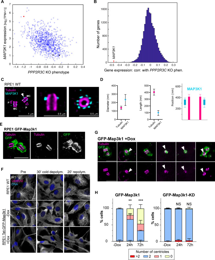
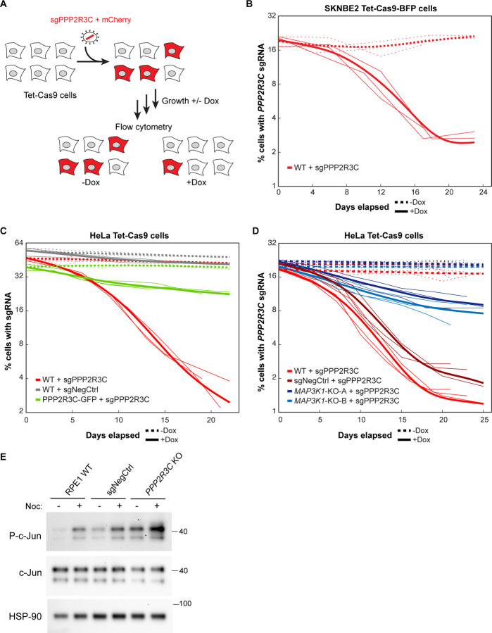
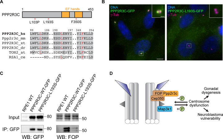
Centrosomes have critical roles in microtubule organization and in cell signaling.1-8 However, the mechanisms that regulate centrosome function are not fully defined, and thus how defects in centrosomal regulation contribute to disease is incompletely understood. From functional genomic analyses, we find here that PPP2R3C, a PP2A phosphatase subunit, is a distal centriole protein and functional partner of centriolar proteins CEP350 and FOP. We further show that a key function of PPP2R3C is to counteract the kinase activity of MAP3K1. In support of this model, MAP3K1 knockout suppresses growth defects caused by PPP2R3C inactivation, and MAP3K1 and PPP2R3C have opposing effects on basal and microtubule stress-induced JNK signaling. Illustrating the importance of balanced MAP3K1 and PPP2R3C activities, acute overexpression of MAP3K1 severely inhibits centrosome function and triggers rapid centriole disintegration. Additionally, inactivating PPP2R3C mutations and activating MAP3K1 mutations both cause congenital syndromes characterized by gonadal dysgenesis.9-15 As a syndromic PPP2R3C variant is defective in centriolar localization and binding to centriolar protein FOP, we propose that imbalanced activity of this centrosomal kinase-phosphatase pair is the shared cause of these disorders. Thus, our findings reveal a new centrosomal phospho-regulatory module, shed light on disorders of gonadal development, and illustrate the power of systems genetics to identify previously unrecognized gene functions.

Keywords: Centriole; PP2A; centrosome; cilia; functional genomics; gonadal dysgenesis; kinase; phosphatase.


Conflict of interest statement

Conflict of interest statement The authors declare no conflicts of interest

Figures

Figure 1.
Figure 1.. PPP2R3C is a centriolar protein linked to FOP and CEP350
A) Hit genes from a genome-wide CRISPR screen for regulators of cilium-dependent Hedgehog signaling are shown, including Ppp2r3c as well as several genes that encode centriolar proteins. B) CRISPR knockout growth phenotypes across >1000 cancer cell lines in the DepMap dataset are shown for PPP2R3C and FOP genes, revealing a strong correlation. C) Histogram showing correlation coefficients calculated between PPP2R3C’s fitness profile across DepMap CRISPR knockout screens and each of >17,000 other genes in the DepMap dataset. CEP350 and FOP are highlighted as the two genes with fitness profiles most highly correlated to that of PPP2R3C. D) Immunofluorescence microscopy analysis of PPP2R3C and centrosomes (marked by γ-tubulin; γ-tub) in wildtype RPE1 cells and PPP2R3C knockout (KO) cells. One of N = 3 representative experiments. Scale bar: 5 μm; inset: 1 μm. E-F) Representative ultra-structured expansion microscopy (U-ExM) images of RPE1 PPP2R3C-GFP cells stained for GFP (yellow) and tubulin (magenta) (E) or wildtype RPE1 cells stained for tubulin (magenta) and FOP (green) (F). Labels indicate the mother centrioles (MC; arrowheads) and the procentrioles (P; arrows). Scale bars: 1 and 0.5 μm (pre-expansion). G) Relative positions of the tubulin wall, PPP2R3C-GFP (yellow) and FOP (green) at the centriole are shown. Quantifications are based on n=38 centrioles from 3 independent experiments (PPP2R3C-GFP) and n=37 centrioles from 3 independent experiments (FOP). Error bars indicate standard deviation. H) Immunofluorescence microscopy analysis of PPP2R3C and centrosomes (marked by γ-tubulin; γ-tub) in wildtype RPE1 cells and FOP KO cells. One of N = 3 representative experiments. Scale bar: 5 μm; inset: 1 μm. I) Proteins captured via anti-GFP immunoprecipitation from wildtype RPE1 cells and from RPE1 cells stably expressing PPP2R3C-GFP were detected by immunoblotting, as indicated. One of N = 3 representative experiments.
Figure 2.
Figure 2.. MAP3K1 is a centrosomal kinase that negatively regulates centrosome function
A) Relationship between expression levels of MAP3K1 and fitness phenotype of PPP2R3C KO across >1000 cancer lines in the DepMap dataset. Red dot indicates SKNBE2 neuroblastoma cells and black dots indicate two diploid RPE1 clones. B) Histogram showing correlation coefficients between the expression level of each gene in the genome across DepMap cancer cell lines and the PPP2R3C KO fitness phenotype in these cells lines. MAP3K1 expression is negatively correlated (higher MAP3K1 expression is associated with greater reduction in fitness upon PPP2R3C KO. C) Representative U-ExM image of RPE1 cells stained for tubulin (magenta) and MAP3K1 (cyan), with nine-fold symmetric MAP3K1 localization revealed in top view (right). MC: mother centriole, arrowhead; P: pro-centriole. Scale bars: 1 and 0.5 µm (pre-expansion). D) Relative positions of the centriole tubulin wall and MAP3K1 were quantified for n=19 centrioles from 2 independent experiments. Error bars indicate standard deviation. E) Representative U-ExM images of RPE1 cells expressing GFP-Map3k1 stained for tubulin and GFP. Scale bar: 1 µm (pre-expansion). F) Immunofluorescence staining shows microtubule organization before, during, and 20 min after cold-induced depolymerization of microtubules. Images show wildtype RPE1 cells or RPE1 cells with Dox-inducible expression of GFP-Map3k1. Scale bar: 10 μm. See also Fig. S2C. G) Representative images of deformed and/or disintegrating centrioles observed in cells over-expressing GFP-Map3k1 for 24–36 hrs. Arrowheads denote centrioles with structural defects. Scale bar: 1 µm (pre-expansion). H) Quantification of centriole number per cell in cells over-expressing GFP-Map3k1 (left) or kinase-dead (KD) Map3k1-GFP for the indicated duration (n > 200 cells per cell line from N = 3 independent experiments. Error bars indicate standard deviation. Asterisks denote significant differences in mean (**, P = 0.0001; ***, P < 0.0001; NS, not significant).
Figure 3.
Figure 3.. PPP2R3C promotes cell fitness by opposing kinase MAP3K1
A) Illustration of flow cytometry-based competitive growth assay in which cells expressing Cas9 are transduced with a lentiviral construct co-expressing an sgRNA targeting a gene of interest (such as PPP2R3C) and mCherry to mark transduced cells. Flow cytometry analysis at successive timepoints allows monitoring of the relative growth of mutant cells relative to untransduced wildtype cells. B) The percentage of mCherry-positive cells expressing sgRNAs targeting PPP2R3C is plotted over time for SKNBE2 Tet-Cas9 cells growth with or without doxycycline (Dox)-mediated Cas9 induction. Thin lines show N = 3 replicate experiments, and thick lines show smoothing spline curves fit to replicates. C) The percentage of mCherry-positive cells is plotted as in (B) for sgRNAs targeting PPP2R3C or a non-targeting control (sgNegCtrl) in wildtype HeLa Tet-Cas9 cells or HeLa Tet-Cas9 cells stably expressing a sgRNA-resistant PPP2R3C-GFP transgene. D) The percentage of mCherry-positive cells is plotted as in (B) for sgRNAs targeting PPP2R3C introduced into wildtype HeLa Tet-Cas9 cells, HeLa Tet-Cas9 cells expressing a non-targeting sgRNA, or clonal MAP3K1 KO HeLa Tet-Cas9 cell lines. See also Figure S3A–B. E) Immunoblot analysis of phosphorylated c-Jun (P-c-Jun) and total c-Jun levels in whole cell lysates prepared from the indicated cells grown for 1 hr in the presence or absence of 2 μg/ml Nocodazole (Noc). HSP90 was used as a loading control. One of N = 3 replicate experiments.
Figure 4.
Figure 4.. A human pathogenic variant in PPP2R3C impairs its centriolar function
A) Top: Illustration of PPP2R3C highlighting two EF hand domains and localization of three pathogenic variants observed in individuals with myo-ectodermal gonadal dysgenesis (MEGD) syndrome. Bottom: alignment of PPP2R3C sequence showing human (hs), mouse (mm), frog (Xenopus tropicalis; xt), zebrafish (Danio rerio; dr), and plant (Arabidopsis thaliana; at) homologs. Residues mutated in MEGD are shown in red, and conserved residues are highlighted gray. B) Immunofluorescence microscopy analysis of wildtype and L193S forms PPP2R3C-GFP stably expressed in RPE1 cells. Centrosomes are marked by γ-tubulin; γ-tub). One of N = 3 representative experiments. Scale bar: 5 μm; inset: 1 μm. C) Proteins captured via anti-GFP immunoprecipitation from wildtype RPE1 cells and from RPE1 cells stably expressing wildtype or L193S forms of PPP2R3C-GFP were detected by immunoblotting, as indicated. One of N = 3 representative experiments. D) Model for PPP2R3C and MAP3K1 function, indicating association of PPP2R3C with FOP and CEP350 and its localization at the centriole distal torus. MAP3K1 kinase and PP2A-PPP2R3C phosphatase have opposing roles, with excess MAP3K1 or loss of PPP2R3C promoting gonadal dysgenesis and fitness defects, including in cancers such as neuroblastoma that are characterized by high MAP3K1 expression.

References

    1. Chavali P.L., Putz M., and Gergely F. (2014). Small organelle, big responsibility: the role of centrosomes in development and disease. Philos Trans R Soc Lond B Biol Sci 369. 10.1098/rstb.2013.0468. - DOI - PMC - PubMed
    1. Breslow D.K., and Holland A.J. (2019). Mechanism and Regulation of Centriole and Cilium Biogenesis. Annu Rev Biochem 88, 691–724. 10.1146/annurev-biochem-013118-111153. - DOI - PMC - PubMed
    1. Nigg E.A., and Holland A.J. (2018). Once and only once: mechanisms of centriole duplication and their deregulation in disease. Nat Rev Mol Cell Biol 139, 663. 10.1038/nrm.2017.127. - DOI - PMC - PubMed
    1. Nigg E.A., and Raff J.W. (2009). Centrioles, centrosomes, and cilia in health and disease. Cell 139, 663–678. 10.1016/j.cell.2009.10.036. - DOI - PubMed
    1. Douanne T., Stinchcombe J.C., and Griffiths G.M. (2021). Teasing out function from morphology: Similarities between primary cilia and immune synapses. J Cell Biol 220, e202102089. 10.1083/jcb.202102089. - DOI - PMC - PubMed

Publication types