[Preprint]. 2024 Apr 4:2024.04.03.588010.
doi: 10.1101/2024.04.03.588010.

Pseudomonas aeruginosa surface motility and invasion into competing communities enhances interspecies antagonism

Affiliations

Pseudomonas aeruginosa surface motility and invasion into competing communities enhances interspecies antagonism

Andrea Sánchez-Peña et al. bioRxiv. .

Update in

Abstract

Chronic polymicrobial infections involving Pseudomonas aeruginosa and Staphylococcus aureus are prevalent, difficult to eradicate, and associated with poor health outcomes. Therefore, understanding interactions between these pathogens is important to inform improved treatment development. We previously demonstrated that P. aeruginosa is attracted to S. aureus using type IV pili-mediated chemotaxis, but the impact of attraction on S. aureus growth and physiology remained unknown. Using live single-cell confocal imaging to visualize microcolony structure, spatial organization, and survival of S. aureus during coculture, we found that interspecies chemotaxis provides P. aeruginosa a competitive advantage by promoting invasion into and disruption of S. aureus microcolonies. This behavior renders S. aureus susceptible to P. aeruginosa antimicrobials. Conversely, in the absence of type IV pilus motility, P. aeruginosa cells exhibit reduced invasion of S. aureus colonies. Instead, P. aeruginosa builds a cellular barrier adjacent to S. aureus and secretes diffusible, bacteriostatic antimicrobials like 2-heptyl-4-hydroxyquinoline-N-oxide (HQNO) into the S. aureus colonies. P. aeruginosa reduced invasion leads to the formation of denser and thicker S. aureus colonies with significantly increased HQNO-mediated lactic acid fermentation, a physiological change that could complicate the effective treatment of infections. Finally, we show that P. aeruginosa motility modifications of spatial structure enhance competition against S. aureus. Overall, these studies build on our understanding of how P. aeruginosa type IV pili-mediated interspecies chemotaxis mediates polymicrobial interactions, highlighting the importance of spatial positioning in mixed-species communities.

Keywords: Pseudomonas aeruginosa; Staphylococcus aureus; biofilms; polymicrobial; spatial organization; type IV pili motility.


Conflict of interest statement

COMPETING INTERESTS The authors have declared no competing interests.

Figures
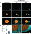
Figure 1.
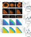
Figure 1.. Type IV pili are necessary for P. aeruginosa invasion into S. aureus colonies.
Resonant scanning confocal live imaging of S. aureus and P. aeruginosa. A. Representative micrographs of WT S. aureus (pseudocolored orange) in monoculture or in coculture with P. aeruginosa (pseudocolored cyan; WT or TFP-deficient mutant ΔpilA). B. Quantification of P. aeruginosa single-cell invasion into S. aureus colonies t ~7 h in mono- or coculture with P. aeruginosa (WT or ΔpilA). At least four biological replicates with two technical replicates each were analyzed. Each data point represents one technical replicate. Statistical significance was determined by a Mann-Whitney’s test. **** = P < 0.0001. C. Zoomed micrograph of S. aureus colony edge in coculture with P. aeruginosa (WT or ΔpilA). White arrows indicate dispersed S. aureus cells. S. aureus: pCM29 PsarAP1-sgfp; P. aeruginosa: chromosomal PA1/04/03-mCherry.
Figure 2.
Figure 2.. P. aeruginosa type IV pili motility-mediated invasion influences the architecture of S. aureus colonies independently of P. aeruginosa-secreted antimicrobials.
Analysis of S. aureus colony edge disruption and thickness. A. Representative resonant scanning confocal micrographs of the whole colony (top row) or Galvano scanner colony edge micrographs of WT S. aureus (orange) in monoculture or coculture with P. aeruginosa (not shown; WT or ΔpilA) t ~24 h shown from the top (middle row) of the colony or the side (bottom row). The micrographs in A (bottom row) show the colonies on the Z-plane and demonstrate how the height was quantified. B & C. Quantification of S. aureus whole colony area at t ~24 h (B), or height at the edge of S. aureus (Sa) colony (μm) at t ~24 h (C) in monoculture or coculture with P. aeruginosa (WT, ΔpilA, ΔAMB (bacteriostatic antimicrobials; HQNO, pyoverdine, and pyochelin), ΔlasA, ΔAMC (complete antimicrobials; HQNO, pyoverdine, pyochelin, and LasA), or ΔpilA ΔAMC). D & E. Representative BiofilmQ heatmaps of local surface roughness (D) and cell packing analysis (E) at S. aureus colony edge in mono- or coculture with P. aeruginosa (WT or ΔpilA). Three biological replicates with two technical replicates each were analyzed in D & E. F. The number of invading P. aeruginosa (Pa) single cells found inside S. aureus was quantified at t ~7 h in mono- or coculture with the P. aeruginosa strains described above. At least four biological replicates with two technical replicates each were analyzed per condition in B, C, and F. Each data point represents one technical replicate. Statistical significance was determined by Kruskal-Wallis followed by Dunn’s Multiple Comparisons Test. n.s. = not significant; * = P < 0.05; ** = P < 0.01; *** = P < 0.001.
Figure 3.
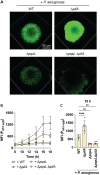
Figure 3.. Increased S. aureus cell packing enhances HQNO-mediated S. aureus fermentation.
S. aureus lactate fermentation (Pldh1-sgfp) was measured in the presence of the indicated P. aeruginosa strains. A. Representative resonant scanning confocal micrographs of S. aureus fermentation in coculture with P. aeruginosa (WT, ΔpilA, ΔpqsL, or ΔpqsL ΔpilA) t = 18 h, S. aureus channel only. B. Mean fluorescence intensity (MFI, fluorescence / colony volume) was quantified over time. C. MFI at 18 h. Data represent the mean and standard deviation from three biological replicates with two technical replicates per condition. Statistical analyses were performed at 18 h using one-way ANOVA followed by Dunnett’s Multiple Comparisons Test comparing each condition to +WT P. aeruginosa. ** = P < 0.01; **** = P < 0.0001.
Figure 4.
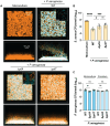
Figure 4.. P. aeruginosa type IV pili motility is necessary for competition against S. aureus in artificial sputum media.
Resonant scanning confocal imaging of S. aureus and P. aeruginosa under static conditions in artificial sputum media, with CFU quantification. A. Representative images of resonant scanning confocal micrographs of WT S. aureus (pseudocolored orange) in monoculture or in coculture with P. aeruginosa (pseudocolored cyan; WT, ΔpilA, or ΔpilT) t ~22 h. S. aureus (B) or P. aeruginosa (C) CFU quantification in monoculture or in coculture with P. aeruginosa (WT, ΔpilA, or ΔpilT) (B), or in mono- or coculture with S. aureus (C) at t = 24 h. The CFUs/well in Y-axes are portrayed as log10 transformed. Three biological replicates with one technical replicate each were analyzed, and the mean and standard deviation are shown. Each data point represents one biological replicate. Statistical significance was determined by one-way ANOVA followed by Dunnett’s Multiple Comparisons Test. n.s. = not significant; * = P < 0.05; **** = P < 0.0001.
Figure 5.
Figure 5.. P. aeruginosa type IV pili motility is necessary for disruption and competition against pre-formed S. aureus biofilms.
Resonant scanning confocal imaging of S. aureus and P. aeruginosa under static conditions in artificial sputum media, with CFU quantification at late time points. P. aeruginosa was added to S. aureus pre-formed biofilms at 5 h. A. Representative images of resonant scanning confocal micrographs of WT S. aureus (pseudocolored orange) in monoculture or in coculture with P. aeruginosa (pseudocolored cyan; WT, ΔpilA, or ΔpilT) t ~26 h. The insets show ~3X zoomed images at 30 μm from the base of the coverslip. S. aureus (B) or P. aeruginosa (C) CFU quantification in mono- or in coculture with P. aeruginosa (WT, ΔpilA, or ΔpilT) (B), or in mono- or coculture with S. aureus (C) at t ~29 h. The CFUs/well in Y-axes are portrayed as log10 transformed. Five biological replicates with one technical replicate each were analyzed, and the mean and standard deviation are shown. Statistical significance was determined by one-way ANOVA followed by Dunnett’s Multiple Comparisons Test. n.s. = not significant; * = P < 0.05; *** = P < 0.001; **** = P < 0.0001.
Figure 6.
Figure 6.. Model for motility-driven interspecies competition.
We propose that P. aeruginosa type IV pili motility-mediated attraction towards, invasion, and disruption of S. aureus colonies promotes the diffusion of antimicrobials (AM) to maximize interspecies competition. Lack of motility affects S. aureus spatial organization and physiology in a manner that promotes coexistence.

References

    1. Strassmann J.E., Gilbert O.M., and Queller D.C., Kin discrimination and cooperation in microbes. Annu Rev Microbiol, 2011. 65: p. 349–67. - PubMed
    1. Filkins L.M. and O’Toole G.A., Cystic fibrosis lung infections: polymicrobial, complex, and hard to treat. PLoS Pathog, 2015. 11(12): p. e1005258. - PMC - PubMed
    1. Little W., Black C., and Smith A.C., Clinical implications of polymicrobial synergism effects on antimicrobial susceptibility. Pathogens, 2021. 10(2). - PMC - PubMed
    1. Limoli D.H. and Hoffman L.R., Help, hinder, hide and harm: what can we learn from the interactions between Pseudomonas aeruginosa and Staphylococcus aureus during respiratory infections? Thorax, 2019. 74(7): p. 684–692. - PMC - PubMed
    1. Jean-Pierre F., et al. , Community composition shapes microbial-specific phenotypes in a cystic fibrosis polymicrobial model system. Elife, 2023. 12. - PMC - PubMed

Publication types