

TOB1 modulates neutrophil phenotypes to influence gastric cancer progression and immunotherapy efficacy
- PMID: 38617839
- PMCID: PMC11010640
- DOI: 10.3389/fimmu.2024.1369087
TOB1 modulates neutrophil phenotypes to influence gastric cancer progression and immunotherapy efficacy
Abstract
Introduction: The ErbB-2.1(TOB1) signaling transducer protein is a tumor-suppressive protein that actively suppresses the malignant phenotype of gastric cancer cells. Yet, TOB1 negatively regulates the activation and growth of different immune cells. Understanding the expression and role of TOB1 in the gastric cancer immune environment is crucial to maximize its potential in targeted immunotherapy.
Methods: This study employed multiplex immunofluorescence analysis to precisely delineate and quantify the expression of TOB1 in immune cells within gastric cancer tissue microarrays. Univariate and multivariate Cox analyses were performed to assess the influence of clinical-pathological parameters, immune cells, TOB1, and double-positive cells on the prognosis of gastric cancer patients. Subsequent experiments included co-culture assays of si-TOB1-transfected neutrophils with AGS or HGC-27 cells, along with EdU, invasion, migration assays, and bioinformatics analyses, aimed at elucidating the mechanisms through which TOB1 in neutrophils impacts the prognosis of gastric cancer patients.
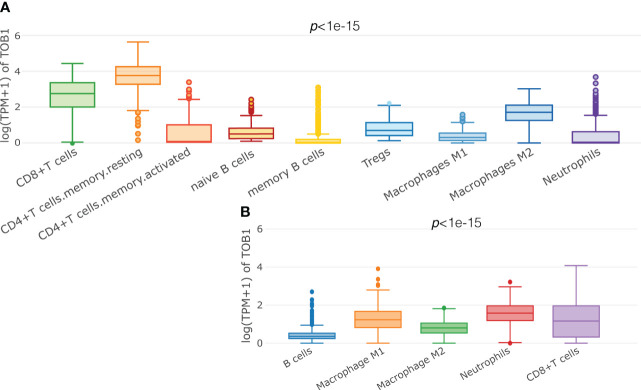
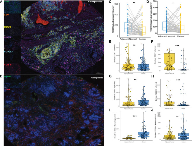
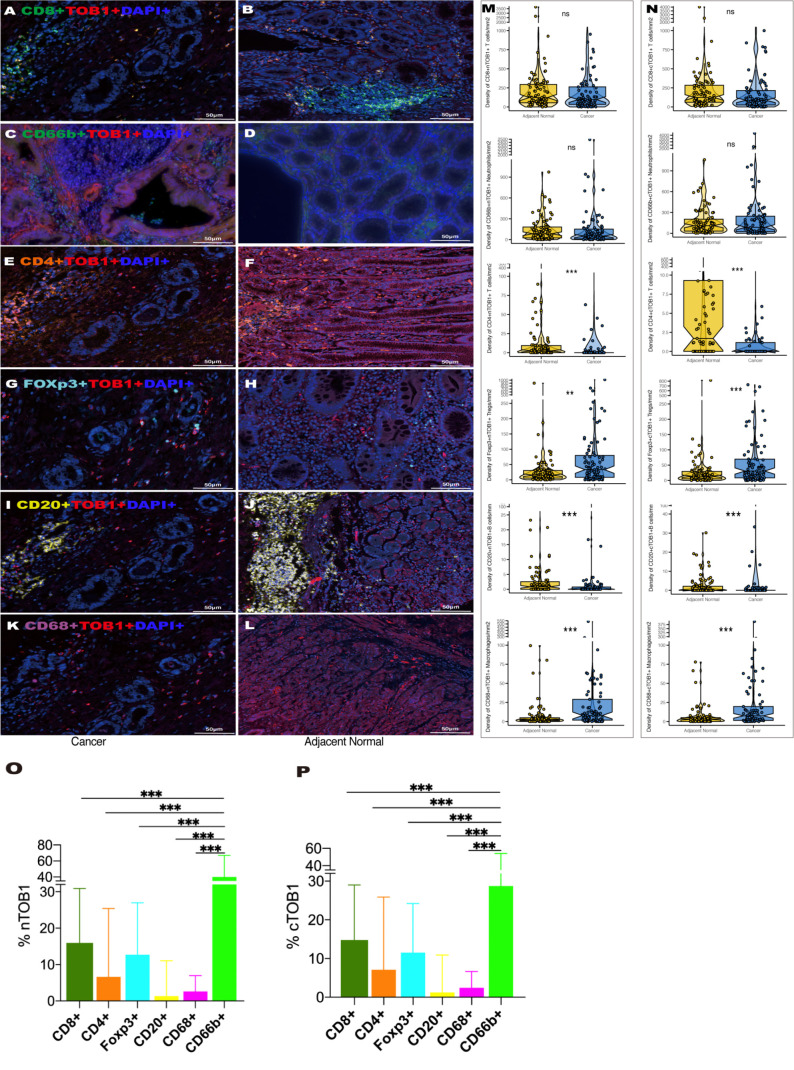
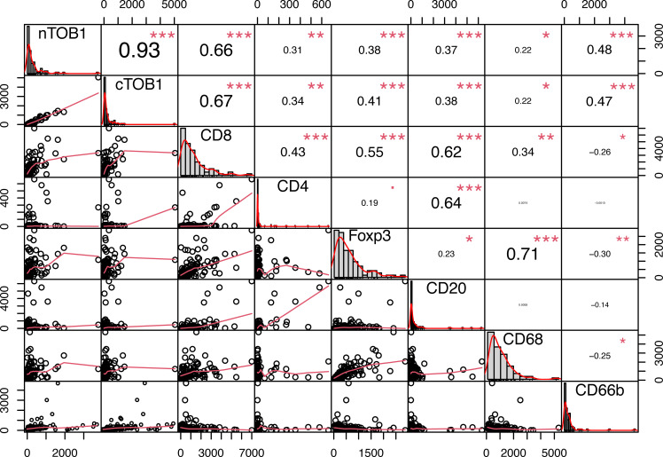
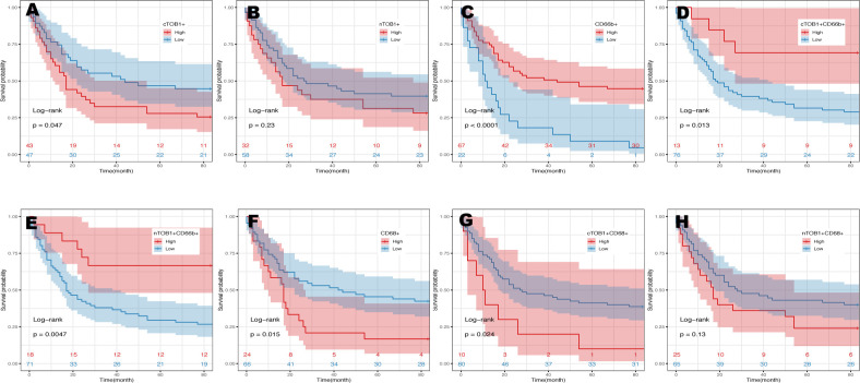
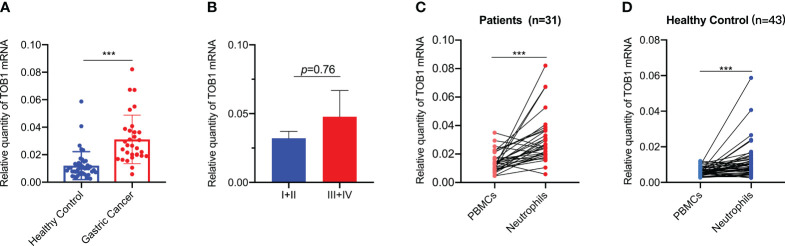
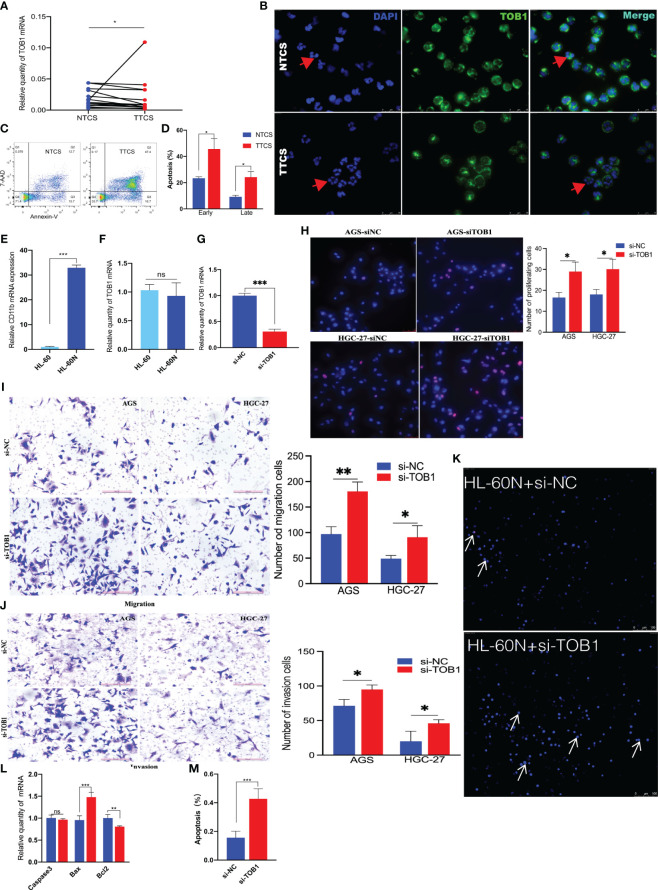
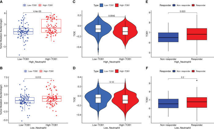
Results: We remarkably revealed that TOB1 exhibits varying expression levels in both the nucleus (nTOB1) and cytoplasm (cTOB1) of diverse immune cell populations, including CD8+ T cells, CD66b+ neutrophils, FOXP3+ Tregs, CD20+ B cells, CD4+ T cells, and CD68+ macrophages within gastric cancer and paracancerous tissues. Significantly, TOB1 was notably concentrated in CD66b+ neutrophils. Survival analysis showed that a higher density of cTOB1/nTOB1+CD66b+ neutrophils was linked to a better prognosis. Subsequent experiments revealed that, following stimulation with the supernatant of tumor tissue culture, the levels of TOB1 protein and mRNA in neutrophils decreased, accompanied by enhanced apoptosis. HL-60 cells were successfully induced to neutrophil-like cells by DMSO. Neutrophils-like cells with attenuated TOB1 gene expression by si-TOB1 demonstrated heightened apoptosis, consequently fostering a malignant phenotype in AGS and HCG-27 cells upon co-cultivation. The subsequent analysis of the datasets from TCGA and TIMER2 revealed that patients with high levels of TOB1 combined neutrophils showed better immunotherapy response.
Discussion: This study significantly advances our comprehension of TOB1's role within the immune microenvironment of gastric cancer, offering promising therapeutic targets for immunotherapy in this context.
Keywords: TOB1; gastric cancer; immune cells; immunotherapy; neutrophils apoptosis.
Copyright © 2024 Zhang, Li, Chen, Huang, Lin, Pi, Hao, Wang, Liang, Fu and Yu.
Conflict of interest statement
The authors declare that the research was conducted in the absence of any commercial or financial relationships that could be construed as a potential conflict of interest.
Figures
Similar articles
-
B7-H3 and CD47 co-expression in gastric cancer is a predictor of poor prognosis and potential targets for future dual-targeting immunotherapy.J Cancer Res Clin Oncol. 2023 Dec;149(18):16609-16621. doi: 10.1007/s00432-023-05408-4. Epub 2023 Sep 16. J Cancer Res Clin Oncol. 2023. PMID: 37715830 Free PMC article.
-
Reprogramming of hsa_circ_0008719 and its target miR-3615 induced by TOB1 in exosomes of gastric cancer cells: a contribution to inhibit gastric cancer progression.Sci Rep. 2025 Jul 25;15(1):27089. doi: 10.1038/s41598-025-12789-8. Sci Rep. 2025. PMID: 40715337 Free PMC article.
-
Calcium overload via PVT1 reprograms neutrophil fate and constrains gastric cancer progression.J Transl Med. 2025 Aug 7;23(1):878. doi: 10.1186/s12967-025-06912-6. J Transl Med. 2025. PMID: 40775351 Free PMC article.
-
Systemic treatments for metastatic cutaneous melanoma.Cochrane Database Syst Rev. 2018 Feb 6;2(2):CD011123. doi: 10.1002/14651858.CD011123.pub2. Cochrane Database Syst Rev. 2018. PMID: 29405038 Free PMC article.
-
The tumor immune composition of mismatch repair deficient and Epstein-Barr virus-positive gastric cancer: A systematic review.Cancer Treat Rev. 2024 Jun;127:102737. doi: 10.1016/j.ctrv.2024.102737. Epub 2024 Apr 20. Cancer Treat Rev. 2024. PMID: 38669788
Cited by
-
A literature review of recent advances in gastric cancer treatment: exploring the cross-talk between targeted therapies.Cancer Cell Int. 2025 Jan 24;25(1):23. doi: 10.1186/s12935-025-03655-8. Cancer Cell Int. 2025. PMID: 39856676 Free PMC article. Review.
-
Comparison of immune checkpoint inhibitors in combination with chemotherapy versus chemotherapy alone in the first-line treatment of advanced gastric cancer patients with low PD-L1 expression: a systematic review and meta-analysis.Ther Adv Med Oncol. 2025 May 8;17:17588359251336627. doi: 10.1177/17588359251336627. eCollection 2025. Ther Adv Med Oncol. 2025. PMID: 40351322 Free PMC article.
-
The heterogeneous roles of neutrophils in gastric cancer: scaffold or target?Cell Mol Biol Lett. 2025 Jun 16;30(1):71. doi: 10.1186/s11658-025-00744-4. Cell Mol Biol Lett. 2025. PMID: 40524157 Free PMC article. Review.
-
Target neutrophil heterogeneity and plasticity in cancer.J Hematol Oncol. 2025 Aug 12;18(1):79. doi: 10.1186/s13045-025-01731-0. J Hematol Oncol. 2025. PMID: 40797279 Free PMC article. Review.
References
-
- Shitara K, Van Cutsem E, Bang YJ, Fuchs C, Wyrwicz L, Lee KW, et al. . Efficacy and safety of pembrolizumab or pembrolizumab plus chemotherapy vs chemotherapy alone for patients with first-line, advanced gastric cancer: the KEYNOTE-062 phase 3 randomized clinical trial. JAMA Oncol. (2020) 6:1571–80. doi: 10.1001/jamaoncol.2020.3370 - DOI - PMC - PubMed
Publication types
MeSH terms
Substances
LinkOut - more resources
Full Text Sources
Medical
Molecular Biology Databases
Research Materials
Miscellaneous