Review
. 2024 Mar;85(2):327-344.
doi: 10.3348/jksr.2022.0171. Epub 2024 Feb 15.

The Parathyroid Gland: An Overall Review of the Hidden Organ for Radiologists

Review

The Parathyroid Gland: An Overall Review of the Hidden Organ for Radiologists

Suho Kim et al. J Korean Soc Radiol. 2024 Mar.

Abstract

Parathyroid glands are small endocrine glands that regulate calcium metabolism by producing parathyroid hormone (PTH). These are located at the back of the thyroid gland. Typically, four glands comprise the parathyroid glands, although their numbers may vary among individuals. Parathyroid diseases are related to parathyroid gland dysfunction and can be caused by problems with the parathyroid gland itself or abnormal serum calcium levels arising from renal disease. In recent years, as comprehensive health checkups have become more common, abnormal serum calcium levels are often found incidentally in blood tests, after which several additional tests, including a PTH test, ultrasonography (US), technetium-99m sestamibi parathyroid scan, single-photon-emission CT (SPECT)/CT, four-dimensional CT (4D-CT), and PET/CT, are performed for further evaluation. However, the parathyroid gland remains an organ less familiar to radiologists. Therefore, the normal anatomy, pathophysiology, imaging, and clinical findings of the parathyroid gland and its associated diseases are discussed here.

부갑상선은 부갑상선 호르몬(parathyroid hormone; 이하 PTH)을 생성하여 칼슘 대사를 조절하는 작은 내분비선으로 구성되어 있다. 일반적으로 갑상선 뒤에 4개의 부갑상선이 위치해 있으나 개수 또는 위치는 개인차가 있으며 4개보다 많거나 적은 경우들이 있다. 부갑상선 질환은 부갑상선 기능 장애와 관련이 있으며, 부갑상선 자체의 문제 또는 신장질환으로 인한 비정상적인 혈청 칼슘 수치로 인해 발생할 수 있다. 최근 건강검진이 보편화되면서 우연히 비정상적으로 높은 혈청 칼슘 값이 발견되어 PTH 검사, 초음파, 테크네튬-99m 세스타미비 부갑상선 스캔, 단일광자방출단층촬영/컴퓨터단층촬영(SPECT/CT), 4차원 컴퓨터단층촬영(4D-CT), 그리고 양전자방출단층촬영/컴퓨터단층촬영(PET/CT) 등의 추가적인 검사가 시행된다. 그러나 부갑상선은 여전히 영상의학과 의사에게 익숙하지 않은 기관이다. 이 종설에서 부갑상선의 해부학, 병태생리, 영상 및 임상 소견에 대해 알아보고자 한다.

Keywords: PET/CT; Parathyroid Gland; Tc-99m Sestamibi Scan; Ultrasonography.


Conflict of interest statement

Conflicts of Interest: The authors have no potential conflicts of interest to disclose.

Figures

Fig. 1
Fig. 1. Schematic of orthotopic and ectopic parathyroid glands, with ectopic parathyroid adenomas most commonly located in the thymus, paraesophageal or retroesophageal space, and the thyroid.
Fig. 2
Fig. 2. Atypical parathyroid tumor at the left superior parathyroid gland in a 60-year-old female with primary hyperparathyroidism: transverse and color Doppler US images show a 1.5 cm-sized heterogeneous isoto hypoechoic mass (arrowhead) with a polar vessel sign at the posterior aspect of the left thyroid gland, while the 99mTc-MIBI scan shows a lesion with residual tracer uptake (arrow) in the left thyroid gland.
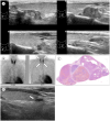
Fig. 3
Fig. 3. Solitary adenoma at the left inferior parathyroid gland in a 63-year-old female with primary hyperparathyroidism.
A. Transverse, sagittal and color doppler US images show a markedly hypoechoic nodule (arrow) with residual parathyroid sign (arrowhead) and polar vessel sign at the posterior aspect of the left thyroid lower pole, and 99mTc-MIBI scan shows a lesion (arrow) with residual uptake of the tracer in the region of the left thyroid gland. B. Histopathology reveals a well-circumscribed mass with thin fibrous capsule and reduced stromal adipocytes (hematoxylin and eosin stain) attached to the thyroid tissue (arrow). Compressed nonneoplastic parathyroid tissue (arrowhead) containing 40%–50% fat component is seen at the edge, possibly corresponding to the residual parathyroid sign at US.
Fig. 4
Fig. 4. Solitary adenoma with misinterpreted ultrasonography in an 82-year-old female with primary hyperparathyroidism.
A. Transverse, sagittal and color doppler US images show hypoechoic masses (arrows) with peripheral vascularity at the posterior aspect of the both thyroid upper poles, which were initially thought to be parathyroid lesions. B. 99mTc-MIBI scan shows a lesion (arrow) with residual uptake of the tracer in the region of the left thyroid gland; 11C-methionine PET/CT also shows hot uptake (arrow) at the posterosuperior aspect to the left thyroid upper pole, but no uptake (arrowhead) at the right upper pole. The right lesion is a thyroid lesion.
Fig. 5
Fig. 5. Parathyroid carcinoma at the right superior parathyroid gland in a 64-year-old female with primary hyperparathyroidism.
A. Transverse, sagittal and color doppler US images show a hypoechoic mass (arrowhead) with irregular margin, internal echogenic calcification and increased vascularity at the posterior aspect of the right thyroid mid-pole, 99mTc-MIBI scan shows a lesion (arrow) with prolonged uptake of the tracer in the region of the right thyroid gland, and 99mTc-MIBI SPECT/CT shows hot uptake and calcification in the lesion (arrow). B. Histopathology shows a poorly circumscribed, multilobular mass with invasion of thyroid gland (arrow) (hematoxylin and eosin stain) and vascular invasion (arrowhead) on immunohistochemical staining for CD31.
Fig. 6
Fig. 6. Solitary adenoma with false-negative US, MIBI scan and SPECT/CT in a 69-year-old female with primary hyperparathyroidism.
A. US images show a partially cystic isoechoic mass (arrow) with scanty flow signals at the inferior aspect of the left thyroid gland; 99mTc-MIBI scan and 99mTc SPECT/CT are negative with no tracer uptake, whereas non-enhanced CT shows a high attenuated mass (arrow) in the left thyroid gland. B. 11C-methionine PET/CT shows a nodular lesion (arrowhead) with hot uptake at the posterior aspect of the left thyroid gland upper pole, and US images reveal an oval marked hypoechoic mass (crosses) with a feeding vessel at the posterosuperior aspect to the left thyroid upper pole.
Fig. 7
Fig. 7. Multiple hyperplasia of all four parathyroid glands in a 46-year-old male with tertiary hyperparathyroidism: sagittal US images (A) showing multiple masses (crosses) of variable size and mixed echogenicity in the posterior and inferior aspects of both thyroid glands, while 99mTc-MIBI scan (B) shows all four parathyroid lesions (arrows) with residual tracer uptake in both thyroid glands, histopathology (C) shows nodular growth without a definite rim of compressed normal parathyroid tissue (hematoxylin and eosin staining), and follow-up sagittal US image (D) after one year shows the preserved remaining left inferior parathyroid gland (arrow) at the inferior aspect of the left thyroid gland.
Fig. 8
Fig. 8. Parathyroid cyst at the left inferior parathyroid gland in a 54-year-old female who presented with anterior neck swelling: US images show an anechoic cystic mass (cross) at the inferior aspect of the left thyroid gland, and neck CT images performed at other hospitals show a cystic mass (arrows) in the left infrathyroidal and paratracheal areas.

Similar articles

References

    1. Johnson NA, Carty SE, Tublin ME. Parathyroid imaging. Radiol Clin North Am. 2011;49:489–509. vi. - PubMed
    1. Walker MD, Silverberg SJ. Primary hyperparathyroidism. Nat Rev Endocrinol. 2018;14:115–125. - PMC - PubMed
    1. Wilhelm SM, Wang TS, Ruan DT, Lee JA, Asa SL, Duh QY, et al. The American Association of Endocrine Surgeons guidelines for definitive management of primary hyperparathyroidism. JAMA Surg. 2016;151:959–968. - PubMed
    1. Wang C. The anatomic basis of parathyroid surgery. Ann Surg. 1976;183:271–275. - PMC - PubMed
    1. Taterra D, Wong LM, Vikse J, Sanna B, Pękala P, Walocha J, et al. The prevalence and anatomy of parathyroid glands: a meta-analysis with implications for parathyroid surgery. Langenbecks Arch Surg. 2019;404:63–70. - PMC - PubMed