

Extrapulmonary Tuberculosis Presenting As Stage V Bilateral Cervical Lymphadenitis With Cortical Cerebral Watershed Infarct Along With Maxillary and Sphenoid Sinusitis
- PMID: 38618370
- PMCID: PMC11009437
- DOI: 10.7759/cureus.56055
Extrapulmonary Tuberculosis Presenting As Stage V Bilateral Cervical Lymphadenitis With Cortical Cerebral Watershed Infarct Along With Maxillary and Sphenoid Sinusitis
Abstract
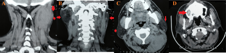
Extrapulmonary tuberculosis is an infrequently reported condition. However, in endemic settings, it contributes to a significant number of cases. The most common site of extrapulmonary tuberculosis is the lymph nodes. Herein, an exceedingly rare case of extrapulmonary tuberculosis presenting as bilateral cervical lymphadenitis with external cerebral watershed infarct along with sphenoid and maxillary sinusitis in an Indian male is presented. A detailed literature search revealed that a case with all these clinical conditions together has never been reported to date. A diagnostic workup supported by radiometric investigations helped in the diagnosis, and timely management was initiated.
Keywords: antituberculous chemotherapy; cervical lymphadenitis; extrapulmonary; lymphadenitis; maxillary sinusitis; scrofula; sphenoid sinusitis; tuberculosis; watershed infarct.
Copyright © 2024, Yadav et al.
Conflict of interest statement
The authors have declared that no competing interests exist.
Figures


References
-
- Watershed cerebral infarction - a case report. Hanvitha M, Kumar EAA. https://www.iaimjournal.com/storage/2020/11/iaim_2020_0711_07.pdf Int Arch Integr Med. 2020;7:44–55.
-
- Border zone infarcts: pathophysiologic and imaging characteristics. Mangla R, Kolar B, Almast J, Ekholm SE. Radiographics. 2011;31:1201–1214. - PubMed
Publication types
LinkOut - more resources
Full Text Sources
Research Materials