

Rotator Cuff Tears: Correlation Between Clinical Examination, Magnetic Resonance Imaging and Arthroscopy
- PMID: 38618461
- PMCID: PMC11009554
- DOI: 10.7759/cureus.56065
Rotator Cuff Tears: Correlation Between Clinical Examination, Magnetic Resonance Imaging and Arthroscopy
Abstract
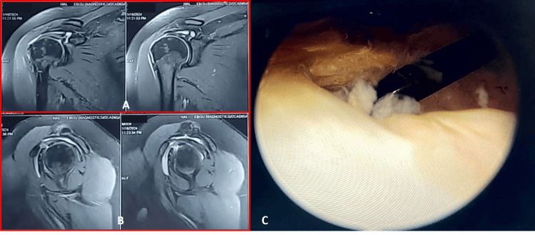
Background Arthroscopy in diagnosing a rotator cuff injury has surgical and anaesthesia-related risks. Magnetic resonance imaging (MRI), a non-invasive procedure, is expensive, and lacks dynamic components making it less favourable. Clinical examination narrows the diagnosis, but lacks diagnostic accuracy due to overlap of clinical signs and symptoms. We aimed to determine the diagnostic accuracy of clinical examination and MRI in rotator cuff tears by correlating it with arthroscopy. Methods This prospective, cross-sectional validation study included patients (N=28) with shoulder pain with clinical characteristics suggestive of rotator cuff tears. Clinical diagnoses and MRI were done preoperatively, following which each patient underwent arthroscopic surgery. Shoulder arthroscopy findings were correlated with those of clinical examination and MRI. Results The mean age of patients was 50.21±9.66 years, with 60.71% being males. Clinical examination was 100% sensitive and 73.8% specific for detecting rotator cuff tears. MRI was 92.85% sensitive and 98.8% specific in detecting rotator cuff tears. Shoulder MRI demonstrated a higher agreement with arthroscopy than clinical results for subscapularis, supraspinatus, infraspinatus, teres, and biceps tendon appearance. Conclusion MRI results in identifying rotator cuff pathologies are comparable with arthroscopy. Clinical examination findings are variable due to an examiner's bias and therefore its diagnostic scope is limited. However, clinical examination with MRI together might accurately identify the rotator cuff injury.
Keywords: arthroscopy; magnetic resonance imaging; physical examination; rotator cuff; shoulder pain.
Copyright © 2024, Gowda et al.
Conflict of interest statement
The authors have declared that no competing interests exist.
Figures
Similar articles
-
Diagnostic performance of a 3D double-echo steady-state sequence at 3 T using radial reformats for detecting and grading rotator cuff tears: a pilot diagnostic accuracy study with magnetic resonance imaging and arthroscopic correlation.Acta Radiol. 2023 Oct;64(10):2768-2776. doi: 10.1177/02841851231190359. Epub 2023 Aug 21. Acta Radiol. 2023. PMID: 37603569
-
Needle Diagnostic Arthroscopy and Magnetic Resonance Imaging of the Shoulder Have Comparable Accuracy With Surgical Arthroscopy: A Prospective Clinical Trial.Arthroscopy. 2021 Jul;37(7):2090-2098. doi: 10.1016/j.arthro.2021.03.006. Epub 2021 Mar 30. Arthroscopy. 2021. PMID: 33798653
-
The frequency of subscapularis tears in arthroscopic rotator cuff repairs: A retrospective study comparing magnetic resonance imaging and arthroscopic findings.Int J Shoulder Surg. 2011 Oct;5(4):90-4. doi: 10.4103/0973-6042.91000. Int J Shoulder Surg. 2011. PMID: 22223958 Free PMC article.
-
Surgery for rotator cuff tears.Cochrane Database Syst Rev. 2019 Dec 9;12(12):CD013502. doi: 10.1002/14651858.CD013502. Cochrane Database Syst Rev. 2019. PMID: 31813166 Free PMC article.
-
Diagnostic Accuracy of Ultrasonography for Rotator Cuff Tears: A Systematic Review and Meta-analysis.Orthop J Sports Med. 2021 Oct 11;9(10):23259671211035106. doi: 10.1177/23259671211035106. eCollection 2021 Oct. Orthop J Sports Med. 2021. PMID: 34660823 Free PMC article. Review.
Cited by
-
Correlation of Preoperative MRI and Shoulder-Specific Tests With Intraoperative Findings in Rotator Cuff Tears.Cureus. 2025 Mar 21;17(3):e80951. doi: 10.7759/cureus.80951. eCollection 2025 Mar. Cureus. 2025. PMID: 40255767 Free PMC article.
References
-
- A systematic review and pooled analysis of the prevalence of rotator cuff disease with increasing age. Teunis T, Lubberts B, Reilly BT, Ring D. J Shoulder Elbow Surg. 2014;23:1913–1921. - PubMed
-
- The effectiveness of diagnostic tests for the assessment of shoulder pain due to soft tissue disorders: a systematic review. Dinnes J, Loveman E, McIntyre L, Waugh N. Health Technol Assess. 2003;7 - PubMed
-
- Arthroscopic evaluation of ultrasonography and magnetic resonance imaging for diagnosis of rotator cuff tear. Frei R, Chladek P, Trc T, Kopecny Z, Kautzner J. https://europepmc.org/article/med/18449121. Ortop Traumatol Rehabil. 2008;10:111–114. - PubMed
LinkOut - more resources
Full Text Sources
Medical