. 2024 Jul;61(4):550-561.
doi: 10.1177/03009858241244849. Epub 2024 Apr 15.

Tissue and cellular tropism of Eptesicus fuscus gammaherpesvirus in big brown bats, potential role of pulmonary intravascular macrophages

Affiliations

Tissue and cellular tropism of Eptesicus fuscus gammaherpesvirus in big brown bats, potential role of pulmonary intravascular macrophages

Ursula G Perdrizet et al. Vet Pathol. 2024 Jul.

Abstract

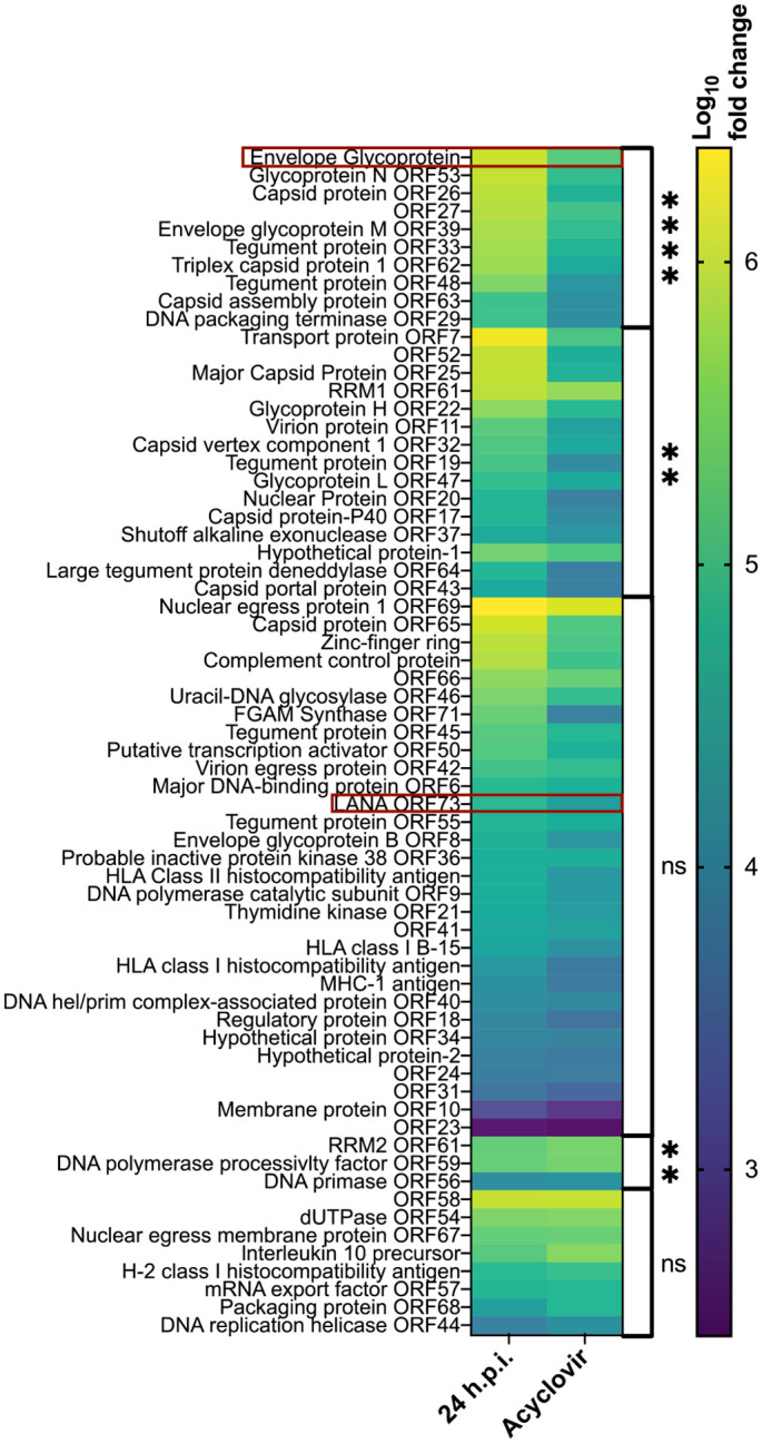
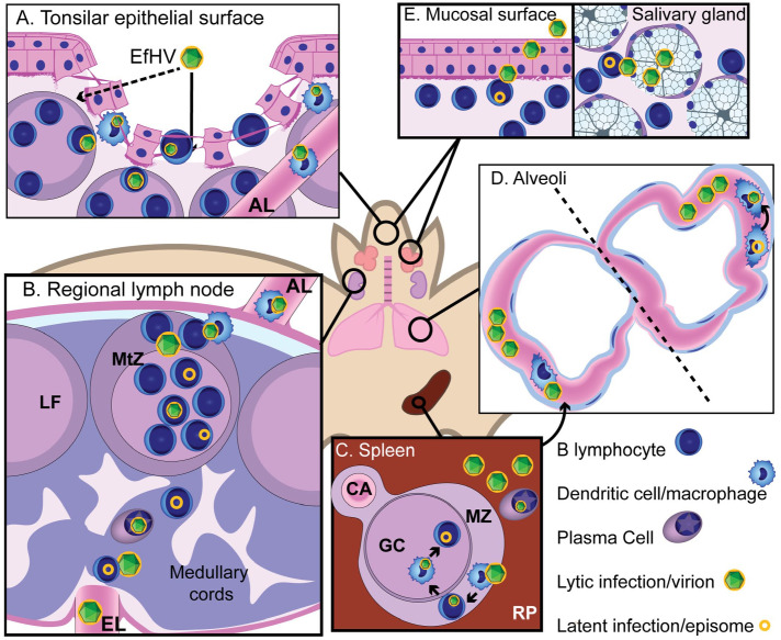
Gammaherpesviruses (γHVs) are recognized as important pathogens in humans but their relationship with other animal hosts, especially wildlife species, is less well characterized. Our objectives were to examine natural Eptesicus fuscus gammaherpesvirus (EfHV) infections in their host, the big brown bat (Eptesicus fuscus), and determine whether infection is associated with disease. In tissue samples from 132 individual big brown bats, EfHV DNA was detected by polymerase chain reaction in 41 bats. Tissues from 59 of these cases, including 17 from bats with detectable EfHV genomes, were analyzed. An EfHV isolate was obtained from one of the cases, and electron micrographs and whole genome sequencing were used to confirm that this was a unique isolate of EfHV. Although several bats exhibited various lesions, we did not establish EfHV infection as a cause. Latent infection, defined as RNAScope probe binding to viral latency-associated nuclear antigen in the absence of viral envelope glycoprotein probe binding, was found within cells of the lymphoid tissues. These cells also had colocalization of the B-cell probe targeting CD20 mRNA. Probe binding for both latency-associated nuclear antigen and a viral glycoprotein was observed in individual cells dispersed throughout the alveolar capillaries of the lung, which had characteristics of pulmonary intravascular macrophages. Cells with a similar distribution in bat lungs expressed major histocompatibility class II, a marker for antigen presenting cells, and the existence of pulmonary intravascular macrophages in bats was confirmed with transmission electron microscopy. The importance of this cell type in γHVs infections warrants further investigation.

Keywords: Eptesicus fuscus; Patagivirus vespertilionid gammaherpesvirus 3; bats; chiroptera; gammaherpesvirus; pulmonary intravascular macrophage.


Conflict of interest statement

Declaration of Conflicting InterestsThe author(s) declared no potential conflicts of interest with respect to the research, authorship, and/or publication of this article.

Figures

Figure 1.
Figure 1.
Epithelial dysplasia and intranuclear inclusion bodies, trachea, big brown bat. EfHV/SK/02/2020 was isolated from this bat. Case 126. Hematoxylin and Eosin (HE). Lower right inset: higher magnification demonstrating intranuclear inclusion bodies (arrows). HE. Upper left inset: serial section of the trachea negative for Eptesicus fuscus gammaherpesvirus gp52 and LANA. gp52 and LANA in situ hybridization.
Figure 2.
Figure 2.
Heatmap of the log10 fold change of Eptesicus fuscus gammaherpesvirus (EfHV) genes relative to 4°C infection control. Gene expression was measured at 24 hours post-infection (h.p.i.) with or without acyclovir. They are ordered by gene expression for those that were inhibited by acyclovir at the top and those that were potentiated at the bottom, followed by a P-value comparing the 2 groups, then by fold change at 24 h.p.i. (highest to lowest). Red boxes indicate target genes for in situ hybridization probes. Statistical significance of the comparisons between the acyclovir treated and untreated cells at 24 h.p.i. are indicated to the right of the graph (ns, not significant; ****P < .0001; and **P < .01). RRM1, ribonucleoside-diphosphate reductase large subunit; RRM2, ribonucleoside-diphosphate reductase small subunit; LANA, latency-associated nuclear antigen.
Figure 3.
Figure 3.
Eptesicus fuscus gammaherpesvirus (EfHV) infection in cell culture and big brown bats. (a, b) Eptesicus fuscus kidney cell line 3b (EfK3b) cells. LANA and gp52 in situ hybridization (ISH). (a) No probe binding was observed in uninfected cells. (b) Colocalization of EfHV LANA (red) and gp52 (teal) was observed in cells infected with EfHV. (c, d) Splenic white pulp from big brown bat 123. LANA and CD20 ISH. (c) Colocalization of LANA (red) and CD20 (teal) probes. (d) Higher magnification of the area surrounded by a dashed rectangle in (c). (e–i) Lung, big brown bat, case 129. (e) Hematoxylin and eosin. (f) Serial section demonstrating intense probe binding for LANA (red) and gp52 (teal). LANA and gp52 ISH. (g) Higher magnification showing colocalization of these probes in cells within alveolar capillaries. LANA and gp52 ISH. (h) Serial section of lung in (e). Cells with major histocompatibility (MHC) class II immunolabeling (brown), as indicated by arrows, displayed similar pattern to the ISH in (f) and (g). MHC class II immunohistochemistry. (i) Electron photomicrograph of lung tissue from the same blocks as shown in (e–h) demonstrating a pulmonary intravascular macrophage outlined by a dashed line. RBC, red blood cell. Scale bar 2 µm.
Figure 4.
Figure 4.
Proposed pathogenesis for Eptesicus fuscus gammaherpesvirus (EfHV). (A) New host contacts saliva containing virus primed for infecting B lymphocytes and dendritic cells (DC). Exposure to virions could occur through gaps in the epithelium of the tonsillar crypts (arrow) or transcytosis (dashed arrow). Infection of B lymphocytes and DCs occur. (B) DCs migrate to regional lymph node via the afferent lymphatics and infect follicular B cells and eventually memory B cells in the germinal center. Further systemic spread occurs by reactivation of latently infected B cells during differentiation into plasma cells. Dissemination is possibly by infected B cells or cell-free viremia. (C) The primary site of latency, the splenic white pulp, is colonized by infection of marginal zone macrophages then marginal zone B cells, follicular dendritic cells, and finally germinal center B cells through cell to cell spread. Differentiation of splenic B cells into plasma cells results in reactivation of gammaherpesvirus infection. (D) Pulmonary intravascular macrophages (PIMs) could be involved in the viral cycle in a number of ways: phagocytosing virus or virus-infected cells following splenic reactivation, becoming infected during phagocytosis, or being directly involved in productive infection, either via latently infected migratory monocytes, which then differentiate into PIMs, or resident PIMs that reactivate virus. (E) Virus is shed from epithelial or glandular cells that are infected by B cells. B cells are infected by migrating macrophages or cell-free virus produced from a myeloid cell, or they reactivate virus following latent infection. Abbreviations: AL, afferent lymphatic; CA, central arteriole; EfHV, Eptesicus fuscus gammaherpesvirus; EL, efferent lymphatic; GC, germinal center; LF, lymphoid follicle; MtZ, mantle zone; MZ, marginal zone; RP, red pulp.

Similar articles

Cited by

References

    1. Ackermann M. Pathogenesis of gammaherpesvirus infections. Vet Microbiol. 2006;113(3–4):211–222. - PubMed
    1. Banerjee A, Rapin N, Miller M, et al. Generation and characterization of Eptesicus fuscus (big brown bat) kidney cell lines immortalized using the Myotis polyomavirus large T-antigen. J Virol Methods. 2016;237:166–173. - PMC - PubMed
    1. Blank H, Davis C, Collins C. Electron microscopy for the diagnosis of cutaneous viral infections. Br J Dermatol. 1970;83(1):69–80. - PubMed
    1. Bolger AM, Lohse M, Usadel B. Trimmomatic: a flexible trimmer for Illumina sequence data. Bioinforma Oxf Engl. 2014;30(15):2114–2120. - PMC - PubMed
    1. Bordet E, Maisonnasse P, Renson P, et al. Porcine alveolar macrophage-like cells are pro-inflammatory pulmonary intravascular macrophages that produce large titers of porcine reproductive and respiratory syndrome virus. Sci Rep. 2018;8:10172. - PMC - PubMed

Publication types

LinkOut - more resources