. 2024 Mar 29:(205):10.3791/66707.
doi: 10.3791/66707.

Light-Sheet Imaging to Reveal Cardiac Structure in Rodent Hearts

Affiliations

Light-Sheet Imaging to Reveal Cardiac Structure in Rodent Hearts

Milad Almasian et al. J Vis Exp. .

Abstract

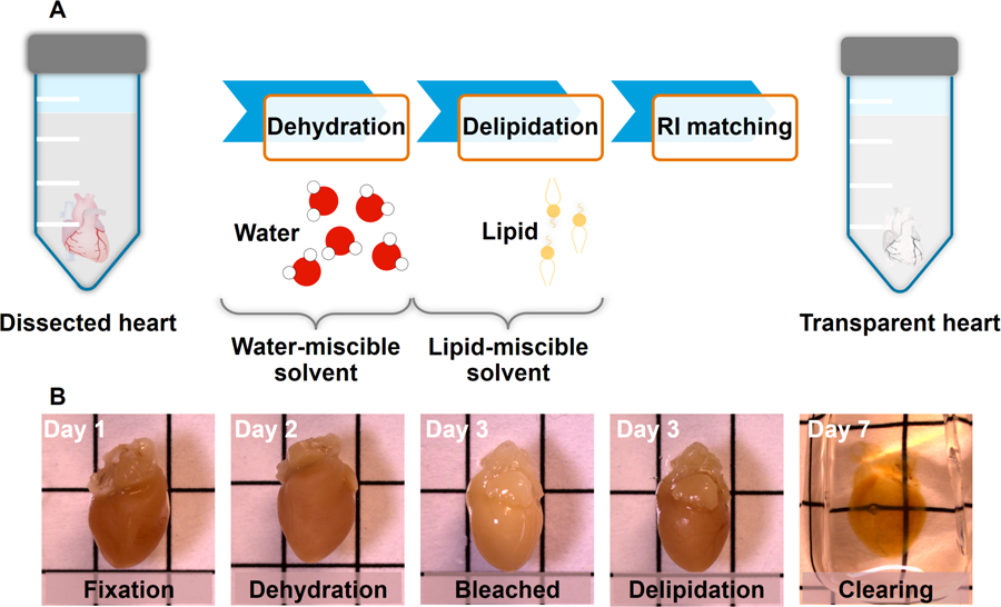
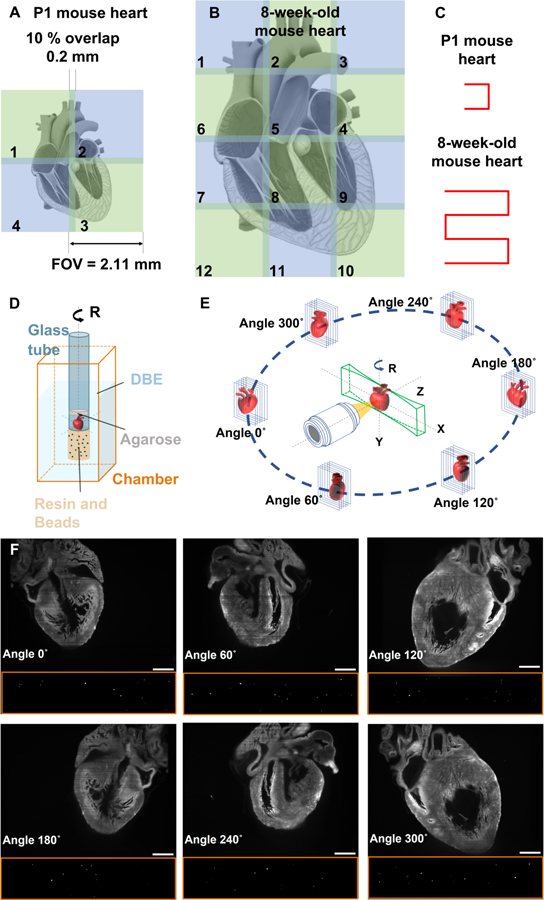
Light-sheet microscopy (LSM) plays a pivotal role in comprehending the intricate three-dimensional (3D) structure of the heart, providing crucial insights into fundamental cardiac physiology and pathologic responses. We hereby delve into the development and implementation of the LSM technique to elucidate the micro-architecture of the heart in mouse models. The methodology integrates a customized LSM system with tissue clearing techniques, mitigating light scattering within cardiac tissues for volumetric imaging. The combination of conventional LSM with image stitching and multiview deconvolution approaches allows for the capture of the entire heart. To address the inherent trade-off between axial resolution and field of view (FOV), we further introduce an axially swept light-sheet microscopy (ASLM) method to minimize out-of-focus light and uniformly illuminate the heart across the propagation direction. In the meanwhile, tissue clearing methods such as iDISCO enhance light penetration, facilitating the visualization of deep structures and ensuring a comprehensive examination of the myocardium throughout the entire heart. The combination of the proposed LSM and tissue clearing methods presents a promising platform for researchers in resolving cardiac structures in rodent hearts, holding great potential for the understanding of cardiac morphogenesis and remodeling.


Conflict of interest statement

DISCLOSURES:

The authors have no conflict of interest to disclose.

Figures

Figure 1:
Figure 1:. Tissue clearing process.
(A) The hydrophobic method involves tissue dehydration, lipid extraction, and refractive index matching using dibenzyl ether (DBE) as the organic solvent. (B) Submerge the heart in a mixture of 4% formaldehyde (PFA) solution and dehydrate it with a gradient of methanol and deionized (DI) water mixtures. Bleach the heart using 5% H2O2 in methanol at 4 °C. Delipidate the heart with dichloromethane (DCM) and methanol solution until the sample sinks to the bottom of the tube. Lastly, incubate the heart in DBE in order to obtain the desired refractive index. Grid lines 5 mm.
Figure 2:
Figure 2:. Schematic of axially swept light-sheet microscopy.
(A) Customized 3D printed chamber, (B) chamber magnet, (C) clamp holder, and (D) ASLM system. Abbreviations: CL: cylindrical lens; ETL: electronic tunable lens; IL: illumination lens; DL: detection lens; FW: filter wheel; TL: tube lens. This figure has been modified from.
Figure 3:
Figure 3:. Image stitching and multiview deconvolution.
(A-B) Image stitching is used to cover the entire mouse hearts, with each tile having a 10% FOV overlap with its adjacent tiles. (C) Movement pattern of the mouse heart for image stitching. (D) Fluorescent beads are affixed to the end of the glass tube to facilitate image registration, while the heart is positioned inside the glass tube containing DBE, enabling concurrent imaging of both the mouse heart and fluorescent beads. (E) Multiview deconvolution involves the acquisition of images from six distinct perspectives, each gathering images from unique directions. (F) Raw data of P1 mouse heart and beads at different angles of 0°, 60°, 120°, 180°, 240°, and 300°. Scale bars: 500 μm. This figure has been modified from.
Figure 4:
Figure 4:. Block diagram of ASLM system.
Utilizing DAQ card for synchronizing ETL and sCMOS camera. The housing of the ETL driver has been opened for soldering of signal and ground wires to the PCB.
Figure 5:
Figure 5:. LabView control panel.
LabVIEW control panel for generating triggers to synchronize ETL and activated pixel of sCMOS camera.
Figure 6:
Figure 6:. Synchronizing the focus of light-sheet with activated pixels.
(A) The identification of the starting and ending points of the light-sheet scanning range is achieved by configuring the appropriate voltage for the ETL trigger. (B) The synchronous result of fluorescent beads is evident along the diagonal of the image. (C-F) The non-uniform spatial resolution across the entire FOV arises from the asynchronous operation of the ETL with the sCMOS camera. The ETL initiates scanning (C) later or (D) earlier than the activated pixel. (E-F) The focal area is not parallel to the diagonal of the image, indicating an incompatibility between scanning and activation speeds. ETL scans (E) faster or (F) slower than the sweeping of active pixels in sCMOS. Scale bar: 200 µm.
Figure 7:
Figure 7:. Troubleshooting of ASLM.
(A) The light-sheet is placed exactly at the focal distance of the detection lens. The ETL scans the focus of the light-sheet along the propagation direction while synchronizing with the activated pixel of the sCMOS sensor. (B) XY view of fluorescent beads when the ETL is correctly aligned and synchronized. (C-D) Results of fluorescent beads on the focal plane and out-of-focus in cross-sectional images, indicating the misalignment between the ETL scanning and laser propagation. Scale bars: 200 µm and 10 µm in the inset.
Figure 8:
Figure 8:. Light-sheet imaging of mouse hearts.
(A) Volume-rendered image of the multiview deconvolution P1 mouse heart highlighting the trabeculation within the ventricular cavity. (B) Cross-sectional images of the 8-week-old mouse heart generated by conventional LSM (left) and ASLM (right). Images are presented as raw data. (C) Combination of Image stitching and ASLM for imaging an 8-week-old mouse heart, comprising 12 tiles arranged in 3 tiles horizontally and 4 tiles vertically. Scale bar: 500 µm. Abbreviations: LV: left ventricle, LA: left atrium, RV: right ventricle, and RA: right atrium. This figure has been modified from.

References

    1. Sadek H & Olson EN Toward the goal of human heart regeneration. Cell Stem Cell 26, 7–16 (2020). - PMC - PubMed
    1. Porrello ER et al. Transient regenerative potential of the neonatal mouse heart. Science 331, 1078–1080 (2011). - PMC - PubMed
    1. Stelzer EHKK et al. Light sheet fluorescence microscopy. Nat Rev Methods Prim 1, 73 (2021).
    1. Girkin JM & Carvalho MT The light-sheet microscopy revolution. J Opt 20, 053002 (2018).
    1. Power RM & Huisken J A guide to light-sheet fluorescence microscopy for multiscale imaging. Nat Methods 14, 360–373 (2017). - PubMed

Publication types

LinkOut - more resources