. 2024 Jun 1;212(11):1706-1713.
doi: 10.4049/jimmunol.2300649.

Iron Is Critical for Mucosal-Associated Invariant T Cell Metabolism and Effector Functions

Affiliations

Iron Is Critical for Mucosal-Associated Invariant T Cell Metabolism and Effector Functions

Eimear K Ryan et al. J Immunol. .

Abstract

Mucosal-Associated Invariant T (MAIT) cells are a population of innate T cells that play a critical role in host protection against bacterial and viral pathogens. Upon activation, MAIT cells can rapidly respond via both TCR-dependent and -independent mechanisms, resulting in robust cytokine production. The metabolic and nutritional requirements for optimal MAIT cell effector responses are still emerging. Iron is an important micronutrient and is essential for cellular fitness, in particular cellular metabolism. Iron is also critical for many pathogenic microbes, including those that activate MAIT cells. However, iron has not been investigated with respect to MAIT cell metabolic or functional responses. In this study, we show that human MAIT cells require exogenous iron, transported via CD71 for optimal metabolic activity in MAIT cells, including their production of ATP. We demonstrate that restricting iron availability by either chelating environmental iron or blocking CD71 on MAIT cells results in impaired cytokine production and proliferation. These data collectively highlight the importance of a CD71-iron axis for human MAIT cell metabolism and functionality, an axis that may have implications in conditions where iron availability is limited.


Conflict of interest statement

The authors have no financial conflicts of interest.

Figures
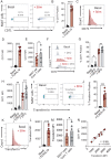
FIGURE 1.
FIGURE 1.
MAIT cells increase CD71 and transferrin uptake upon activation. (AD) Flow cytometric dot plots, scatterplots, and representative flow cytometric histograms showing CD71 expression in MAIT cells, basal or stimulated for 18 h with anti-CD3/CD28 TCR beads and IL-18. (E) Scatterplot comparing CD71 expression on MAIT cells and conventional T cells (non-MAIT CD3+ cells) after 18-h stimulation with anti-CD3/CD28 TCR beads and IL-18. (FH) Flow cytometric dot plots, scatterplots, and representative flow cytometric histograms showing CD71 expression in MAIT cells, basal or stimulated for 18 h with anti-CD3/CD28 TCR beads and IL-18, in the absence or presence of the iron chelator, DFO. (IL) Flow cytometric dot plots, scatterplots, and representative flow cytometric histograms showing transferrin content in MAIT cells, basal or stimulated for 18 h with anti-CD3/CD28 TCR beads and IL-18. (M) Scatterplot comparing transferrin uptake by MAIT cells and conventional T cells (non-MAIT CD3+ cells) after 18-h stimulation with anti-CD3/CD28 TCR beads and IL-18. (N) Correlation plot showing the relationship between CD71 expression and transferrin uptake in activated MAIT cells (18 h with anti-CD3/CD28 TCR beads and IL-18). *p < 0.05, **p < 0.01, ***p < 0.001, ****p < 0.0001.
FIGURE 2.
FIGURE 2.
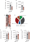
MAIT cells store and use iron. (A and B) Scatterplot showing protein copy number of the H and L chain subunits of ferritin in IL-2 expanded MAIT cells, basal or stimulated for 18 h with anti-CD3/CD28 TCR beads and IL-18. (C) Heatmap of iron-related protein content in IL-2 expanded MAIT cells, basal or stimulated for 18 h with anti-CD3/CD28 TCR beads and IL-18. (D) Pie graph showing proportional pathway analysis based on iron-related protein content in MAIT cells (analysis performed with Panther). (EG) Estimated iron, heme, and iron-sulfur cluster atoms extrapolated from proteome of IL-2 expanded MAIT cells, basal or stimulated for 18 h with anti-CD3/CD28 TCR beads and IL-18. *p < 0.05.
FIGURE 3.
FIGURE 3.
Iron restriction alters MAIT cell metabolism. (AD) Representative flow cytometric histograms and scatterplots showing puromycin incorporation in MAIT cells, basal or stimulated for 18 h with anti-CD3/CD28 TCR beads and IL-18, in the absence or presence of the iron chelator, DFO. (E) Scatterplot showing percentage dependency on glucose metabolism in MAIT cells stimulated for 18 h with anti-CD3/CD28 TCR beads and IL-18, in the presence or absence of DFO. (F) Scatterplot showing percentage dependency on fatty acid oxidation and amino acid oxidation in MAIT cells stimulated for 18 h with anti-CD3/CD28 TCR beads and IL-18, in the presence or absence of DFO. (G and H) Scatterplots showing the MFI of MitoTracker Green and MitoTracker Deep Red, depicting mitochondrial mass and mitochondrial membrane potential, respectively, in MAIT cells stimulated with anti-CD3/CD28 TCR beads and IL-18 for 18 h, in the presence or absence of DFO. (I and J) Representative Seahorse trace and scatterplot showing oxygen consumption rates (OCRs) in MAIT cells stimulated with anti-CD3/CD28 TCR beads and IL-18 for 24 h, in the presence or absence of DFO. (K) Scatterplot showing the mitochondrial capacity (after FCCP treatment) in MAIT cells stimulated with anti-CD3/CD28 TCR beads and IL-18 for 24 h, in the presence or absence of DFO. (L) Scatterplot showing the impact of an anti-CD71 mAb with blocking activity on ATP production in MAIT cells stimulated with anti-CD3/CD28 TCR beads and IL-18 for 18 h. *p < 0.05, **p < 0.01, ***p < 0.001, ****p < 0.0001.
FIGURE 4.
FIGURE 4.
MAIT cells require iron for their functional responses. (A) Representative flow cytometry histogram and scatterplot showing IFN-γ levels (MFI) in MAIT cells stimulated with anti-CD3/CD28 TCR beads and IL-18 for 18 h, in the absence or presence of the iron chelator, DFO, measured by flow cytometry. (BD) Scatterplots showing IFN-γ, IL-17, and IL-2 secreted protein levels (normalized to assay) in IL-2 expanded MAIT cells stimulated with anti-CD3/CD28 TCR beads and IL-18 for 18 h, in the absence or presence of DFO, as measured by ELISA. (E and F) Scatterplots showing the impact of an anti-CD71 mAb with blocking activity on IFN-γ and IL-26 secreted protein levels (normalized to assay) in IL-2 expanded MAIT cells stimulated with anti-CD3/CD28 TCR beads and IL-18 for 18 h, as measured by ELISA. (G and H) Line graph showing IFN-γ or granzyme B levels (percentage of parent population) in MAIT cells cocultured at 1:1 ratio with THP-1 cells alone or prepulsed with fixed E. coli (DH5α) for 18 h, in the absence or presence of the anti-CD71 mAb with blocking activity, as measured by flow cytometry. (I) Scatterplot showing the impact of the ATP synthase inhibitor, oligomycin, on MAIT cell proliferative capacity (during a 5-d IL-2–mediated MAIT cell expansion culture), whereby the MFI of the intracellular dye CellTrace Violet decreases with each cycle of cell division. (J and K) Scatterplots showing the impact of long-term iron depletion via DFO and alternative iron repletion via Iron(II) sulfate heptahydrate (FeSO4 • 7H2O) on MAIT cell proliferation and viability during a 5-d IL-2–mediated MAIT cell expansion culture. *p < 0.05, **p < 0.01, ***p < 0.001.

References

    1. Godfrey, D. I., Koay H. F., McCluskey J., Gherardin N. A.. 2019. The biology and functional importance of MAIT cells. Nat. Immunol. 20: 1110–1128. - PubMed
    1. Le Bourhis, L., Martin E., Péguillet I., Guihot A., Froux N., Coré M., Lévy E., Dusseaux M., Meyssonnier V., Premel V., et al. . 2010. Antimicrobial activity of mucosal-associated invariant T cells. Nat. Immunol. 11: 701–708. - PubMed
    1. van Wilgenburg, B., Scherwitzl I., Hutchinson E. C., Leng T., Kurioka A., Kulicke C., de Lara C., Cole S., Vasanawathana S., Limpitikul W., et al. . 2016. MAIT cells are activated during human viral infections. Nat. Commun. 7: 11653. - PMC - PubMed
    1. Treiner, E., Duban L., Bahram S., Radosavljevic M., Wanner V., Tilloy F., Affaticati P., Gilfillan S., Lantz O.. 2003. Selection of evolutionarily conserved mucosal-associated invariant T cells by MR1. Nature 422: 164–169. - PubMed
    1. Kjer-Nielsen, L., Patel O., Corbett A. J., Le Nours J., Meehan B., Liu L., Bhati M., Chen Z., Kostenko L., Reantragoon R., et al. . 2012. MR1 presents microbial vitamin B metabolites to MAIT cells. Nature 491: 717–723. - PubMed

MeSH terms