. 2024 May 1;151(9):dev202386.
doi: 10.1242/dev.202386. Epub 2024 May 9.

Piezo1 and Piezo2 collectively regulate jawbone development

Affiliations

Piezo1 and Piezo2 collectively regulate jawbone development

Xuguang Nie et al. Development. .

Abstract

Piezo1 and Piezo2 are recently reported mechanosensory ion channels that transduce mechanical stimuli from the environment into intracellular biochemical signals in various tissues and organ systems. Here, we show that Piezo1 and Piezo2 display a robust expression during jawbone development. Deletion of Piezo1 in neural crest cells causes jawbone malformations in a small but significant number of mice. We further demonstrate that disruption of Piezo1 and Piezo2 in neural crest cells causes more striking defects in jawbone development than any single knockout, suggesting essential but partially redundant roles of Piezo1 and Piezo2. In addition, we observe defects in other neural crest derivatives such as malformation of the vascular smooth muscle in double knockout mice. Moreover, TUNEL examinations reveal excessive cell death in osteogenic cells of the maxillary and mandibular arches of the double knockout mice, suggesting that Piezo1 and Piezo2 together regulate cell survival during jawbone development. We further demonstrate that Yoda1, a Piezo1 agonist, promotes mineralization in the mandibular arches. Altogether, these data firmly establish that Piezo channels play important roles in regulating jawbone formation and maintenance.

Keywords: Jawbone; Mouse; Neural crest cell; Piezo1; Piezo2.


Conflict of interest statement

Competing interests The authors declare no competing or financial interests.

Figures
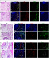
Fig. 1.
Fig. 1.
Piezo1 and Piezo2 expression during jawbone development. (A-D) Piezo1 and Alp co-localization in the mandibular arches, with a histological image illustrating the morphology for each panel (A,C). Dashed lines outline the bone-forming zone. DAPI indicates nuclei. (E-H) Piezo1 and Piezo2 colocalization in the mandibular arches, with a histological image illustrating the morphology for each panel (E,G). Dashed lines outline the areas of mineralization. (I-L) Piezo1 and Alp co-staining in the mandibles of control and Piezo1 CKO mice, with a histological image illustrating the morphology for each panel (I,K). Note, the absence of Piezo1 in the periosteum (arrows) and periodontal ligament of the CKO mouse (J). Dashed line indicates alveolar bone. N=3 for each stage. Avb, alveolar bone; dm, dental mesenchyme; eo, enamel organ; gv, gingival; mc, Meckel's cartilage; md, mandible; ms, masseter; pdl, periodontal ligament; tb, tooth bud; th, tooth; wk, week. Scale bars: 50 µm (A-D); 100 µm [E-L (bar in J applies to L)].
Fig. 2.
Fig. 2.
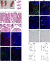
The phenotype of Piezo1 CKO mice. (A) Gross appearance of control and CKO mice at 3 weeks. (B) Alcian Blue and Alizarin Red staining of the mandibles (n=2). (C-E) H&E staining of mandibular and maxillary sections. Note that a small cleft is observed in the anterior palate of a growth-retarded mouse (asterisk). (F,G) Two-dimensional µCT images of the lower jaws (F) and quantification of lamellar bone thickness (G) at 6 weeks (n=3, bilateral analysis). (H,I) µCT (n=3) and histology of adult (12-week-old) mice (n=3). (J) TRAP staining of adult mice (n=5). (K) Alveolar bone and lamellar bone thicknesses of the mandibles measured by µCT images (n=3, bilateral analysis). (L) Quantification of TRAP+ cells (n=5). (M-O) Piezo1 and Piezo2 co-staining showing the upregulation of Piezo2 in Piezo1 CKO mice, as indicated by increased number of Piezo2+ cells in bone lining cells (n=6). Dashed lines outline the alveolar bone. Data are mean±s.d. *P<0.05; **P<0.01; ns, not significant (unpaired t-test). Avb, alveolar bone; GR, growth retardation; ic, incisor; LB, lamellar bone; M1, first molar; M2, second molar; md, mandible; MO, malocclusion; mx, maxilla; ns, nasal septum; pdl, periodontal ligament; th, tooth. Scale bars: 300 µm (C-J); 50 µm (N,O).
Fig. 3.
Fig. 3.
The phenotype of Piezo1 and Piezo2 double CKO mice. (A) Gross appearance of mice at the newborn stage. (B) Alizarin Red and Alcian Blue staining of the mandibles for each genotype (n=2). (C-F) H&E staining at the newborn stage showing vasodilation, malformed tongue muscle, attenuated bone formation and cleft palates in dCKO mice (n=3). (G,H) Immunofluorescence of Alp, showing reduced number of osteogenic cells in dCKO mice. (I,J) Col1a1 and neurofilament double staining. (K,L) β-Catenin staining reveals reduced Wnt signaling in the Alp+ cells. (M,N) Immunofluorescence of α-SMA, showing defective smooth muscle layers and vasodilation in dCKO mice. (O,P) TRAP staining reveals comparable numbers of osteoclasts in the two genotypes. (Q) Quantification of Alp+, Col1a1+, β-catenin+ and TRAP+ cells (n=6 for each). Data are mean±s.d. *P<0.05; **P<0.01; ns, not significant (unpaired t-test). dCKO, double CKO; md, mandible; NF, neurofilament; pl, palate; SMA, smooth muscle actin; th, tooth; ton, tongue; vsd, vasodilation. Images in I and N are inverted to match the structure orientation. Scale bars: 50 µm (I-L); 100 µm others.
Fig. 4.
Fig. 4.
Deletion of Piezo1 and Piezo2 causes excessive apoptosis and Yoda1 promotes mineralization in the first pharyngeal arches. (A,B) Proliferation examination by PHH3 staining at E15.5 (n=6). (C,D) TUNEL at E15.5 (n=6). (E) TUNEL and Alp double staining in postnatal day (P)0 mice (n=3). (F) Quantification of proliferation, and apoptosis at E15 and P0, revealing excessive apoptosis in the jawbones of CKO mice (n=6). (G) TUNEL for single and dCKO mice at the newborn stage (n=6). (H) Mandibular slices of the E13.5 stage in culture with and without Yoda1 for 4 days (n=6). (I) Quantification of mineralization areas, showing increased mineralization in Yoda1-treated samples. (J-L) Quantification and immunofluorescence of β-catenin in cultured mandibular slices (n=6). Dashed line indicates the bone-forming zone. Data are mean±s.d. *P<0.05; **P<0.01; ***P<0.001; ns, not significant (unpaired t-test for two groups and one-way ANOVA with post-hoc test for multiple groups). dp, dental mesenchyme; eo, enamel organ; mc, Meckel's cartilage; md, mandible; mda, mandibular arch; Phh3, Phospho-Histone H3; smg, submandibular gland; th, tooth; veh, vehicle. Scale bars: 50 µm.

References

    1. Alfaqeeh, S. A. and Tucker, A. S. (2013). The slice culture method for following development of tooth germs in explant culture. J. Vis. Exp., e50824. - PMC - PubMed
    1. Alper, S. L. (2017). Genetic diseases of PIEZO1 and PIEZO2 dysfunction. Curr. Top. Membr. 79, 97-134. 10.1016/bs.ctm.2017.01.001 - DOI - PubMed
    1. Bagriantsev, S. N., Gracheva, E. O. and Gallagher, P. G. (2014). Piezo proteins: regulators of mechanosensation and other cellular processes. J. Biol. Chem. 289, 31673-31681. 10.1074/jbc.R114.612697 - DOI - PMC - PubMed
    1. Beech, D. J. and Kalli, A. C. (2019). Force sensing by piezo channels in cardiovascular health and disease. Arterioscler. Thromb. Vasc. Biol. 39, 2228-2239. 10.1161/ATVBAHA.119.313348 - DOI - PMC - PubMed
    1. Cahalan, S. M., Lukacs, V., Ranade, S. S., Chien, S., Bandell, M. and Patapoutian, A. (2015). Piezo1 links mechanical forces to red blood cell volume. eLife 4, e07370. 10.7554/eLife.07370 - DOI - PMC - PubMed

Publication types

LinkOut - more resources