

Status of alternative angiogenic pathways in glioblastoma resected under and after bevacizumab treatment
- PMID: 38619734
- PMCID: PMC11052834
- DOI: 10.1007/s10014-024-00481-0
Status of alternative angiogenic pathways in glioblastoma resected under and after bevacizumab treatment
Erratum in
-
Correction: Status of alternative angiogenic pathways in glioblastoma resected under and after bevacizumab treatment.Brain Tumor Pathol. 2024 Oct;41(3-4):155. doi: 10.1007/s10014-024-00485-w. Brain Tumor Pathol. 2024. PMID: 38713373 Free PMC article. No abstract available.
Abstract
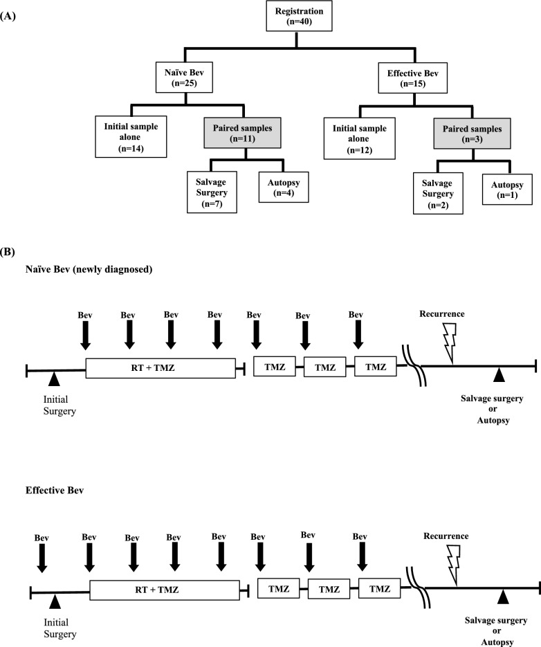
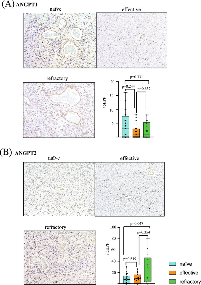
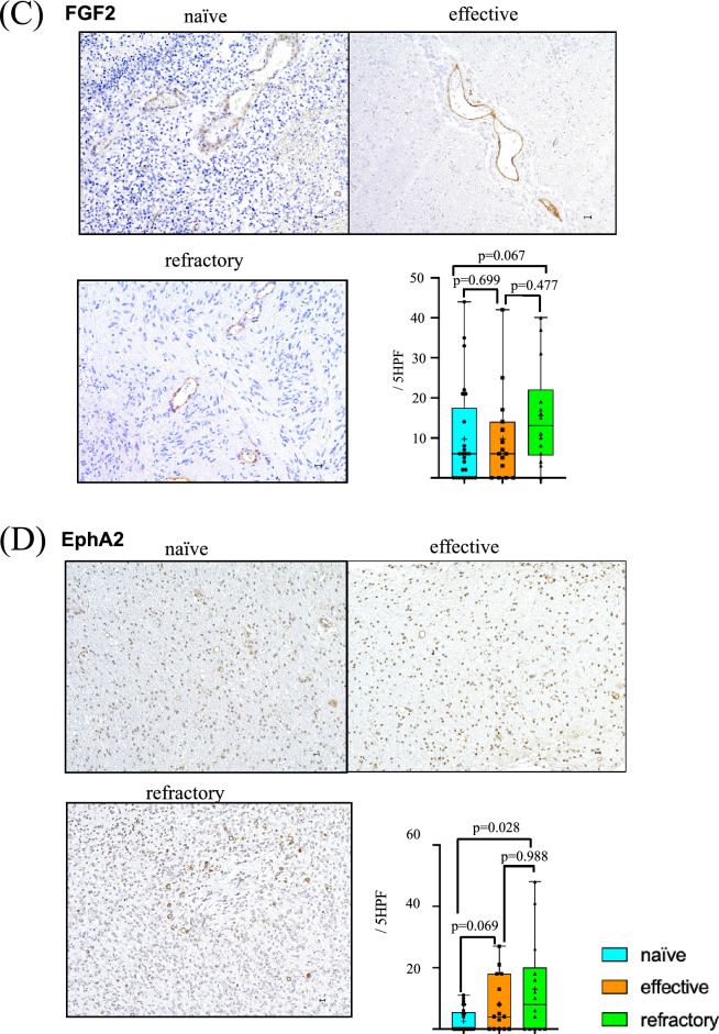
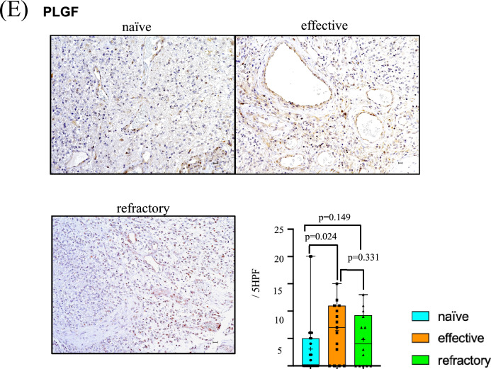
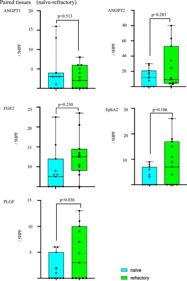
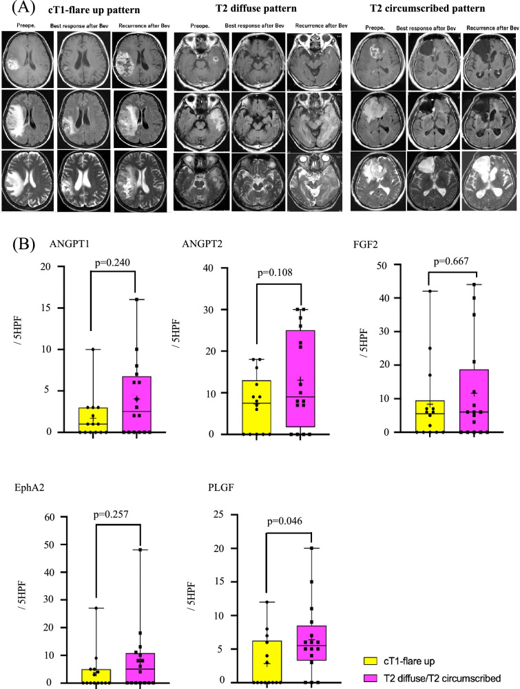
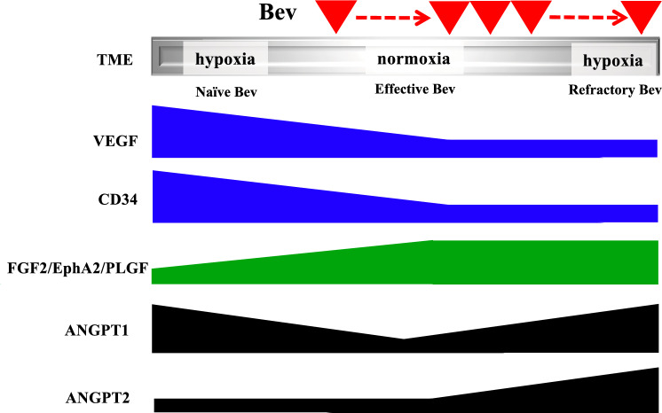
Glioblastoma multiforme (GBM) acquires resistance to bevacizumab (Bev) treatment. Bev affects angiogenic factors other than vascular endothelial growth factor (VEGF), which are poorly understood. We investigated changes in angiogenic factors under and after Bev therapy, including angiopoietin-1 (ANGPT1), angiopoietin-2 (ANGPT2), placental growth factor (PLGF), fibroblast growth factor 2, and ephrin A2 (EphA2). Fifty-four GBM tissues, including 28 specimens from 14 cases as paired specimens from the same patient obtained in three settings: initial tumor resection (naïve Bev), tumors resected following Bev therapy (effective Bev), and recurrent tumors after Bev therapy (refractory Bev). Immunohistochemistry assessed their expressions in tumor vessels and its correlation with recurrent MRI patterns. PLGF expression was higher in the effective Bev group than in the naïve Bev group (p = 0.024) and remained high in the refractory Bev group. ANGPT2 and EphA2 expressions were higher in the refractory Bev group than in the naïve Bev group (p = 0.047 and 0.028, respectively). PLGF expression was higher in the refractory Bev group compared with the naïve Bev group for paired specimens (p = 0.036). PLGF was more abundant in T2 diffuse/circumscribe patterns (p = 0.046). This is the first study to evaluate angiogenic factors other than VEGF during effective and refractory Bev therapy in patient-derived specimens.
Keywords: Alternative angiogenesis factor; Bevacizumab; Glioblastoma; Vascular endothelial growth factor.
© 2024. The Author(s).
Conflict of interest statement
The authors have no relevant financial or non-financial interests to disclose.
Figures
Similar articles
-
Persistent restoration to the immunosupportive tumor microenvironment in glioblastoma by bevacizumab.Cancer Sci. 2019 Feb;110(2):499-508. doi: 10.1111/cas.13889. Epub 2018 Dec 21. Cancer Sci. 2019. PMID: 30467920 Free PMC article.
-
CD44 expression in the tumor periphery predicts the responsiveness to bevacizumab in the treatment of recurrent glioblastoma.Cancer Med. 2021 Mar;10(6):2013-2025. doi: 10.1002/cam4.3767. Epub 2021 Feb 5. Cancer Med. 2021. PMID: 33543833 Free PMC article.
-
Noninvasive Characterization of Tumor Angiogenesis and Oxygenation in Bevacizumab-treated Recurrent Glioblastoma by Using Dynamic Susceptibility MRI: Secondary Analysis of the European Organization for Research and Treatment of Cancer 26101 Trial.Radiology. 2020 Oct;297(1):164-175. doi: 10.1148/radiol.2020200978. Epub 2020 Jul 28. Radiology. 2020. PMID: 32720870 Clinical Trial.
-
New Directions in Anti-Angiogenic Therapy for Glioblastoma.Neurotherapeutics. 2017 Apr;14(2):321-332. doi: 10.1007/s13311-016-0510-y. Neurotherapeutics. 2017. PMID: 28083806 Free PMC article. Review.
-
Clinical outcomes in recurrent glioblastoma with bevacizumab therapy: An analysis of the literature.J Clin Neurosci. 2017 Oct;44:101-106. doi: 10.1016/j.jocn.2017.06.070. Epub 2017 Jul 12. J Clin Neurosci. 2017. PMID: 28711289 Free PMC article. Review.
Cited by
-
Towards Effective Treatment of Glioblastoma: The Role of Combination Therapies and the Potential of Phytotherapy and Micotherapy.Curr Issues Mol Biol. 2024 Dec 19;46(12):14324-14350. doi: 10.3390/cimb46120859. Curr Issues Mol Biol. 2024. PMID: 39727987 Free PMC article. Review.
-
Recent Treatment Strategies and Molecular Pathways in Resistance Mechanisms of Antiangiogenic Therapies in Glioblastoma.Cancers (Basel). 2024 Aug 27;16(17):2975. doi: 10.3390/cancers16172975. Cancers (Basel). 2024. PMID: 39272834 Free PMC article. Review.
-
Comparative analyses of erythroblast transformation specific-1 related gene expression before and after neoadjuvant bevacizumab therapy for newly diagnosed glioblastoma.Int J Clin Exp Pathol. 2024 Oct 15;17(10):346-359. doi: 10.62347/GQWP4029. eCollection 2024. Int J Clin Exp Pathol. 2024. PMID: 39544713 Free PMC article.
-
Angiogenesis in Glioblastoma-Treatment Approaches.Cells. 2025 Mar 11;14(6):407. doi: 10.3390/cells14060407. Cells. 2025. PMID: 40136656 Free PMC article. Review.
-
Liposomal honokiol enhance the anti-tumor effect of bevacizumab in glioblastoma by inhibiting autophagy.Future Sci OA. 2025 Dec;11(1):2546232. doi: 10.1080/20565623.2025.2546232. Epub 2025 Aug 13. Future Sci OA. 2025. PMID: 40802280 Free PMC article.
References
MeSH terms
Substances
Grants and funding
LinkOut - more resources
Full Text Sources
Medical
Miscellaneous
