. 2024 Jul:511:63-75.
doi: 10.1016/j.ydbio.2024.04.004. Epub 2024 Apr 15.

Dyrk1a is required for craniofacial development in Xenopus laevis

Affiliations

Dyrk1a is required for craniofacial development in Xenopus laevis

H Katherine Johnson et al. Dev Biol. 2024 Jul.

Abstract

Loss of function variations in the dual specificity tyrosine-phosphorylation-regulated kinase 1 A (DYRK1A) gene are associated with craniofacial malformations in humans. Here we characterized the effects of deficient DYRK1A in craniofacial development using a developmental model, Xenopus laevis. Dyrk1a mRNA and protein were expressed throughout the developing head and both were enriched in the branchial arches which contribute to the face and jaw. Consistently, reduced Dyrk1a function, using dyrk1a morpholinos and pharmacological inhibitors, resulted in orofacial malformations including hypotelorism, altered mouth shape, slanted eyes, and narrower face accompanied by smaller jaw cartilage and muscle. Inhibition of Dyrk1a function resulted in misexpression of key craniofacial regulators including transcription factors and members of the retinoic acid signaling pathway. Two such regulators, sox9 and pax3 are required for neural crest development and their decreased expression corresponds with smaller neural crest domains within the branchial arches. Finally, we determined that the smaller size of the faces, jaw elements and neural crest domains in embryos deficient in Dyrk1a could be explained by increased cell death and decreased proliferation. This study is the first to provide insight into why craniofacial birth defects might arise in humans with variants of DYRK1A.

Keywords: Craniofacial; DYRK1A; Xenopus laevis.


Figures

Fig. 1.
Fig. 1.. DECIPHER analysis of phenotypes in patients with DYRK1A gene variants.
A) Pie chart showing that 62.5% (green) of patients identified with loss of function DYRK1A sequence variants had a craniofacial difference (n = 25/40 patients). B) 64.3% (blue) of patients identified with loss of function DYRK1A Copy number variants (CNVs) had some type of craniofacial phenotype (n = 18/28). C) Craniofacial differences reported in patients with sequence or CNV variants if DYRK1A.
Fig. 2.
Fig. 2.. Dyrk1a expression in the craniofacial regions of X. laevis embryos.
A) Representative frontal and lateral views (anterior to left) of embryos showing dyrk1a mRNA expression using in situ hybridization at three time points. i, ii) stage 25 (n = 27, 2 experiments). iii, iv) stage 32 (n = 29, 2 experiments). v, vi) stage 35 (n = 30, 2 experiments). B) Representative embryos showing Dyrk1a protein expression by immunofluorescence. All images are sagittal sections (anterior to the left) with Dyrk1a labeled green and DAPI labeled blue. Representatives are shown from 18 to 20 embryos in 2 experiments. i-iv) stage 25. v-viii) stage 32. ix-xii) stage 40. C) Plot of relative expression of dyrk1a.L mRNA and Dyrk1a.L protein generated in Xenbase.org ((http://www.xenbase.org/, RRID:SCR_003280 (Fisher et al., 2023)). D) Ventral view of a representative embryo labeled with Dyrk1a antibody at stage 43 (anterior is to the top, Dyrk1a = green and DAPI = blue). Based on 20 embryos in 2 biological replicates. Abbreviations: ves = vesicle, ba = branchial arches, bas = branchial arches fg = foregut, noto = notochord, ov = otic vesicle, st. = stage, cg = cement gland.
Fig. 3.
Fig. 3.. Decreased Dyrk1a function causes craniofacial differences.
Ai) Schematic showing dyrk1a morpholino injection. Aii-v) Frontal views of representative embryos injected with increasing concentrations of dyrk1a morpholinos. Bi) Schematic showing Dyrk1a inhibitor treatments. Bii-ix) Frontal views of representative embryos treated with increasing concentrations of INDY or harmine. C) Quantification of the intercanthal distance (distance between the eyes) based on 60 embryos in 3 biological replicates (ANOVA, TUKEY posthoc tests, all p values < 0.001). C) Schematic showing DYRK1A in vitro activity assay protocol to assess effectiveness of Dyrk1a knockdown tools. E) Western blots showing phosphorylated LIN52 on serine residue 28. Vinculin was used as a loading control. Fi-ii) Representative tissue sections through the head in control (i) and INDY treated embryos (ii). Based on 10 embryos in 2 biological replicates. Abbreviations: MO = morpholino.
Fig. 4.
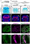
Fig. 4.. Dyrk1a in jaw cartilage and muscle development.
A) Schematic of the injection protocol, 10 ng of MO was injected into the embryos. B) Schematic of the exposure protocol, embryos were exposed to 25 μM of INDY. Ci-iii) Alcian blue labeling of cartilages of representative embryos (ventral views) injected with dyrk1a morpholinos or treated with INDY. Based on examination of 40 embryos (2 biological replicates). Abbreviations: cg = cement gland, ch = ceratohyal, ir = infrarostral, Mk = Meckel’s, bc = branchial cartilages, MO = morpholino. Civ-vii) Muscle specific labeling (12/101) of representative embryos (ventral views) injected with dyrk1a morpholinos or treated with INDY. Based on examination of 36 embryos (2 biological replicates). Abbreviations; qha = quadratohyoangularis, oh = orbitohyoideus, gh = geniohyoidues, ih = interhyoidues. Di-iii) Double labeling of a representative tissue section through the head. Dyrk1a is labeled green and Collagen II is labeled pink. Collagen II labels cells of the craniofacial cartilage. Based on 20 embryos (2 biological replicates). Ei-iii) Double labeling of a representative tissue whole mount embryo of the head. Dyrk1a is labeled green and muscle (12/101) is labeled pink. Based on 20 embryos (2 biological replicates).
Fig. 5.
Fig. 5.. Decreased Dyrk1a function results in changes to craniofacial regulators and sox10-GFP expression domain.
A) Schematic of the experimental plan. B) Results of RT-qPCR showing expression levels of craniofacial regulators. Levels are shown relative to controls set to 1 (blue line). Two biological replicates were performed and variation between the two is represented by the error bar (standard error). Each biological replicate is an average of 3 technical replicates. Fold changes are sox9 = 0.69, pax3 = 0.38, alx4 = 0.65, and rarg = 0.49 fold, gbx2 = 0.82 and crabp2 = 2.5. C) i) Lateral view of the head, anterior to the left, of a representative sox10-GFP transgenic embryo exposed to control DMSO. ii) Magnified image of the anterior branchial arch sox10 expression domain in (i) outlined in white. iii) Lateral view of the head, anterior to the left, of a representative sox10-GFP transgenic embryo exposed to INDY. Iv) Magnified image of the anterior branchial arch sox10 expression domain in iii outlined in white. v) Quantification of the area of most anterior sox10 positive branchial arch relative to the area of the head (n = 8). The asterisk indicates statistical difference based on student t-test (p = 0.911E-05). Abbreviations: ba = branchial arch, bas = branchial arches, Cont = control, ov = otic vesicle.
Fig. 6.
Fig. 6.. Decreased Dyrk1a function alters cell survival and proliferation.
A) Schematic shows the experimental design where fluorescein labeled morpholinos (dyrk1a or control, 5ng/embryo) were injected in one cell at the 2-cell stage and then embryos were fixed, sectioned and labeled. Bi-iii) Representative sections showing CC3 labeling (pink), Control morpholino (green) and DAPI counterstain. Biv-vi) Representative sections showing CC3 labeling (pink), dyrk1a morpholino (green) and DAPI counterstain. Bvii) Quantification of 20 embryos (2 replicates) showing a statistical difference between labeling on the dyrk1a morpholino injected sides verses the CC3 labeling on the control morpholino injected side (ttest, *p < 0.001). Ci-vi) Representative sections showing PH3 labeling (pink), Control morpholino (green) and DAPI counterstain (blue). Cvii) Quantification of 23 embryos (2 replicates) showing no statistical difference between PH3 labeling on the dyrk1a morpholino injected sides verses the labeling on the control morpholino injected side (ttest, *p = 0.263). D) Schematic of the experimental design where embryos were treated with INDY (25 μm) or Harmine (50 μM) and fixed, sectioned and labeled. Ei-vi) Representative sections from embryos, treated with INDY or Harmine, showing CC3 labeling (green), and DAPI counterstain CC3 (green). Evii) Quantification of 23 embryos (2 replicates) shows statistical differences in CC3 labeling between control and INDY and control and Harmine (Kruskal-Wallis One Way Analysis of Variance on Ranks p < 0.001, TUKEY posthoc tests, control vs INDY * = p < 0.001 and control vs harmine * = p < 0.001). Fi-vi) Representative sections from embryos, treated with INDY or Harmine, showing PH3 labeling (green), and DAPI counterstain. Fvii) Quantification of 20 embryos (2 replicates) shows statistical differences in PH3 labeling between control and INDY and control and Harmine (Kruskal-Wallis One Way Analysis of Variance on Ranks p < 0.001, TUKEY posthoc tests, control vs INDY * = p < 0.001 and control vs harmine * = p < 0.001). Abbreviations: MO = morpholino.

Update of

References

    1. Adams DS, Uzel SG, Akagi J, Wlodkowic D, Andreeva V, Yelick PC, Devitt-Lee A, Pare JF, Levin M, 2016. Bioelectric signalling via potassium channels: a mechanism for craniofacial dysmorphogenesis in KCNJ2-associated Andersen-Tawil Syndrome. J. Physiol 594, 3245–3270. - PMC - PubMed
    1. Alkobtawi M, Ray H, Barriga EH, Moreno M, Kerney R, Monsoro-Burq AH, Saint-Jeannet JP, Mayor R, 2018. Characterization of Pax3 and Sox10 transgenic Xenopus laevis embryos as tools to study neural crest development. Dev. Biol 444 (Suppl. 1), S202–s208. - PMC - PubMed
    1. Ananthapadmanabhan V, Shows KH, Dickinson AJ, Litovchick L, 2023. Insights from the protein interaction Universe of the multifunctional “Goldilocks” kinase DYRK1A. Front. Cell Dev. Biol 11, 1277537. - PMC - PubMed
    1. Arron JR, Winslow MM, Polleri A, Chang CP, Wu H, Gao X, Neilson JR, Chen L, Heit JJ, Kim SK, Yamasaki N, Miyakawa T, Francke U, Graef IA, Crabtree GR, 2006. NFAT dysregulation by increased dosage of DSCR1 and DYRK1A on chromosome 21. Nature 441, 595–600. - PubMed
    1. Asher JH Jr., Sommer A, Morell R, Friedman TB, 1996. Missense mutation in the paired domain of PAX3 causes craniofacial-deafness-hand syndrome. Hum. Mutat 7, 30–35. - PubMed

Publication types

MeSH terms

LinkOut - more resources