. 2024 Jul 11;73(8):1280-1291.
doi: 10.1136/gutjnl-2023-329686.

Recurrent RhoGAP gene fusion CLDN18-ARHGAP26 promotes RHOA activation and focal adhesion kinase and YAP-TEAD signalling in diffuse gastric cancer

Affiliations

Recurrent RhoGAP gene fusion CLDN18-ARHGAP26 promotes RHOA activation and focal adhesion kinase and YAP-TEAD signalling in diffuse gastric cancer

Feifei Zhang et al. Gut. .

Abstract

Objective: Genomic studies of gastric cancer have identified highly recurrent genomic alterations impacting RHO signalling, especially in the diffuse gastric cancer (DGC) histological subtype. Among these alterations are interchromosomal translations leading to the fusion of the adhesion protein CLDN18 and RHO regulator ARHGAP26. It remains unclear how these fusion constructs impact the activity of the RHO pathway and what is their broader impact on gastric cancer development. Herein, we developed a model to allow us to study the function of this fusion protein in the pathogenesis of DGC and to identify potential therapeutic targets for DGC tumours with these alterations.

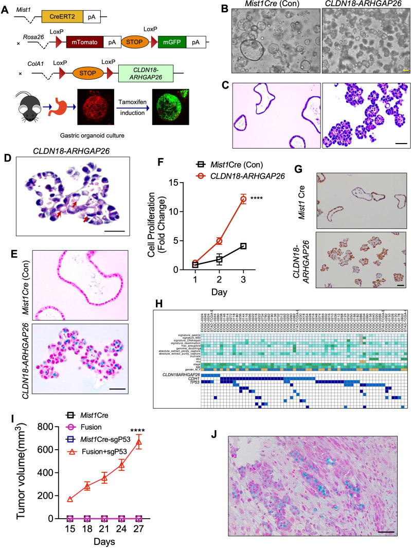
Design: We built a transgenic mouse model with LSL-CLDN18-ARHGAP26 fusion engineered into the Col1A1 locus where its expression can be induced by Cre recombinase. Using organoids generated from this model, we evaluated its oncogenic activity and the biochemical effects of the fusion protein on the RHOA pathway and its downstream cell biological effects in the pathogenesis of DGC.

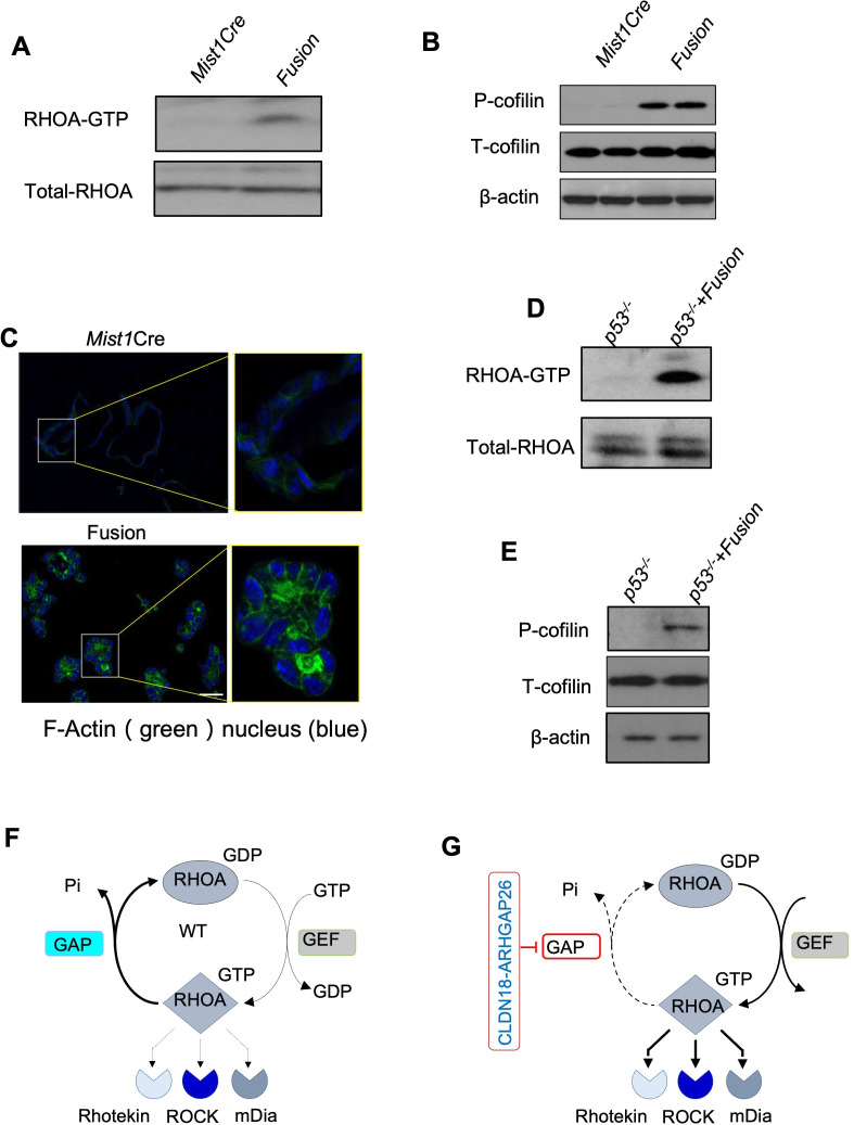
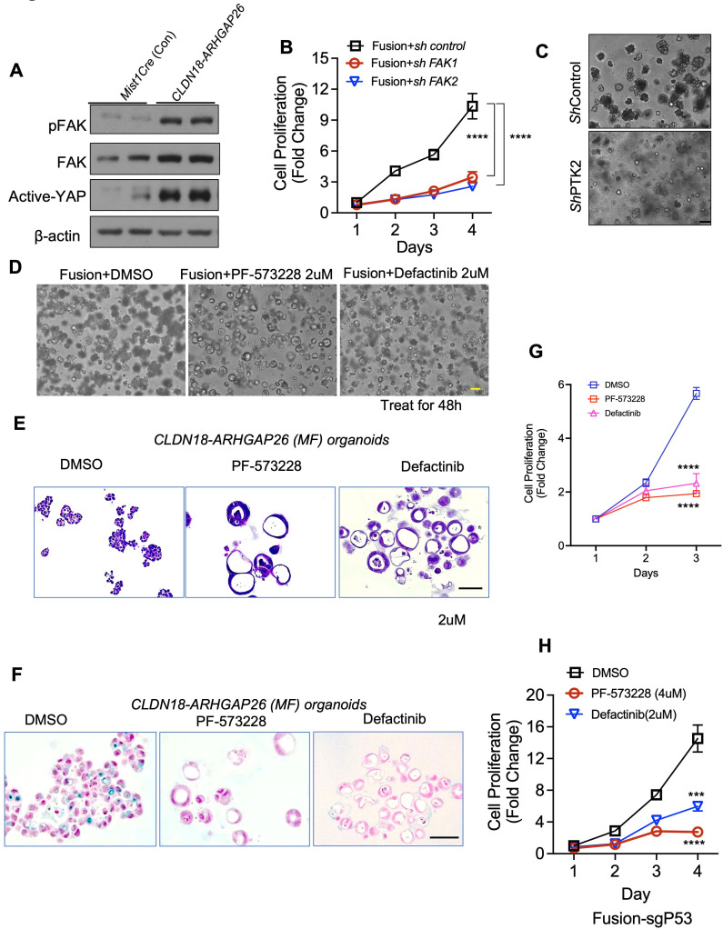
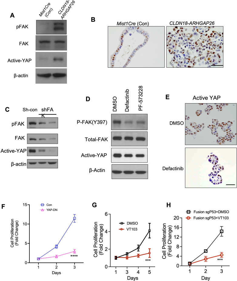
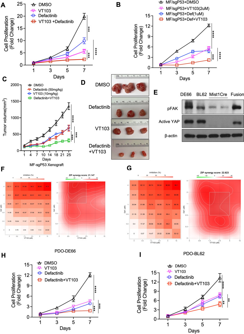
Results: We demonstrated that induction of CLDN18-ARHGAP26 expression in gastric organoids induced the formation of signet ring cells, characteristic features of DGC and was able to cooperatively transform gastric cells when combined with the loss of the tumour suppressor geneTrp53. CLDN18-ARHGAP26 promotes the activation of RHOA and downstream effector signalling. Molecularly, the fusion promotes activation of the focal adhesion kinase (FAK) and induction of the YAP pathway. A combination of FAK and YAP/TEAD inhibition can significantly block tumour growth.

Conclusion: These results indicate that the CLDN18-ARHGAP26 fusion is a gain-of-function DGC oncogene that leads to activation of RHOA and activation of FAK and YAP signalling. These results argue for further evaluation of emerging FAK and YAP-TEAD inhibitors for these deadly cancers.

Keywords: gastric cancer; molecular oncology.


Conflict of interest statement

Competing interests: AJB has received funding from Merck, Novartis, Bayer and Repare, is the advisor to Earli and HelixNano and is now employed by the Novartis Institutes of Biomedical Research. AJB and HZ are co-founders and equity holders in Signet Therapeutics. CJD is an advisory board member for Deciphera Pharmaceuticals, Mirati Therapeutics, Reactive Biosciences, Revolution Medicines and SHY Therapeutics; has received research funding support from Boragen, Deciphera Pharmaceuticals, Mirati Therapeutics, Revolution Medicines and SpringWorks Therapeutics; and has consulted for Day One Biotherapeutics, Eli Lilly, Jazz Therapeutics, Ribometrix, Sanofi and Turning Point Therapeutics. All other authors do not report conflicts.

Figures

Figure 1
Figure 1
CLDN18-ARHGAP26 fusion induces abnormal organoid morphologies and signet ring cells. (A) Schematic for the generation of mice with distinct genotypes, including the tomato-GFP reporter allele; bottom: representative stack confocal image of gastric organoids with Mist1CreERT2-R26mTmG 48 hours after tamoxifen (2 µM) induction in vitro. Representative images of (B) phase contrast and (C) H&E for gastric organoids with annotated genotypes after 4 weeks following in vitro tamoxifen induction. Scale bar=100 µm. (D) Representative higher-magnification image showing signet ring cells in CLDN18-ARHGAP26 fusion organoids following tamoxifen induction. Scale bar=50 µm. (E) Alcian blue staining of paraffin sections of the indicated organoids. Scale bar=100 µm. (F) In vitro proliferation (CellTiter-Glo) of Mist1Cre and CLDN18-ARHGAP26 organoids. Data are mean±SD. ****p<0.0001, two-way ANOVA (CLDN18-ARHGAP26 vs Mist1Cre). (G) Representative images of Ki67 staining of Mist1Cre and CLDN18-ARHGAP organoids. Scale bar=100 µm. (H) Analyses of co-occurrence of Trp53 mutation and CLDN18-ARHGAP fusion. (I) Tumour volumes following NSG flank implantation of organoids with annotated genotypes, showing tumour formation only by CLDN18-ARHGAP26 fusion organoids. Data are mean±SEM. ****p<0.0001, two-way ANOVA (CLDN18-ARHGAP26 fusion with P53 knockout vs other genotypes). (J) Representative image of Alcian blue staining for the CLDN18-ARHGAP26 fusion with Tp53 knockout tumours from panel (I). Scale bar=100 µm. ANOVA, analysis of variance; CLDN18, Claudin18.
Figure 2
Figure 2
CLDN18-ARHGAP26 fusion promotes RHOA activity. (A) Immunoblotting for the RHOA binding of Rhotekin by Rhotekin pulldown assay. (Representative images from three independent experiments). (B) Immunoblotting for p-cofflin in the organoids with annotated genotype. Shown are representative images from three independent experiments. (C) Representative immunofluorescence images for F-actin in organoids from mice with annotated genotypes. Phalloidin (in green) was used to visualise F-actin, DAPI (in blue) for the nucleus. Scale bar=50 µm. (D) Immunoblotting for the Rhotekin-RBD pulldown assay with the Tp53−/− organoids engineered with lentiviral Control or CLDN18-ARHGAP26 fusion. Shown are (representative images from three independent experiments). (E) Immunoblots of P53−/− organoids engineered with lentiviral vector or CLDN18-ARHGAP26 plasmid (representative images from three independent experiments). Comparison of WT (F) and CLDN18-ARHGAP26 fusion (G) biochemical properties. CLDN18, Claudin18; DAPI, 4′,6-diamidino-2-phenylindole; GAP, GTPase activating domain; WT, widetype.
Figure 3
Figure 3
CLDN18-ARHGAP26 induces activation of focal adhesion kinase. (A) Immunoblotting for the organoids with annotated genotype. (Representative images from three independent experiments). (B) In vitro proliferation (CellTiter-Glo) of CLDN18-ARHGAP26 organoids infected with control shRNA or targeting Ptk2. Data are mean± SD. ****p<0.0001, two-way ANOVA. (C) Representative images of phase contrast for gastric organoids with annotated genotypes scale bar=50 µm. Representative images of phase contrast (D) and H&E staining (E) for CLDN18-ARHGAP26 organoids treated with DMSO, PF-573228 (2 µM) or defactinib (2 µM) for 48 hours. Scale bar=100 µm. (F) Alcian blue staining of paraffin sections of the CLDN18-ARHGAP26 organoids treated with DMSO, PF-573228 (2 µM) or defactinib (2 µM) for 48 hours. Scale bar=100 µm. (G) In vitro proliferation (CellTiter-Glo) of CLDN18-ARHGAP26 organoids treated with DMSO, PF-573228 (2 µM) or defactinib (2 µM). Data are mean±SD. ****p<0.0001, two-way ANOVA (PF-573228 or defactinib vs DMSO) (H) In vitro proliferation (CellTiter-Glo) of CLDN18-ARHGAP26 with Tp53 knockout organoids treated with DMSO or FAK inhibitors. Data are mean±SD. ***p<0.001, PF-573228 vs DMSO; ****p<0.0001, defactinib vs DMSO; two-way ANOVA. ANOVA, analysis of variance; CLDN18, Claudin18; DMSO, dimethyl sulfoxide; FAK, focal adhesion kinase; pFAK, phosphorylated focal adhesion kinase.
Figure 4
Figure 4
YAP is a potent downstream target of FAK in CLDN18-ARHGAP26. (A) Immunoblotting for the Mist1Cre organoids and the CLDN18-ARHGAP26 organoids. (Representative images from three independent experiments). (B) Representative images of active-YAP staining for the organoids with annotated genotype. Scale bar=100 µm. (C) Immunoblotting for the CLDN18-ARHGAP26 organoids engineered with shControl or shPTK2 virus (n=3 independent experiments). (D) Immunoblotting for the CLDN18-ARHGAP26 organoids treated with DMSO, PF-573228 (2 µM) or defactinib (2 µM) for 48 hours (n=3 independent experiments). (E) Representative images of active-YAP staining for the paraffin sections of the CLDN18-ARHGAP26 organoids treated with DMSO or FAK inhibitor defactinib (2 µM). Scale bar=100 µm. (F) In vitro proliferation (CellTiter-Glo) of CLDN18-ARHGAP26 organoids infected with control or YAP-DN expressing virus. Data are mean±SD. ****p<0.0001, two-way ANOVA (YAP-DN(S94A) vs DMSO). (G) In vitro proliferation (CellTiter-Glo) of CLDN18-ARHGAP26 organoids treated with DMSO or TEAD inhibitor VT103 (2 µM). Data are mean±SD. ***p<0.0001, two-way ANOVA (VT103 vs DMSO). (H) In vitro proliferation (CellTiter-Glo) of CLDN18-ARHGAP26 with p53 knockout organoids treated with DMSO or TEAD inhibitor VT103 (2 µM). Data are mean±SD. ***p<0.001, two-way ANOVA (VT103 vs DMSO). ANOVA, analysis of variance; CLDN18, Claudin18; DMSO, dimethyl sulfoxide; FAK, focal adhesion kinase; pFAK, phosphorylated focal adhesion kinase.
Figure 5
Figure 5
Dual inhibition of FAK and YAP/TEAD shows significant synergistic effects both in vitro and in vivo. (A) In vitro proliferation (CellTiter-Glo) of CLDN18-ARHGAP26 with p53 knockout organoids treated with DMSO control, TEAD inhibitor VT103 (2 µM), defactinib (1 µM) or the combination. Data are mean±SD. ***p<0.001, ****p<0.0001, two-way ANOVA. (B) In vitro proliferation (CellTiter-Glo) of CLDN18-ARHGAP26 with p53 knockout (MF/sgP53) organoids treated with DMSO control, TEAD inhibitor VT103 (2 µM), defactinib (1 µM) or the combination. Data are mean±SD. ***p<0.001, ****p<0.0001, two-way ANOVA. (C) Tumour growth curve for CLDN18-ARHGAP26 with Tp53 knockout organoids xenograft tumours (n=8–10) treated with vehicle control, defactinib (50 mg/kg, once a day), VT103 (10 mg/kg, once a day) or the combination. Day 1 means the first treatment of the drugs. Data are mean±SEM. ***p<0.001, ****p<0.0001, two-way ANOVA. (D) Representative tumour images of drug treatments from panel (C). (E) Representative immunoblotting images for the patient-derived organoids with the Mist1Cre (M) and CLDN18-ARHGAP26 fusion (MF) organoids as controls (n=3 independent experiments). (F) DE66 patient-derived organoids (PDO-DE66) were treated with defactinib (0.2 μM to 4 µM) or VT103 (0.6 μM to 10 µM) alone or together for 7 days. Viability in the treatment groups was normalised to DMSO control. The inhibition rate was shown (left). Analysis of the synergistic effect in defactinib and VT103 combination was performed by SynergyFinder using zero interaction potency (ZIP) model. The inhibition rate was used to calculate ZIP synergy score. The box indicates the most synergistic area (right). Representatives of two independent experiments were shown. (G) BL62 patient-derived organoids (PDO-BL62) were treated with defactinib (0.2 μM to 4 µM) or VT103 (0.6 μM to 10 µM) alone or together for 5 days. Analysis of the synergistic effect in defactinib and VT103 combination was the same as in panel (F). In vitro proliferation (CellTiter-Glo) of PDO-DE66 (H) or PDO-BL62 (I) organoids were treated with DMSO, TEAD inhibitor VT103 (1 µM), defactinib (2 µM) or the combination. Data are mean± SD. **p<0.01, ***p<0.001, ****p<0.0001, two-way ANOVA. ANOVA, analysis of variance; CLDN18, Claudin18; DMSO, dimethyl sulfoxide; FAK, focal adhesion kinase; pFAK, phosphorylated focal adhesion kinase.

References

    1. Sung H, Ferlay J, Siegel RL, et al. Global cancer statistics 2020: GLOBOCAN estimates of incidence and mortality worldwide for 36 cancers in 185 countries. CA Cancer J Clin 2021;71:209–49. 10.3322/caac.21660 - DOI - PubMed
    1. Lauren P. The two histological main types of gastric carcinoma: diffuse and so-called intestinal-type carcinoma. An attempt at a histo-clinical classification. Acta Pathol Microbiol Scand 1965;64:31–49. 10.1111/apm.1965.64.1.31 - DOI - PubMed
    1. Kakiuchi M, Nishizawa T, Ueda H, et al. Recurrent gain-of-function mutations of RHOA in diffuse-type gastric carcinoma. Nat Genet 2014;46:583–7. 10.1038/ng.2984 - DOI - PubMed
    1. Guilford P, Hopkins J, Harraway J, et al. E-cadherin germline mutations in familial gastric cancer. Nature 1998;392:402–5. 10.1038/32918 - DOI - PubMed
    1. Richards FM, McKee SA, Rajpar MH, et al. Germline E-cadherin gene (CDH1) mutations predispose to familial gastric cancer and colorectal cancer. Hum Mol Genet 1999;8:607–10. 10.1093/hmg/8.4.607 - DOI - PubMed

Publication types

MeSH terms