. 2024 Apr 15;15(1):3186.
doi: 10.1038/s41467-024-47439-6.

Sm-like protein Rof inhibits transcription termination factor ρ by binding site obstruction and conformational insulation

Affiliations

Sm-like protein Rof inhibits transcription termination factor ρ by binding site obstruction and conformational insulation

Nelly Said et al. Nat Commun. .

Abstract

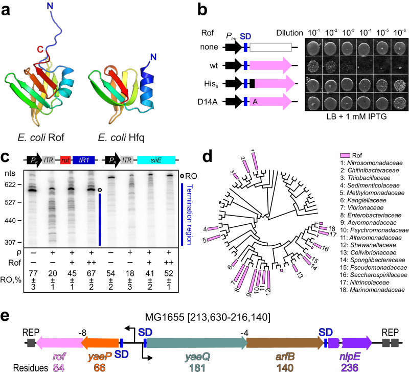
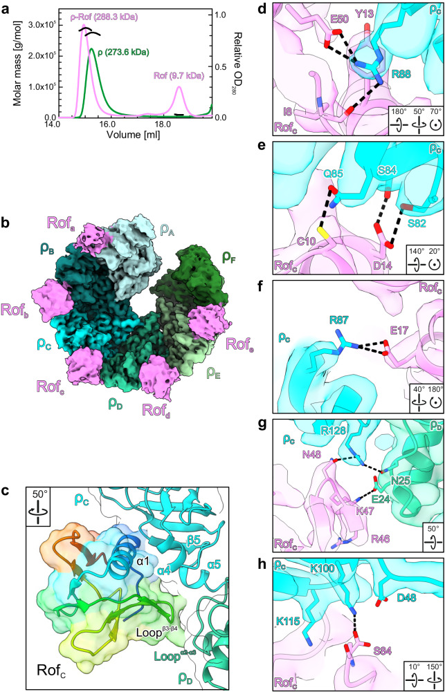
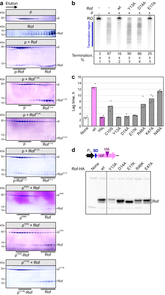
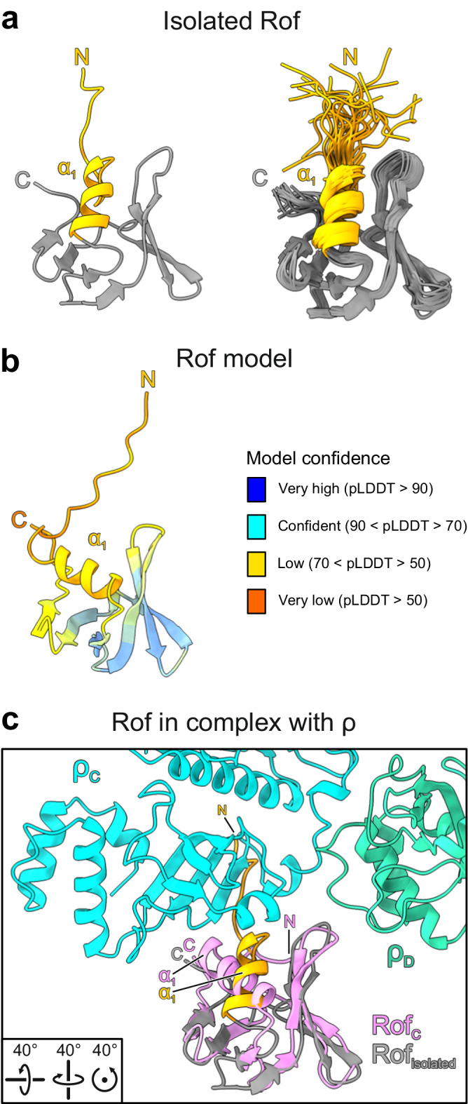
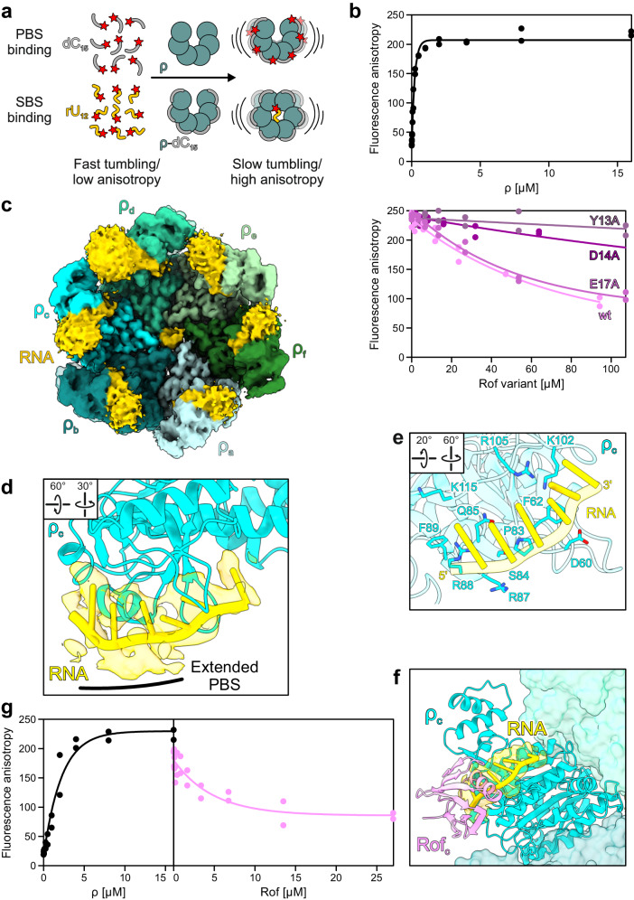
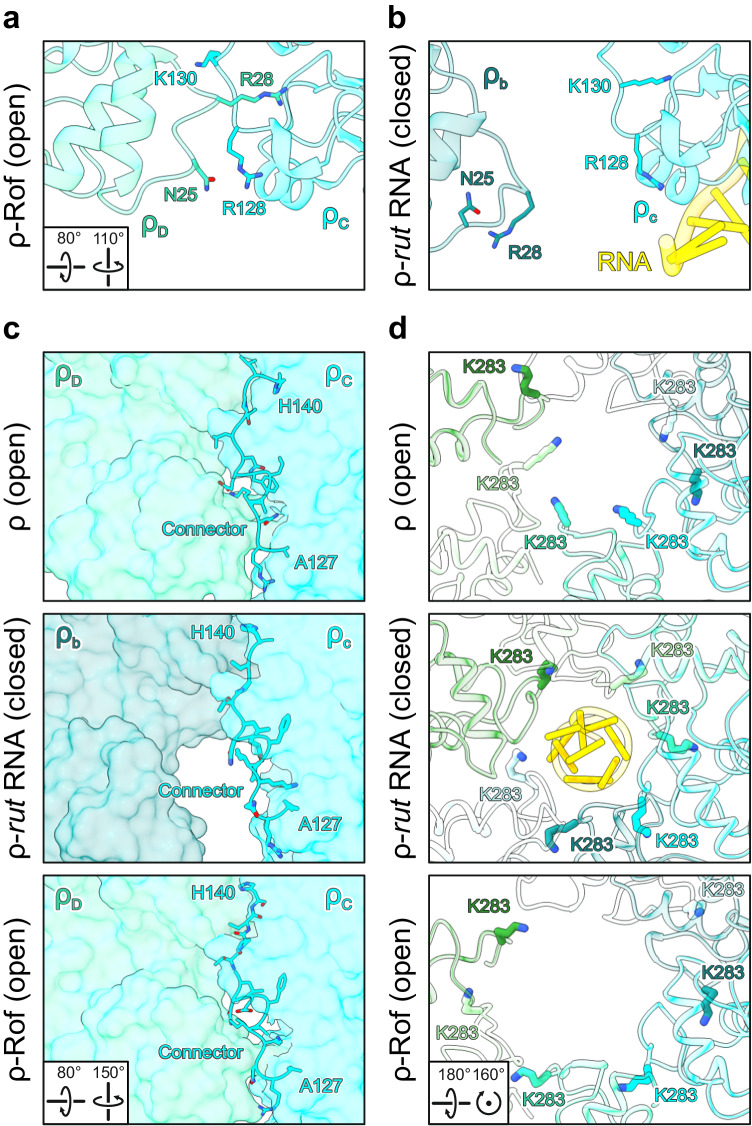
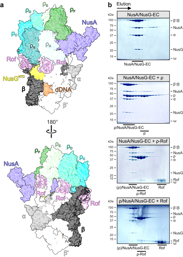
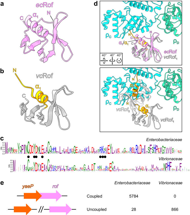
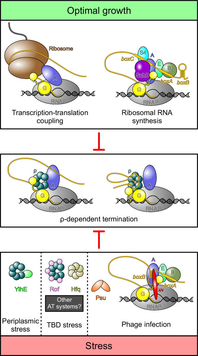
Transcription termination factor ρ is a hexameric, RNA-dependent NTPase that can adopt active closed-ring and inactive open-ring conformations. The Sm-like protein Rof, a homolog of the RNA chaperone Hfq, inhibits ρ-dependent termination in vivo but recapitulation of this activity in vitro has proven difficult and the precise mode of Rof action is presently unknown. Here, our cryo-EM structures of ρ-Rof and ρ-RNA complexes show that Rof undergoes pronounced conformational changes to bind ρ at the protomer interfaces, undercutting ρ conformational dynamics associated with ring closure and occluding extended primary RNA-binding sites that are also part of interfaces between ρ and RNA polymerase. Consistently, Rof impedes ρ ring closure, ρ-RNA interactions and ρ association with transcription elongation complexes. Structure-guided mutagenesis coupled with functional assays confirms that the observed ρ-Rof interface is required for Rof-mediated inhibition of cell growth and ρ-termination in vitro. Bioinformatic analyses reveal that Rof is restricted to Pseudomonadota and that the ρ-Rof interface is conserved. Genomic contexts of rof differ between Enterobacteriaceae and Vibrionaceae, suggesting distinct modes of Rof regulation. We hypothesize that Rof and other cellular anti-terminators silence ρ under diverse, but yet to be identified, stress conditions when unrestrained transcription termination by ρ may be detrimental.


Conflict of interest statement

The authors declare no competing interests.

Figures

Fig. 1
Fig. 1. Rof structure, functions, conservation and genetic organization.
a Structural comparison of E. coli Rof (left) and Hfq (right). Proteins are shown in rainbow colors from N-termini (N; blue) to C-termini (C; red). b Cells transformed with plasmids expressing wild-type (wt) Rof and Rof variants under the control of an IPTG-inducible Ptrc promoter or an empty vector were grown overnight in LB supplemented with carbenicillin at 32 °C. Serial 10-fold dilutions were plated on LB-carbenicillin in the presence of IPTG and incubated overnight at 32 °C. A set representative of five independent experiments is shown. Shine-Dalgarno (SD) elements are indicated by blue bars. c Rof inhibits ρ-dependent termination in vitro. Top, DNA templates encoding the rut+ λ tR1 (left) or rut- siiE leader region (right) downstream of the λ PR promoter and a C-less cassette that allows for the formation of synchronized, radiolabeled ECs halted 26 nts downstream of the transcription start site. Bottom, representative single-round transcription reactions. Halted ECs were incubated with ρ and/or Rof (where indicated), restarted by addition of NTPs, and subsequently quenched. Reactions were analyzed on 5% urea-acrylamide gels. Positions of the full-length run-off (RO) RNA and termination regions are indicated, as are the sizes of the molecular weight markers generated by γ32P-labeling of pBR322 MspI fragments. The fractions of run-off RNA represent the mean ± SD of three independent experiments. Source data are provided as a Source Data file. d Rof distribution on the phylogenetic tree of Pseudomonadota. Each leaf represents one family. The height of the bar indicates the fraction of Rof in each family, with the highest one being 80%. e E. coli yae locus (MG1655 genome coordinates 213,630-216,140); the length of each ORF is indicated below the sequence, the nlpE gene is interrupted to save space. The arfB and rof genes lack SD elements and overlap with the respective upstream ORFs, yaeP and yaeQ, by 4 and 8 nts. The “insulating” REP elements are shown by dark gray boxes.
Fig. 2
Fig. 2. ρ-Rof interaction.
a SEC-MALS of isolated ρ (green) and ρ in the presence of Rof (violet). The average molar masses of the peak fractions are indicated. b CryoEM reconstruction of the ρ6-ADP-Rof5 complex. ρ, different shades of cyan and green; Rof, violet. ρ adopts an open ring conformation and Rof proteins bind between the NTDs of neighboring ρ protomers. c Close-up view of the ρ-Rof interface. Rotation symbols in this and (dh) views are relative to (b). Rof is shown in rainbow colors from N-terminus (blue) to C-terminus (red). Rof binds ρ via its N-terminal helix α1, the β3-β4 loop and the C-terminus. dh Details of the ρ-Rof interaction. Interacting residues are shown as sticks with atoms colored by type; carbon, as the respective protein subunit; nitrogen, blue; oxygen, red. Black dashed lines, hydrogen bonds or salt bridges. Regions of the ρ6-ADP-Rof5 cryoEM reconstruction are shown as semi-transparent surfaces.
Fig. 3
Fig. 3. Effects of Rof and ρ variants.
a Analytical SEC runs, monitoring the interaction between the indicated ρ and Rof variants. For each run, the same fractions were analyzed by SDS-PAGE. First and second panel, SEC runs of isolated ρ and Rof, respectively. Third panel, binding of ρwt to Rofwt. Panels 4–6, binding of indicated Rof variants to ρwt. Panels 7–9, binding of indicated ρ variants to Rofwt. Experiments were performed three times independently with similar results. Source data are provided as a Source Data file. b Effects of Rof variants on ρ-dependent termination in vitro. Assays were done using the λ tR1 template as in Fig. 1c. Termination efficiency values represent means ± SD of three independent experiments. Source data are provided as a Source Data file. c Lag times of growth for IA227 transformed with Ptrc plasmids expressing different Rof variants or none. n = 4 biologically independent samples. Source data are provided as a Source Data file. d Production of Rof variants containing a single HA-tag inserted after residue 57 in IA227 cells. Control experiments demonstrated that the expression of HA-tagged Rof is toxic. Rof expression following the induction with 1 mM IPTG was determined by Western blotting with anti-HA antibodies (Millipore Sigma). Experiments were performed three times independently with similar results. Source data are provided as a Source Data file.
Fig. 4
Fig. 4. Rof conformational changes upon ρ binding.
a Structure of ecRof in isolation. Representative structure (left) and structural ensemble (right) of ecRof determined by NMR spectroscopy (PDB ID: 1SG5). The N-termini (N) and helix α1 are colored in orange. C, C-termini. The N-termini of isolated ecRof show high flexibility and are oriented away from the protein’s core. b AlphaFold model of ecRof. Rof is colored according to the AlphaFold model confidence score (pLDDT). The pLDDT score indicates structural flexibility in the N-terminal region, including helix α1. c Isolated ecRof (gray) superimposed on ρ-bound ecRof (violet) in the same view as isolated ecRof in (a). Rotation symbols, view relative to Fig. 2b. In the conformation of isolated ecRof, the N-terminus and helix α1 would sterically interfere with binding to ρ.
Fig. 5
Fig. 5. Effect of Rof on RNA binding by ρ.
a Principles of fluorescence anisotropy assays used in (b) and (g). Isolated fluorescence-labeled (red star) dC15 DNA oligos (gray) or rU12 RNA oligos (gold) exhibit high tumbling rates (low fluorescence anisotropy). In the presence of ρ, dC15 binds to the PBSes of ρ, which reduces its tumbling rate (increased fluorescence anisotropy). In the presence of dC15, rU12 binds to the SBS of ρ, which leads to ρ ring closure. b PBS binding. Top, binding of dC15 to the ρ PBSes in the presence of increasing concentrations of ρ. Bottom, effect of Rof variants on the binding of dC15 to the PBSes. Data of two independent experiments and fits to a single-exponential Hill function (see Methods) are shown. Source data are provided as a Source Data file. c CryoEM reconstruction of the ρ-rut RNA structure. ρ forms a closed ring and is bound by RNA at the NTD of each protomer and at the SBS. Only RNA regions bound at the SBS and at the ρc NTD were modeled. RNA density and model, gold. As originally proposed,, ρ subunits are labeled in opposite directions around open and closed ρ rings (compare to Fig. 2b). We therefore labeled subunits of open ρ with capitals (A-F) and subunits of closed ρ with lower case letters (af). Rotation symbols in this and (d) views are relative to (c). d ρ interaction with RNA at the PBS. Six RNA residues (cartoon) fit to the density observed at ρc (semitransparent surface). e Path of the RNA along the PBS. ρ residues are shown as sticks and labeled. f Structural comparison between ρ-rut RNA and ρ6-ADP-Rof5 complexes. Structures of the complexes were superimposed based on the ρcC subunits. Bound Rof sterically hinders RNA accommodation at the extended PBS. g Ring closure assays. Left, fluorescence anisotropy of 5’-FAM-labeled rU12 in the presence of increasing amounts of ρ. Right, Rof inhibits ρ ring closure. Data of two independent experiments and fits to a single-exponential Hill function (see Methods) are shown. Source data are provided as a Source Data file.
Fig. 6
Fig. 6. Effects of Rof on ρ conformation.
a, b Structural comparison of ρ subunit interactions in structures of open ρ bound to ATP (PDB ID: 6WA8) (a) and closed ρ bound to rut RNA (b). In the open hexamer conformation, residue R28 from one ρ protomer (ρD) is embedded in a pocket of the adjacent protomer (ρC), formed partially by α5. In the closed hexamer conformation, R28 is ejected from that pocket and substituted by K130 of the same protomer (ρc). Concomitantly, contacts between the N-terminal region of ρD and R128 of ρC are broken during ring closure (ρb an ρc in the closed hexamer). Rotation symbols, view relative to Fig. 5c. c Comparison of the NTD-CTD connectors (sticks) in open ρATP (top), the closed ρ-rut RNA complex (middle) and the open ρ6-ADP-Rof5 complex (bottom). Relative to open ρATP, the NTD-CTD connector (residues 127–140) in the closed ρ-rut RNA complex is re-aligned by one residue, most evident by the rearrangement of H140 towards the protomer interface. In the open ρ6-ADP-Rof5 structure, the NTD-CTD connector retains the register observed in the closed ρ-rut RNA complex. Rotation symbols in this and (d) views are relative to Fig. 2b. d Structural comparison of Q-loop conformations in open ρATP (top), the closed ρ-rut RNA complex (middle) and the open ρ6-ADP-Rof5 complex (bottom). In open ρATP, Q-loops adopt a conformation resulting in residues K283 (sticks) pointing towards the central axis of the ρ hexamer, where they could engage in initial contacts to SBS RNA. In the closed ρ-rut RNA complex, K283 residues are embedded in pockets formed in part by the Q-loops of the adjacent ρ protomers. In the open ρ6-ADP-Rof5 complex, the Q-loops adopt a conformation similar to that observed in the closed ρ-rut RNA complex.
Fig. 7
Fig. 7. Rof effects on ρ-EC interactions.
a Rof interferes with ρ binding to ECs. The ρ6-ADP-Rof5 structure was superimposed on the structure of a ρ/NusA/NusG-modified EC (PDB ID: 6Z9P) based on the ρA protomers. RNAP subunits, different shades of gray; NusA, slate blue; NusG, yellow; downstream (d) DNA, brown. Rof sterically interferes with ρ binding to RNAP and Nus factors. b SDS-PAGE analysis of SEC runs, monitoring ρ binding to ECs in the absence and presence of Rof. First panel, NusA/NusG-EC. Second panel, pre-formed NusA/NusG-EC incubated with a three-fold molar excess of ρ hexamer. Third panel, pre-formed NusA/NusG-EC incubated with a three-fold molar excess (relative to ρ hexamer) of ρ-Rof complex. Fourth panel, pre-formed ρ/NusA/NusG-EC incubated with a ten-fold molar excess of Rof (relative to ρ hexamer). Experiments were performed three times independently with similar results. Source data are provided as a Source Data file.
Fig. 8
Fig. 8. Rof conformational changes and conservation.
a, b The structure of one E. coli (ec) Rof protein as observed in the ρ-Rof complexes (a) compared to one monomer from the crystal structure of a V. cholerae (vc) Rof dimer (b; PDB ID: 6JIE). EcRof bound to ρ and vcRof mainly differ in the orientation of the N-termini and the lengths of helices α1. N, N-termini; C, C-termini. c Conservation of Rof sequences in Enterobacteriaceae (top) and Vibrionaceae (bottom). In Enterobacteriacea, two alternative start sites for Rof are possible, generating 84- and 86-residue Rof. The interface residues revealed by the E. coli ρ-Rof complex structures are indicated by black dots. Sequence logos were generated by WebLogo (version 3.7.8). d Superposition of the vcRof on ecRof bound to ρ. Upper panel, superposition of a vcRof monomer on ecRof bound to ρ. Lower panel, superposition of the vcRof dimer on ecRof bound to ρ; for clarity, ecRof is not shown. Rotation symbols, view relative to Fig. 2b. While monomeric vcRof would align without steric conflict, there might be steric hindrance in the interaction between a dimer of vcRof and ρ. e The rof and yaeP genes are coupled in Enterobacteriaceae, but not in Vibrionaceae; see Supplementary Data 1 for more details.
Fig. 9
Fig. 9. ρ regulation under optimal growth and stress conditions.
Top, under optimal growth conditions bulk mRNA synthesis is protected from premature termination by ρ through transcription-translation coupling,. A specific transcription anti-termination complex (rrnTAC) shields the ribosomal RNAs from ρ. Bottom, under stress conditions, when translation is inefficient, ρ activity must be regulated. Phages use different strategies to protect transcription of their own genomes. Similar to the rrnTAC, lambdoid phages rely on specific anti-termination complexes, such as λN-TAC, to shield the nascent transcript from ρ. Phage P4 uses the Psu protein that directly binds to ρ and thereby prevents the formation of termination complexes. To date, three cellular proteins are known to directly bind ρ and inhibit termination, but other stress-specific regulators likely exist. YihE is induced upon periplasmatic stress,, whereas stress conditions under which Hfq and Rof regulate ρ activity remain to be identified. YihE binds the ρ NTD, inhibiting ρ-RNA interactions; the mechanism of ρ regulation by Rof has been determined in this study, and molecular details of Hfq-mediated ρ inhibition remain to be solved,. By directly binding ρ, each of these anti-terminators may interfere with the formation of ρ-dependent termination complexes that assemble in an EC-dependent manner (center left), or RNA-dependent manner (center right),.

Update of

Similar articles

Cited by

References

    1. Sunday, N., Svetlov, D. & Artsimovitch, I. Rho termination factor: one ring to bind them all. in RNA Polymerases as Molecular Motors: On the Road (eds. Landick, R., Strick, T. & Wang, J.) 100–131 (Royal Society of Chemistry, 2021).
    1. Skordalakes E, Berger JM. Structure of the Rho transcription terminator: mechanism of mRNA recognition and helicase loading. Cell. 2003;114:135–146. doi: 10.1016/S0092-8674(03)00512-9. - DOI - PubMed
    1. Thomsen ND, Lawson MR, Witkowsky LB, Qu S, Berger JM. Molecular mechanisms of substrate-controlled ring dynamics and substepping in a nucleic acid-dependent hexameric motor. Proc. Natl Acad. Sci. USA. 2016;113:E7691–E7700. doi: 10.1073/pnas.1616745113. - DOI - PMC - PubMed
    1. Skordalakes E, Berger JM. Structural insights into RNA-dependent ring closure and ATPase activation by the Rho termination factor. Cell. 2006;127:553–564. doi: 10.1016/j.cell.2006.08.051. - DOI - PubMed
    1. Thomsen ND, Berger JM. Running in reverse: the structural basis for translocation polarity in hexameric helicases. Cell. 2009;139:523–534. doi: 10.1016/j.cell.2009.08.043. - DOI - PMC - PubMed

MeSH terms