

Tetrahedral framework nucleic acids/hyaluronic acid-methacrylic anhydride hybrid hydrogel with antimicrobial and anti-inflammatory properties for infected wound healing
- PMID: 38622128
- PMCID: PMC11018755
- DOI: 10.1038/s41368-024-00290-3
Tetrahedral framework nucleic acids/hyaluronic acid-methacrylic anhydride hybrid hydrogel with antimicrobial and anti-inflammatory properties for infected wound healing
Abstract
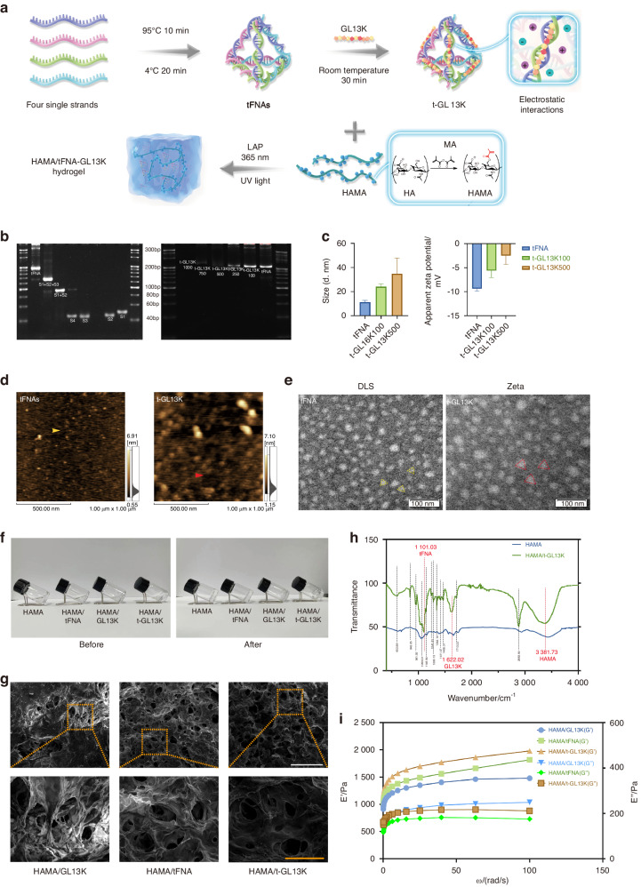
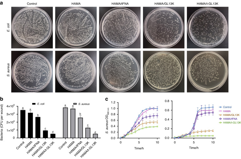
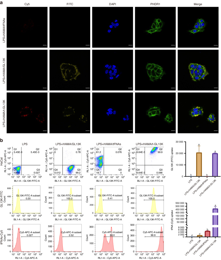
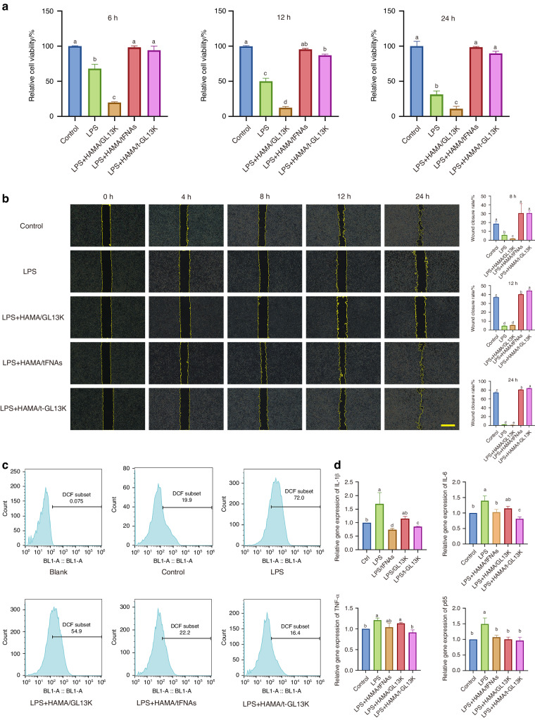
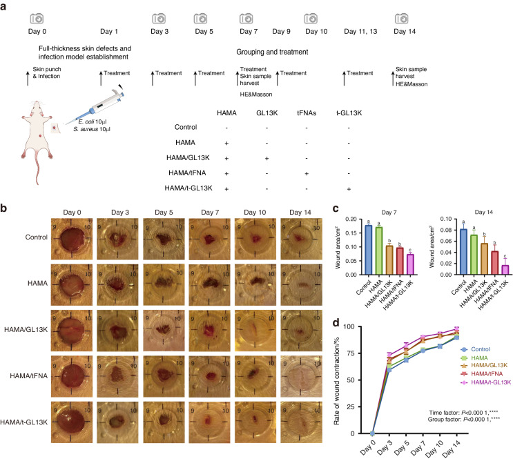
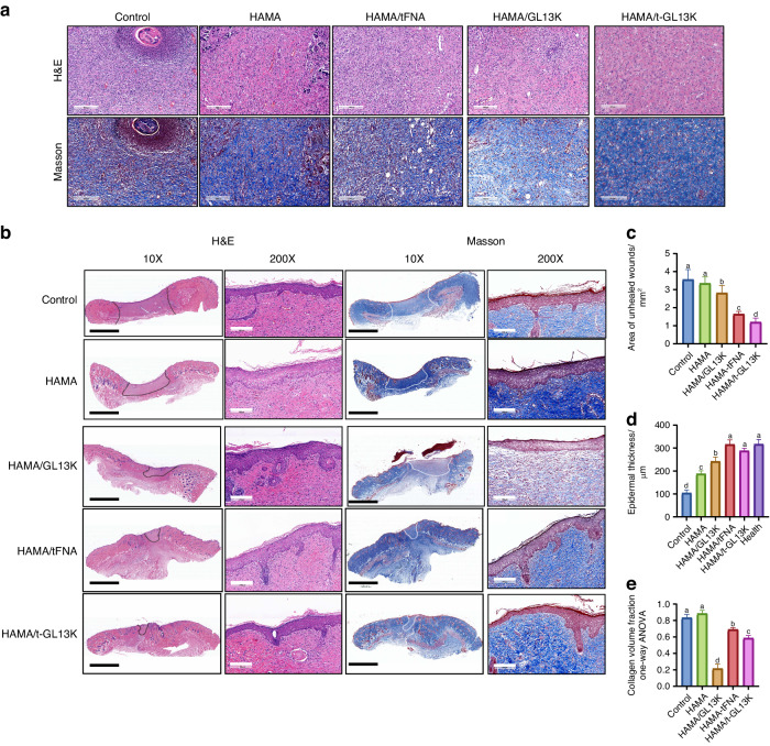
Bacterial resistance and excessive inflammation are common issues that hinder wound healing. Antimicrobial peptides (AMPs) offer a promising and versatile antibacterial option compared to traditional antibiotics, with additional anti-inflammatory properties. However, the applications of AMPs are limited by their antimicrobial effects and stability against bacterial degradation. TFNAs are regarded as a promising drug delivery platform that could enhance the antibacterial properties and stability of nanodrugs. Therefore, in this study, a composite hydrogel (HAMA/t-GL13K) was prepared via the photocross-linking method, in which tFNAs carry GL13K. The hydrogel was injectable, biocompatible, and could be instantly photocured. It exhibited broad-spectrum antibacterial and anti-inflammatory properties by inhibiting the expression of inflammatory factors and scavenging ROS. Thereby, the hydrogel inhibited bacterial infection, shortened the wound healing time of skin defects in infected skin full-thickness defect wound models and reduced scarring. The constructed HAMA/tFNA-AMPs hydrogels exhibit the potential for clinical use in treating microbial infections and promoting wound healing.
© 2024. The Author(s).
Conflict of interest statement
The authors declare no competing interests.
Figures
Similar articles
-
Current Understanding and Translational Prospects of Tetrahedral Framework Nucleic Acids.JACS Au. 2025 Feb 10;5(2):486-520. doi: 10.1021/jacsau.4c01170. eCollection 2025 Feb 24. JACS Au. 2025. PMID: 40017737 Free PMC article. Review.
-
Injectable thermo-sensitive and wide-crack self-healing hydrogel loaded with antibacterial anti-inflammatory dipotassium glycyrrhizate for full-thickness skin wound repair.Acta Biomater. 2022 Apr 15;143:203-215. doi: 10.1016/j.actbio.2022.02.041. Epub 2022 Mar 1. Acta Biomater. 2022. PMID: 35245682
-
An injectable and light curable hyaluronic acid composite gel with anti-biofilm, anti-inflammatory and pro-healing characteristics for accelerating infected wound healing.Int J Biol Macromol. 2023 Dec 31;253(Pt 5):127190. doi: 10.1016/j.ijbiomac.2023.127190. Epub 2023 Oct 5. Int J Biol Macromol. 2023. PMID: 37802452
-
Chitosan and hyaluronic-based hydrogels could promote the infected wound healing.Int J Biol Macromol. 2023 Mar 31;232:123271. doi: 10.1016/j.ijbiomac.2023.123271. Epub 2023 Jan 14. Int J Biol Macromol. 2023. PMID: 36646352
-
Advances of biomacromolecule-based antibacterial hydrogels and their performance evaluation for wound healing: A review.Int J Biol Macromol. 2024 Nov;279(Pt 4):135577. doi: 10.1016/j.ijbiomac.2024.135577. Epub 2024 Sep 11. Int J Biol Macromol. 2024. PMID: 39270907 Review.
Cited by
-
Hyaluronic Acid in Nanopharmaceuticals: An Overview.Curr Issues Mol Biol. 2024 Sep 20;46(9):10444-10461. doi: 10.3390/cimb46090621. Curr Issues Mol Biol. 2024. PMID: 39329973 Free PMC article. Review.
-
Current Understanding and Translational Prospects of Tetrahedral Framework Nucleic Acids.JACS Au. 2025 Feb 10;5(2):486-520. doi: 10.1021/jacsau.4c01170. eCollection 2025 Feb 24. JACS Au. 2025. PMID: 40017737 Free PMC article. Review.
-
A deformable SIS/HA composite hydrogel coaxial scaffold promotes alveolar bone regeneration after tooth extraction.Bioact Mater. 2024 Dec 10;46:97-117. doi: 10.1016/j.bioactmat.2024.12.008. eCollection 2025 Apr. Bioact Mater. 2024. PMID: 39760069 Free PMC article.
-
Tetrahedral Framework Nucleic Acid Relieves Sepsis-Induced Intestinal Injury by Regulating M2 Macrophages.Cell Prolif. 2025 May;58(5):e13803. doi: 10.1111/cpr.13803. Epub 2025 Jan 22. Cell Prolif. 2025. PMID: 39844345 Free PMC article.
-
Sequential delivery of IL-10 and icariin using nanoparticle/hydrogel hybrid system for prompting bone defect repair.Mater Today Bio. 2024 Dec 3;29:101374. doi: 10.1016/j.mtbio.2024.101374. eCollection 2024 Dec. Mater Today Bio. 2024. PMID: 39717024 Free PMC article.
References
Publication types
MeSH terms
Substances
Grants and funding
LinkOut - more resources
Full Text Sources
Medical
Molecular Biology Databases
