. 2024 Apr 15;24(1):474.
doi: 10.1186/s12885-024-12224-7.

Exercise accelerates recruitment of CD8+ T cell to promotes anti-tumor immunity in lung cancer via epinephrine

Affiliations

Exercise accelerates recruitment of CD8+ T cell to promotes anti-tumor immunity in lung cancer via epinephrine

Sai-Nan Miao et al. BMC Cancer. .

Abstract

Background and purpose: In recent years, there has been extensive research on the role of exercise as an adjunctive therapy for cancer. However, the potential mechanisms underlying the anti-tumor therapy of exercise in lung cancer remain to be fully elucidated. As such, our study aims to confirm whether exercise-induced elevation of epinephrine can accelerate CD8+ T cell recruitment through modulation of chemokines and thus ultimately inhibit tumor progression.

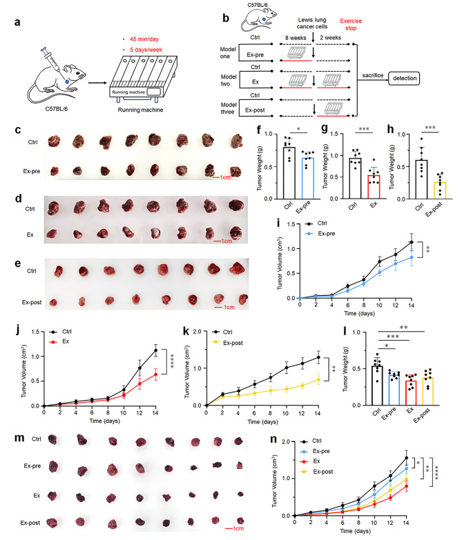
Method: C57BL/6 mice were subcutaneously inoculated with Lewis lung cancer cells (LLCs) to establish a subcutaneous tumor model. The tumor mice were randomly divided into different groups to performed a moderate-intensity exercise program on a treadmill for 5 consecutive days a week, 45 min a day. The blood samples and tumor tissues were collected after exercise for IHC, RT-qPCR, ELISA and Western blot. In addition, another group of mice received daily epinephrine treatment for two weeks (0.05 mg/mL, 200 µL i.p.) (EPI, n = 8) to replicate the effects of exercise on tumors in vivo. Lewis lung cancer cells were treated with different concentrations of epinephrine (0, 5, 10, 20 µM) to detect the effect of epinephrine on chemokine levels via ELISA and RT-qPCR.

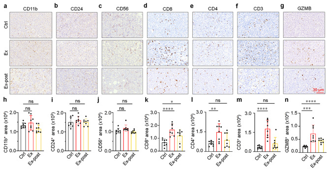
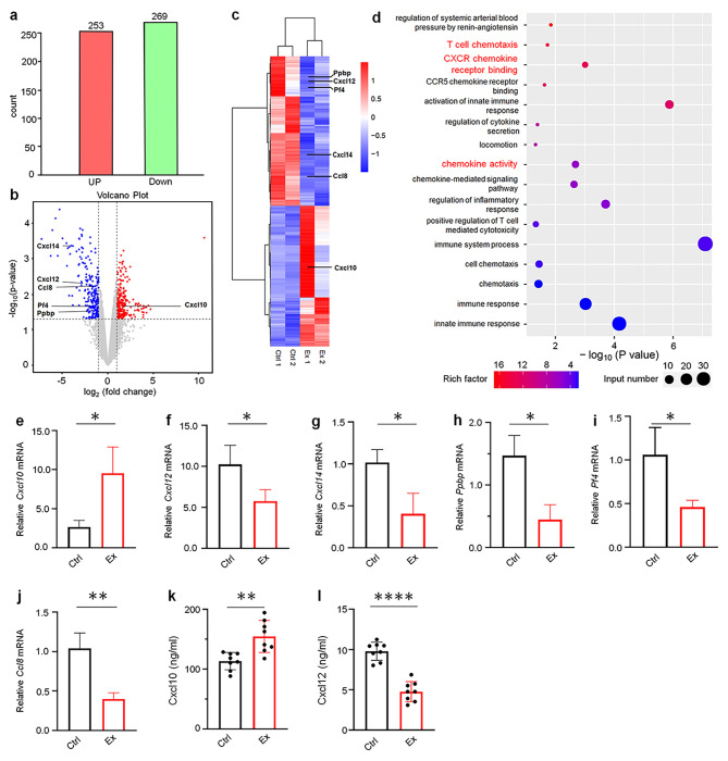
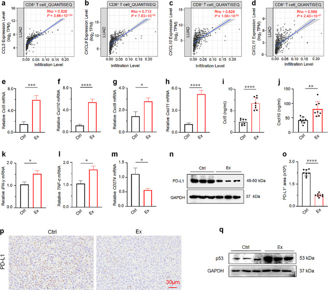
Results: This study reveals that both pre- and post-cancer exercise effectively impede the tumor progression. Exercise led to an increase in EPI levels and the infiltration of CD8+ T cell into the lung tumor. Exercise-induced elevation of EPI is involved in the regulation of Ccl5 and Cxcl10 levels further leading to enhanced CD8+ T cell infiltration and ultimately inhibiting tumor progression.

Conclusion: Exercise training enhance the anti-tumor immunity of lung cancer individuals. These findings will provide valuable insights for the future application of exercise therapy in clinical practice.

Keywords: Ccl5; Cxcl10; Epinephrine; Exercise; Lung cancer.


Conflict of interest statement

The authors declare no competing interests.

Figures

Fig. 1
Fig. 1
Exercise can delay tumor progression. a The forced-treadmill-running model (exercise) is illustrated. b Experimental grouping and design: Ctrl (control group), mice with free movement for 8 weeks before inoculation and 2 weeks after tumor inoculation (n = 8); Ex-pre (exercise before tumor inoculation), mice running for 8 weeks before tumor inoculation (n = 8); Ex: (exercise before and after tumor inoculation), mice that run for 8 weeks before inoculation and 2 weeks after tumor inoculation (n = 8); Ex-post (exercise after tumor inoculation), mice running for 2 weeks after tumor inoculation (n = 8). 45 min/day, 5 days/week. Solid red lines indicate exercise treatment, and dashed black lines indicate no exercise treatment. c-e The brightfield images display tumors from the three exercise models. f Comparison of tumor weight in the Ex-pre and Ctrl mice at the end of the exercise intervention (n = 8). g Comparison of tumor weight in the Ex and Ctrl mice at the end of the exercise intervention (n = 8). h Comparison of tumor weight in the Ex-post and Ctrl mice at the end of the exercise intervention (n = 8). i-k Mean of tumor volume changes after tumor formation in the three exercise models (n = 8). l Comparison of tumor weight in the Ctrl, Ex-pre, Ex and Ex-post mice at the end of the exercise intervention (n = 8). m The brightfield images display tumors from Ctrl, Ex-pre, Ex and Ex-post mice models. n Mean of tumor volume changes after tumor formation in the Ctrl, Ex-pre, Ex and Ex-post mice models (n = 8). Results of f-k are presented as mean ± SEM. l and n were performed using one-way analysis of variance (ANOVA). Statistical analysis was performed using two-tailed unpaired t tests. *p < 0.05, **p < 0.01, ***p < 0.001
Fig. 2
Fig. 2
Exercise promotes infiltration of CD8, CD4, CD3+ T cell. a-g IHC staining of CD11b, CD24, CD56, CD8, CD4, CD3 and GZMB in the tumor in each group (n = 8). Scale bar: 30 μm. h-n Quantitative analysis of CD11b, CD24, CD56, CD8, CD4, CD3 and GZMB IHC staining in indicated groups (n = 8). The results of h-n are presented as mean ± SEM. Statistical analysis was performed using one-way analysis of variance (ANOVA). *p < 0.05, **p < 0.01, ***p < 0.001
Fig. 3
Fig. 3
Transcriptional targets were examined by RNA-seq in tumors after exercise. a The number of differentially expressed genes in the Ex and Ctrl mice were detected by RNA-seq. b Volcano plot of differentially expressed genes in Ex compared with Ctrl tumor tissues. Red dots represent upregulated genes, and blue dots represent downregulated genes. c Heat map of the differentially expressed genes in Ex compared with Ctrl tumor tissues. d Pathway enrichment analysis of significantly upregulated or downregulated genes. e-j RT-qPCR validates the gene expression of Cxcl10, Cxcl12, Cxcl14, Ppbp, Pf4 and Ccl8 in the tumors each group (n = 3) after tumor inoculation. k-l The expression levels of Cxcl10 and Cxcl12 were detected in the tumors in each group by ELISA (n = 8). The results of e-l are presented as mean ± SEM. Statistical analysis was performed using two-tailed unpaired t tests. *p < 0.05, **p < 0.01, ***p < 0.001
Fig. 4
Fig. 4
Exercise promotes CD8+ T cell recruitment with the assistance of Ccl5 and Cxcl10. a-d Correlation between Ccl5, Cxcl9, Cxcl10, and Cxcl11 mRNA level and CD8+ T cell using QUANTISEQ. Data were obtained from TIMER 2.0 (http://timer.comp-genomics.org/). e-h RT-qPCR analyses of mRNA expression of chemokines in the tumors of Ctrl and Ex groups (n = 3). i-j ELISA analysis of serum levels of Cxcl10 and Ccl5 in Ctrl and Ex mice (n = 8). k-m RT-qPCR analyses of mRNA expression of cytokines and PD-L1 in the tumors of Ctrl and Ex mice (n = 3). n Western blot analysis the expression of PD-L1 in the tumors of Ctrl and Ex mice (n = 3). Full-length blots/gels are presented in Fig. 4n of source data. o Automatic quantification of PD-L1 IHC staining in indicated mice tumors (n = 8). p IHC staining of PD-L1 on tumor sections from different groups (n = 8). q Western blot analysis the expression of P53 in the tumors of Ctrl and Ex mice (n = 3). Full-length blots/gels are presented in Fig. 4q of source data. The results of e-m and o are presented as mean ± SEM. Statistical analysis was performed using two-tailed unpaired t tests. *p < 0.05, **p < 0.01, ***p < 0.001
Fig. 5
Fig. 5
Exercise inhibits tumor progression by upregulation of EPI levels. a Serum levels of EPI in Ctrl, Ex and Ex-post groups were detected by ELISA in tumor-bearing mice (n = 8). b-d EPI mice receiving daily injections of EPI (0.5 mg/kg i.p.) (n = 8) and TE mice with daily running for 14 days after tumor inoculation (n = 8) compared with TC mice (n = 8). b Representative images. c Tumor weights of indicated groups. d Tumor volumes of different groups. e IHC staining of CD8, CD4, CD3 and GZMB on tumor sections from indicated groups (n = 8). f Automatic quantification of CD8, CD4, CD3 and GZMB IHC staining in indicated mice tumors (n = 8). Data are presented as the mean ± SEM in each group. Statistical analysis of a was performed using two-tailed unpaired t tests. c, d and f were performed using one-way analysis of variance (ANOVA). *p < 0.05, **p < 0.01, ***p < 0.001
Fig. 6
Fig. 6
EPI regulates changes in chemokine levels. a-f RT-qPCR analyses the mRNA expression of chemokines and cytokines in the tumors of TC and EPI mice (n = 3). g-h ELISA analysis serum levels of Cxcl10 and Ccl5 in TC, TE and EPI treated mice (n = 8). i-j RT-qPCR detect the mRNA levels of Cxcl10 and Ccl5 in LLC cells after treated with different concentrations of EPI (0, 5, 10, 20 µM). k-l Cxcl10 and Ccl5 in the supernatant of LLC cells were measured by ELISA. m RT-qPCR analyses the mRNA expression of CD274 in the tumors of control TC and EPI groups (n = 3). n Western blot analyses PD-L1 in LLC cells after EPI treatment for 24 h at different concentration (0, 5, 10, 20 µM). Full-length blots/gels are presented in Fig. 6n of source data. o IHC staining of PD-L1 on tumor sections from indicated groups (n = 8). p Automatic quantification of PD-L1 IHC staining in indicated mice tumors (n = 8). q Graphical abstract of this study. Exercise-induced elevation of EPI is involved in regulating Ccl5 and Cxcl10 expression, subsequently promoting CD8+ T cells recruitment, and ultimately inhibiting tumor progression. Data are presented as the mean ± SEM in each group. Statistical analysis of a-f and m were performed using two-tailed unpaired t tests. g-l and p were performed using one-way analysis of variance (ANOVA). *p < 0.05, **p < 0.01, ***p < 0.001

References

    1. Siegel RL, Miller KD, Wagle NS, Jemal A. Cancer statistics, 2023. CA Cancer J Clin. 2023;73(1):17–48. doi: 10.3322/caac.21763. - DOI - PubMed
    1. Sung H, Ferlay J, Siegel RL, Laversanne M, Soerjomataram I, Jemal A, Bray F. Global Cancer statistics 2020: GLOBOCAN estimates of incidence and Mortality Worldwide for 36 cancers in 185 countries. Cancer J Clin. 2021;71(3):209–49. doi: 10.3322/caac.21660. - DOI - PubMed
    1. Thandra KC, Barsouk A, Saginala K, Aluru JS, Barsouk A. Epidemiology of lung cancer. Contemp Oncol (Pozn) 2021;25(1):45–52. - PMC - PubMed
    1. Leiter A, Veluswamy RR, Wisnivesky JP. The global burden of lung cancer: current status and future trends. Nat Rev Clin Oncol. 2023;20(9):624–39. doi: 10.1038/s41571-023-00798-3. - DOI - PubMed
    1. Nooreldeen R, Bach H. Current and Future Development in Lung Cancer diagnosis. Int J Mol Sci. 2021;22(16). - PMC - PubMed