Review
. 2024 Apr 15;13(1):22.
doi: 10.1186/s40035-024-00410-3.

The role of the brain renin-angiotensin system in Parkinson´s disease

Affiliations
Review

The role of the brain renin-angiotensin system in Parkinson´s disease

Jose Luis Labandeira-Garcia et al. Transl Neurodegener. .

Abstract

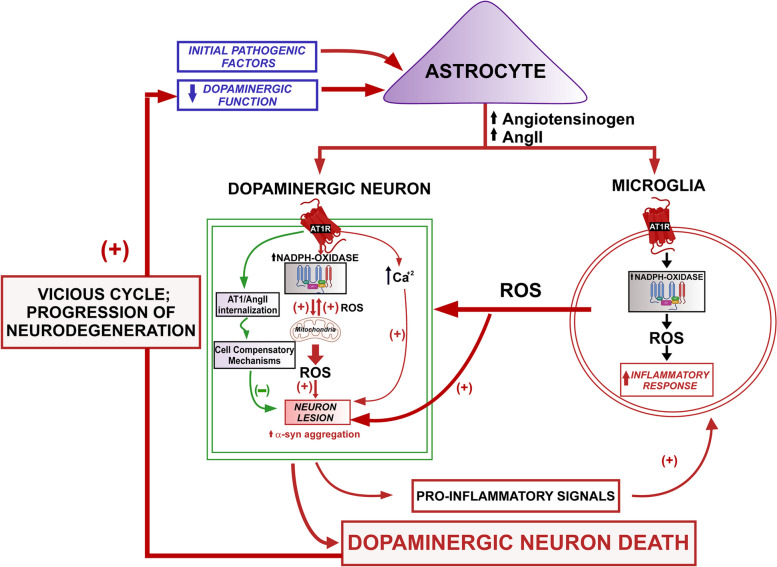
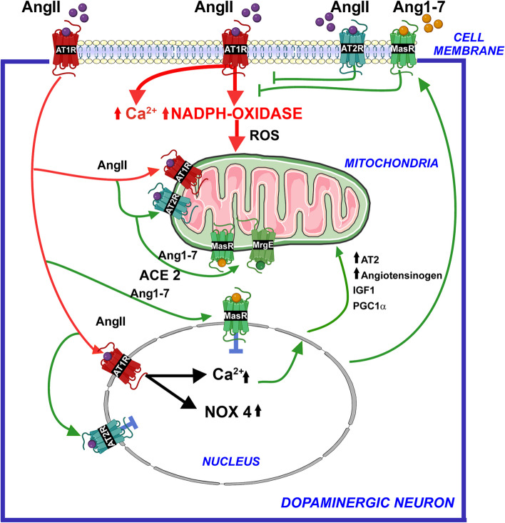
The renin-angiotensin system (RAS) was classically considered a circulating hormonal system that regulates blood pressure. However, different tissues and organs, including the brain, have a local paracrine RAS. Mutual regulation between the dopaminergic system and RAS has been observed in several tissues. Dysregulation of these interactions leads to renal and cardiovascular diseases, as well as progression of dopaminergic neuron degeneration in a major brain center of dopamine/angiotensin interaction such as the nigrostriatal system. A decrease in the dopaminergic function induces upregulation of the angiotensin type-1 (AT1) receptor activity, leading to recovery of dopamine levels. However, AT1 receptor overactivity in dopaminergic neurons and microglial cells upregulates the cellular NADPH-oxidase-superoxide axis and Ca2+ release, which mediate several key events in oxidative stress, neuroinflammation, and α-synuclein aggregation, involved in Parkinson's disease (PD) pathogenesis. An intraneuronal antioxidative/anti-inflammatory RAS counteracts the effects of the pro-oxidative AT1 receptor overactivity. Consistent with this, an imbalance in RAS activity towards the pro-oxidative/pro-inflammatory AT1 receptor axis has been observed in the substantia nigra and striatum of several animal models of high vulnerability to dopaminergic degeneration. Interestingly, autoantibodies against angiotensin-converting enzyme 2 and AT1 receptors are increased in PD models and PD patients and contribute to blood-brain barrier (BBB) dysregulation and nigrostriatal pro-inflammatory RAS upregulation. Therapeutic strategies addressed to the modulation of brain RAS, by AT1 receptor blockers (ARBs) and/or activation of the antioxidative axis (AT2, Mas receptors), may be neuroprotective for individuals with a high risk of developing PD or in prodromal stages of PD to reduce progression of the disease.

Keywords: Angiotensin; Dopamine; NADPH-oxidase; Neurodegeneration; Neuroinflammation; Neuroprotection; Oxidative stress; Parkinson.


Conflict of interest statement

The authors have no competing interests to declare.

Figures

Fig. 1
Fig. 1
The renin-angiotensin system (RAS) is organized into two opposite arms that must be properly balanced: a pro-oxidative/pro-inflammatory axis (in red), mainly formed by Angiotensin II that binds AT1 receptors (AT1R), and an antioxidative/anti-inflammatory axis (in green), mainly formed by Angiotensin II-binding AT2 receptors and Angiotensin 1–7-binding Mas receptors (MasR) or Mas-related G protein-coupled receptors. The enzyme prorenin/renin acting on the precursor protein angiotensinogen produces Angiotensin I, which is converted to Angiotensin II by the angiotensin-converting enzyme (ACE or ACE1). Renin and its precursor prorenin (PR) can also bind specific pro-oxidative PR receptors (PRR). Angiotensin-converting enzyme 2 (ACE2; also known as the major entry receptor for the SARS-COV viruses) plays a major role in balancing both RAS arms, as ACE2 (together with other peptidases such as Neprilysin, NEP) transforms peptides of the pro-inflammatory axis (Angiotensin I and, particularly, Angiotensin II) into peptides of the anti-inflammatory axis (Angiotensin 1–9 and, particularly Angiotensin 1–7)
Fig. 2
Fig. 2
The brain RAS plays a role in the progression of dopaminergic neuron degeneration. Different pathogenic factors may trigger molecular and cellular changes that lead to an initial dysregulation of the brain RAS, or dysregulation of the dopaminergic neuron function leading to decreased dopamine production, which affects the dopamine/RAS interaction in neurons and glial cells. In neurons, a decrease in dopamine level upregulates the angiotensin type-1 (AT1) receptor activity, leading to the recovery of dopamine levels together with overactivation of the NADPH-oxidase-superoxide-mitochondria axis and Ca2+ release, which mediate several key events such as oxidative stress, α-synuclein aggregation, and neuroinflammation involved in the progression of Parkinson's disease (PD). An intraneuronal antioxidative/anti-inflammatory RAS counteracts the effects of the pro-oxidative AT1 receptor overactivation (detailed in Fig. 3). In microglial cells, AT1 receptor upregulation activates the NADPH-oxidase complex, increasing the release of ROS to the extracellular space and the inflammatory response. In astrocytes, a decrease in dopamine level induces an increase in paracrine angiotensinogen/AngII production that can act on neurons and microglial cells. AngII, angiotensin II; AT1, angiotensin type I; ROS, reactive oxygen species
Fig. 3
Fig. 3
The intraneuronal RAS compensates (green lines: neuroprotective mechanisms) for the pro-oxidative effects of plasma membrane AT1 receptor activation by paracrine AngII (red lines: pro-neurodegenerative mechanisms). Internalization of the AT1/Ang II complex to the nucleus and activation of nuclear and mitochondrial receptors by intracellular AngII and Ang 1–7, trigger several mechanisms that protect neurons against AT1-induced oxidative stress during normal cell function. Antioxidative AT2, Mas, and MrgE receptors are more abundant in the mitochondria. In the nucleus, activation of AT1 receptors triggers several compensatory mechanisms, including increased mRNA expression of antioxidative RAS receptors, angiotensinogen, IGF1, and PGC1α. However, an excess of cell membrane AngII/AT1 receptor activity to compensate for dopamine decrease or other pathogenic factors may overwhelm the buffering mechanisms, leading to the progression of dopaminergic degeneration. AngII, angiotensin II; Ang1-7, angiotensin 1–7; AT1, angiotensin type 1; AT2, angiotensin type 2; MAS, Mas receptors; MrgE, Mas-related receptor MrgE; Nox4, NADPH-oxidase 4; ROS, reactive oxygen species

References

    1. Tigerstedt R, Bergman PQ. Niere und kreislauf. Skand Arch Physiol. 1898;8:223–71. doi: 10.1111/j.1748-1716.1898.tb00272.x. - DOI
    1. Nishimura H. Renin-angiotensin system in vertebrates: phylogenetic view of structure and function. Anat Sci Int. 2017;92:215–247. doi: 10.1007/s12565-016-0372-8. - DOI - PubMed
    1. Wong MKS, Takei Y. Molecular and evolutionary perspectives of the renin-angiotensin system from lamprey. Gen Comp Endocrinol. 2018;257:137–142. doi: 10.1016/j.ygcen.2017.01.031. - DOI - PubMed
    1. Lev-Ran A, Porta M. Salt and hypertension: a phylogenetic perspective. Diabetes Metab Res Rev. 2005;21:118–131. doi: 10.1002/dmrr.539. - DOI - PubMed
    1. Ganong WF. Origin of the angiotensin II secreted by cells. Proc Soc Exp Biol Med. 1994;205:213–219. doi: 10.3181/00379727-205-43699A. - DOI - PubMed

Publication types