. 2024 May 7;92(5):e0011324.
doi: 10.1128/iai.00113-24. Epub 2024 Apr 16.

Plasmodium yoelii surface-related antigen (PySRA) modulates the host pro-inflammatory responses via binding to CD68 on macrophage membrane

Affiliations

Plasmodium yoelii surface-related antigen (PySRA) modulates the host pro-inflammatory responses via binding to CD68 on macrophage membrane

Xin Feng et al. Infect Immun. .

Abstract

Malaria, one of the major infectious diseases in the world, is caused by the Plasmodium parasite. Plasmodium antigens could modulate the inflammatory response by binding to macrophage membrane receptors. As an export protein on the infected erythrocyte membrane, Plasmodium surface-related antigen (SRA) participates in the erythrocyte invasion and regulates the immune response of the host. This study found that the F2 segment of P. yoelii SRA activated downstream MAPK and NF-κB signaling pathways by binding to CD68 on the surface of the macrophage membrane and regulating the inflammatory response. The anti-PySRA-F2 antibody can protect mice against P. yoelii, and the pro-inflammatory responses such as IL-1β, TNF-α, and IL-6 after infection with P. yoelii are attenuated. These findings will be helpful for understanding the involvement of the pathogenic mechanism of malaria with the exported protein SRA.

Keywords: CD68; Plasmodium yoelii; inflammatory response; macrophage; surface-related antigen.


Conflict of interest statement

The authors declare no conflict of interest.

Figures

Fig 1
Fig 1
Schematic diagram and polymerase chain reaction (PCR) amplification of PySRA fragments. (A) Schematic diagram of PySRA. The PySRA protein contains 871 aa with a predicted signal peptide (1–24 aa) and a transmembrane domain (853–871 aa). PySRA-F1 fragment (25–298 aa), PySRA-F2 fragment (299–548 aa), and PySRA-F3 fragment (705–852 aa) were constructed for expression. (B) The recombinant protein purification by Coomassie blue-stained SDS-PAGE gels(right) and SDS-PAGE(left) was verified. Recombinant PySRA-F1 (~56 kDa), PySRA-F2 (~52 kDa), and PySRA-F3 (~38 kDa). M: Protein marker.
Fig 2
Fig 2
Anti-PySRA-F2 antibody had protective effect on mice infected with P.y 17XL. (A) The recombinant PySRA could be specifically recognized by the sera of mice immunized with PySRA. M: Protein marker. (B) Detection of antibody titer in serum of mice immunized with recombinant PySRA protein. (C) The parasitemia of immunized mice infected with P.y 17XL. (D) The percent survival of immunized mice infected with P.y 17XL. (E) HE staining results of mice spleen infected with P.y 17XL. The red arrow indicates malarial hemozoin (Scale bars: top, 200 µm; mid, 100 µm; bottom, 20 µm).
Fig 3
Fig 3
PySRA-F2 regulates macrophage-mediated inflammatory responses. (A) The IL-1β pro-inflammatory factor in mice sera was detected by ELISA (*P < 0.05; ns, no significance). (B) The TNF-α pro-inflammatory factor in mice sera was detected by ELISA (*P < 0.05; ns, no significance). (C) The IL-6 pro-inflammatory factor in mice sera was detected by ELISA (*P < 0.05; ns, no significance). (D) PySRA-F2 induced M1 RAW264.7 cells polarization (***P < 0.001). (E) NO secretion from RAW264.7 cells was promoted by PySRA-F2 (**P < 0.01; ***P < 0.001; ns, no significance). (F) PySRA-F2 promoted the mRNA expression of NO in RAW264.7 cells (***P < 0.001). (G) IL-1β transcription levels after PySRA-F2 action on RAW264.7 macrophages (***P < 0.001). (H) TNF-α transcription levels after PySRA-F2 action on RAW264.7 macrophages (***P < 0.001). (I) IL-6 transcription levels after PySRA-F2 action on RAW264.7 macrophages (***P < 0.001). One-way ANOVA was used for the comparison of multiple groups of samples, and then the SNK test was used for pound-for-pair comparison.
Fig 4
Fig 4
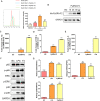
PySRA-F2 binds to RAW264.7 cells. (A) The binding ability of PySRA-F2 and RAW264.7 cells by Flow cytometry (**P < 0.01; ns, no significance). (B) The binding ability of PySRA-F2 and RAW264.7 cells by Western blot. (C) NF-κB and MAPK signaling pathways in RAW264.7 cells were activated by PySRA-F2. M: Protein marker. (D) Statistical analysis of p65, ERK, and p38 protein phosphorylation in macrophages after 15 min stimulation (*P < 0.05; ***P < 0.001). One-way ANOVA was used for the comparison of multiple groups of samples, and then the SNK test was used for pound-for-pair comparison.
Fig 5
Fig 5
CD68 blockade reduces pro-inflammatory responses. (A) To screen the RAW264.7 cell membrane proteins binding to PySRA by Silver staining. 1: Recombinant PySRA-F2 purified protein. 2: The first 200 mM imidazole eluent. 3: The second 200 mM imidazole eluent. 4: RAW264.7 cell membrane proteins. (The red arrows point to bands with differences.) (B) The recombinant protein purification by SDS-PAGE was verified. Recombinant protein of CKAP4 (~38 kDa), and CD68 (~41 kDa). M: Protein marker. (C) PySRA-F2 binds to CD68 molecule in the macrophage membrane protein. (D) The CD68 antibody attenuated the binding of PySRA to macrophages (**P < 0.01). (E) The CD68 antibody attenuated the activization of MAPK and NF-κB signaling pathways (*P < 0.05; **P < 0.01). (F) Anti-CD68 antibody attenuated the level of IL-1β secretion of RAW264.7 cells regulated by PySRA-F2 (*P < 0.05; **P < 0.01). (G) Anti-CD68 antibody attenuated the level of IL-6 secretion of RAW264.7 cells regulated by PySRA-F2 (*P < 0.05; **P < 0.01; ***P < 0.001). (H) Anti-CD68 antibody attenuated the level of TNF-α secretion of RAW264.7 cells regulated by PySRA-F2 (*P < 0.05; **P < 0.01; ns, no significance). One-way ANOVA was used for the comparison of multiple groups of samples, and then the SNK test was used for pound-for-pair comparison.

Similar articles

References

    1. WHO . 2022. World malaria report (2022). World Health Organization, Geneva.
    1. Gowda DC, Wu X. 2018. Parasite recognition and signaling mechanisms in innate immune responses to malaria. Front Immunol 9:3006. doi:10.3389/fimmu.2018.03006 - DOI - PMC - PubMed
    1. Autino B, Corbett Y, Castelli F, Taramelli D. 2012. Pathogenesis of malaria in tissues and blood. Mediterr J Hematol Infect Dis 4:e2012061. doi:10.4084/MJHID.2012.061 - DOI - PMC - PubMed
    1. Anstey NM, Russell B, Yeo TW, Price RN. 2009. The pathophysiology of vivax malaria. Trends Parasitol 25:220–227. doi:10.1016/j.pt.2009.02.003 - DOI - PubMed
    1. Miller LH, Baruch DI, Marsh K, Doumbo OK. 2002. The pathogenic basis of malaria. Nature 415:673–679. doi:10.1038/415673a - DOI - PubMed

Publication types

MeSH terms