

Genes involved in the limited spread of SARS-CoV-2 in the lower respiratory airways of hamsters may be associated with adaptive evolution
- PMID: 38624229
- PMCID: PMC11092350
- DOI: 10.1128/jvi.01784-23
Genes involved in the limited spread of SARS-CoV-2 in the lower respiratory airways of hamsters may be associated with adaptive evolution
Abstract
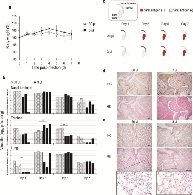
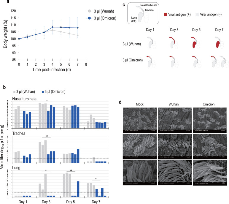
Novel respiratory viruses can cause a pandemic and then evolve to coexist with humans. The Omicron strain of severe acute respiratory syndrome coronavirus 2 has spread worldwide since its emergence in late 2021, and its sub-lineages are now established in human society. Compared to previous strains, Omicron is markedly less invasive in the lungs and causes less severe disease. One reason for this is that humans are acquiring immunity through previous infection and vaccination, but the nature of the virus itself is also changing. Using our newly established low-volume inoculation system, which reflects natural human infection, we show that the Omicron strain spreads less efficiently into the lungs of hamsters compared with an earlier Wuhan strain. Furthermore, by characterizing chimeric viruses with the Omicron gene in the Wuhan strain genetic background and vice versa, we found that viral genes downstream of ORF3a, but not the S gene, were responsible for the limited spread of the Omicron strain in the lower airways of the virus-infected hamsters. Moreover, molecular evolutionary analysis of SARS-CoV-2 revealed a positive selection of genes downstream of ORF3a (M and E genes). Our findings provide insight into the adaptive evolution of the virus in humans during the pandemic convergence phase.IMPORTANCEThe severe acute respiratory syndrome coronavirus 2 (SARS-CoV-2) Omicron variant has spread worldwide since its emergence in late 2021, and its sub-lineages are established in human society. Compared to previous strains, the Omicron strain is less invasive in the lower respiratory tract, including the lungs, and causes less severe disease; however, the mechanistic basis for its restricted replication in the lower airways is poorly understood. In this study, using a newly established low-volume inoculation system that reflects natural human infection, we demonstrated that the Omicron strain spreads less efficiently into the lungs of hamsters compared with an earlier Wuhan strain and found that viral genes downstream of ORF3a are responsible for replication restriction in the lower respiratory tract of Omicron-infected hamsters. Furthermore, we detected a positive selection of genes downstream of ORF3a (especially the M and E genes) in SARS-CoV-2, suggesting that these genes may undergo adaptive changes in humans.
Keywords: animal models; coronavirus; evolution.
Conflict of interest statement
The authors declare no conflict of interest.
Figures
Similar articles
-
Characterization of the SARS-CoV-2 BA.5.5 and BQ.1.1 Omicron variants in mice and hamsters.J Virol. 2023 Sep 28;97(9):e0062823. doi: 10.1128/jvi.00628-23. Epub 2023 Sep 7. J Virol. 2023. PMID: 37676002 Free PMC article.
-
Impact of Reinfection with SARS-CoV-2 Omicron Variants in Previously Infected Hamsters.J Virol. 2023 Jan 31;97(1):e0136622. doi: 10.1128/jvi.01366-22. Epub 2023 Jan 12. J Virol. 2023. PMID: 36633406 Free PMC article.
-
SARS-CoV-2 Omicron variant causes mild pathology in the upper and lower respiratory tract of hamsters.Nat Commun. 2022 Jun 20;13(1):3519. doi: 10.1038/s41467-022-31200-y. Nat Commun. 2022. PMID: 35725735 Free PMC article.
-
Molecular evolution of SARS-CoV-2 from December 2019 to August 2022.J Med Virol. 2023 Jan;95(1):e28366. doi: 10.1002/jmv.28366. J Med Virol. 2023. PMID: 36458547 Free PMC article. Review.
-
Hamsters as a Model of Severe Acute Respiratory Syndrome Coronavirus-2.Comp Med. 2021 Oct 1;71(5):398-410. doi: 10.30802/AALAS-CM-21-000036. Epub 2021 Sep 29. Comp Med. 2021. PMID: 34588095 Free PMC article. Review.
Cited by
-
Characterization of A(H1N1)pdm09 influenza viruses isolated between 2016 and 2019.Npj Viruses. 2025 May 22;3(1):42. doi: 10.1038/s44298-025-00126-9. Npj Viruses. 2025. PMID: 40404816 Free PMC article.
-
SGV-caller: SARS-CoV-2 genome variation caller.Heliyon. 2025 Feb 12;11(4):e42613. doi: 10.1016/j.heliyon.2025.e42613. eCollection 2025 Feb 28. Heliyon. 2025. PMID: 40040978 Free PMC article.
References
-
- Nelson MI, Viboud C, Simonsen L, Bennett RT, Griesemer SB, St George K, Taylor J, Spiro DJ, Sengamalay NA, Ghedin E, Taubenberger JK, Holmes EC. 2008. Multiple reassortment events in the evolutionary history of H1N1 influenza A virus since 1918. PLoS Pathog 4:e1000012. doi:10.1371/journal.ppat.1000012 - DOI - PMC - PubMed
MeSH terms
Grants and funding
- 16H06429, 16K21723, 16H06434, JP22H02521/MEXT | Japan Society for the Promotion of Science (JSPS)
- JP21H02736/MEXT | Japan Society for the Promotion of Science (JSPS)
- JP16K21723, JP16H06432/MEXT | Japan Society for the Promotion of Science (JSPS)
- 22K15469, 21J01036/MEXT | Japan Society for the Promotion of Science (JSPS)
- JP20fk0108281, JP19fk0108113, JP20pc0101047/Japan Agency for Medical Research and Development (AMED)
- JP20fk0108401, JP21fk0108493/Japan Agency for Medical Research and Development (AMED)
- JP23wm0125008, JP223fa627005/Japan Agency for Medical Research and Development (AMED)
- JP19fk018113, JP223fa627002h, 22gm1610010h0001/Japan Agency for Medical Research and Development (AMED)
- JPMJMS2025/MEXT | Japan Science and Technology Agency (JST)
- JPMJCR20H6/MEXT | Japan Science and Technology Agency (JST)
- Takeda Science Foundation (TSF)
LinkOut - more resources
Full Text Sources
Medical
Miscellaneous
