. 2024 May 21;90(5):e0020524.
doi: 10.1128/aem.00205-24. Epub 2024 Apr 16.

Characterization of Amycolatopsis 75iv2 dye-decolorizing peroxidase on O-glycosides

Affiliations

Characterization of Amycolatopsis 75iv2 dye-decolorizing peroxidase on O-glycosides

Silja Välimets et al. Appl Environ Microbiol. .

Abstract

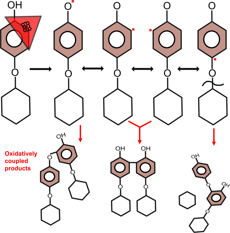
Dye-decolorizing peroxidases are heme peroxidases with a broad range of substrate specificity. Their physiological function is still largely unknown, but a role in the depolymerization of plant cell wall polymers has been widely proposed. Here, a new expression system for bacterial dye-decolorizing peroxidases as well as the activity with previously unexplored plant molecules are reported. The dye-decolorizing peroxidase from Amycolatopsis 75iv2 (DyP2) was heterologously produced in the Gram-positive bacterium Streptomyces lividans TK24 in both intracellular and extracellular forms without external heme supplementation. The enzyme was tested on a series of O-glycosides, which are plant secondary metabolites with a phenyl glycosidic linkage. O-glycosides are of great interest, both for studying the compounds themselves and as potential models for studying specific lignin-carbohydrate complexes. The primary DyP reaction products of salicin, arbutin, fraxin, naringin, rutin, and gossypin were oxidatively coupled oligomers. A cleavage of the glycone moiety upon radical polymerization was observed when using arbutin, fraxin, rutin, and gossypin as substrates. The amount of released glucose from arbutin and fraxin reached 23% and 3% of the total substrate, respectively. The proposed mechanism suggests a destabilization of the ether linkage due to the localization of the radical in the para position. In addition, DyP2 was tested on complex lignocellulosic materials such as wheat straw, spruce, willow, and purified water-soluble lignin fractions, but no remarkable changes in the carbohydrate profile were observed, despite obvious oxidative activity. The exact action of DyP2 on such lignin-carbohydrate complexes therefore remains elusive.

Importance: Peroxidases require correct incorporation of the heme cofactor for activity. Heterologous overproduction of peroxidases often results in an inactive enzyme due to insufficient heme synthesis by the host organism. Therefore, peroxidases are incubated with excess heme during or after purification to reconstitute activity. S. lividans as a production host can produce fully active peroxidases both intracellularly and extracellularly without the need for heme supplementation. This reduces the number of downstream processing steps and is beneficial for more sustainable production of industrially relevant enzymes. Moreover, this research has extended the scope of dye-decolorizing peroxidase applications by studying naturally relevant plant secondary metabolites and analyzing the formed products. A previously overlooked artifact of radical polymerization leading to the release of the glycosyl moiety was revealed, shedding light on the mechanism of DyP peroxidases. The key aspect is the continuous addition, rather than the more common approach of a single addition, of the cosubstrate, hydrogen peroxide. This continuous addition allows the peroxidase to complete a high number of turnovers without self-oxidation.

Keywords: O-glycosides; dye-decolorizing peroxidase; recombinant expression.


Conflict of interest statement

The authors declare no conflict of interest.

Figures

Fig 1
Fig 1
Extracellular production of DyP2 in Streptomyces lividans. Cells were cultured in selective growth medium, and samples were taken at defined time points. The supernatant was harvested by centrifugation, and the activity was measured spectrophotometrically. The activity measurements are an average of three biological replicates in three technical replicates each. Wet cell weight (WCW) was measured to follow bacterial growth. (A) Secretion of DyP2 using the signal peptide from B. subtilis A-type DyP (SP-DyP2). (B) Secretion of DyP2 without signal peptide (DyP2). (C) Ultraviolet-visible spectrum of secreted and purified DyP2. The enzyme was purified from the culture medium after 2 days of production using affinity chromatography. The maximum absorption of the Soret band was at 411 nm.(D) Western blot (WB) analysis of purified SP-DyP2. Sizes 75, 50, and 37 kDa are marked on the ladder (L). The expected size of DyP2 based on theoretical molecular weight calculation after secretion and cleavage of the signal peptide was 52 kDa.
Fig 2
Fig 2
Overview of O-glycosides used in this study. Salicin, arbutin, and fraxin containing ether-linked glucosyl moiety were fully soluble in reaction buffer. Naringin, rutin, and gossypin containing ether-linked neohesperidosyl, rutinosyl, or glucosyl moieties, respectively, were partially soluble in reaction buffer.
Fig 3
Fig 3
UHPLC-MS time course chromatograms in negative mode. (A) Fraxin reaction products and (B) arbutin reaction products. Substrates with a final concentration of 2.6 mM were solubilized in 50 mM sodium acetate buffer at pH 4.5; 0.2 µM enzyme was added, and the reaction was initiated with 200 µM H2O2. The reaction was continued by adding fresh H2O2 after every 3 min for 60 min. The H2O2 turnover per time point is color coded above the figures. Samples were taken at 3, 15, 30, 45, and 60 min. Substrates containing only H2O2 or DyP2 were used as controls. The incubations were performed in duplicate, but only one of the identical replicates is shown. Tentatively annotated products of fraxin and arbutin incubations are listed in Table 1 or 2, respectively.
Fig 4
Fig 4
High-resolution fragmentation of arbutin dimers. Arbutin 15-min incubation time point was analyzed in high-resolution MS IQ-X. By using acetonitrile gradient starting from 1%, the dimers eluted at RTs of 6.11 and 10.61 min corresponded to the dimers at RTs 2.12 and 4.59 min in the UHPLC-MS analysis starting at 5% acetonitrile. Full MS2 was obtained using HCD 35% collision energy, and obtained MS2 fragments were further fragmented in MS3 using CID with 35% collision energy. Dimers at RTs of (A) 6.11 min and (B) 10.61 min have significantly different fragmentation patterns indicating different linkages upon oxidative coupling.
Fig 5
Fig 5
MALDI-TOF MS time course spectra of fraxin and arbutin. Substrates with a final concentration of 2.6 mM were solubilized in 50-mM sodium acetate buffer at pH 4.5; 0.2 µM enzyme was added; and the reaction was initiated with 200 µM H2O2. The reaction was continued by adding fresh H2O2 after every 3 min for 60 min. The H2O2 turnovers per time point are color coded above the figures. Samples were taken at 3, 15, 30, 45, and 60 min. Substrates containing only H2O2 or DyP2 were used as controls. The incubations were performed in duplicate, but only one of the identical replicates is shown. MALDI-TOF MS was measured in positive mode. (A) Fraxin incubation products were partially soluble in buffer. No precipitate was observed after 3 min. The insoluble fractions were observed after 15 min and were separated by centrifugation. The remaining pellet was solubilized in MeOH/DMSO mixture and analyzed. The products are listed in Table 3. (B) Arbutin incubation products were fully soluble in buffer. The products are listed in Table 4 as Na+ adducts.
Fig 6
Fig 6
Carbohydrate analysis of O-glycoside incubations with DyP2. HPAEC time course chromatograms of released glucose after (A) fraxin and (B) arbutin incubations. Substrates with H2O2 or DyP2 only were used as controls. Glucose with defined concentrations were used as standards. The peak eluting at 4.3 min corresponds to unconsumed H2O2, and the peak eluting at 4.6 min corresponds to glucose. The incubations were performed in duplicate, but only one of the identical replicates is shown. (C) Quantification of glucose release from the total substrate. The initial substrate concentration (2.6 mM) was set to 100%. Glucose release was time dependent in both substrate incubations, reaching 14.4% in arbutin and 3.2% in fraxin incubations after 60 min. The average of duplicates is presented. (D) Quantification of glucose release from 0.26 mM arbutin. The initial substrate concentration 2.6 mM was decreased 10 times to 0.26 mM and was set to 100%. After the total substrate consumption in 15 min, the glucose yield was 23.5%. The average of duplicates is presented.
Fig 7
Fig 7
Proposed mechanism of radical mediated glycone release. DyP2 (red triangle) oxidizes the hydroxy group, but the radicals (red squares) can be distributed in the aromatic ring, resulting in many products of different isomers indicated by red arrows. When the radical is at the para position, then it could lead to destabilization of the ether linkage resulting in carbohydrate release.

References

    1. Kim SJ, Shoda M. 1999. Purification and characterization of a novel peroxidase from Geotrichum candidum Dec 1 involved in decolorization of dyes. Appl Environ Microbiol 65:1029–1035. doi:10.1128/AEM.65.3.1029-1035.1999 - DOI - PMC - PubMed
    1. Heinfling A, Martínez MJ, Martínez AT, Bergbauer M, Szewzyk U. 1998. Purification and characterization of peroxidases from the dye-decolorizing fungus Bjerkandera adusta. FEMS Microbiol Lett 165:43–50. doi:10.1111/j.1574-6968.1998.tb13125.x - DOI - PubMed
    1. Hofrichter M, Ullrich R, Pecyna MJ, Liers C, Lundell T. 2010. New and classic families of secreted fungal heme peroxidases. Appl Microbiol Biotechnol 87:871–897. doi:10.1007/s00253-010-2633-0 - DOI - PubMed
    1. Colpa DI, Fraaije MW, van Bloois E. 2014. DyP-type peroxidases: a promising and versatile class of enzymes. J Ind Microbiol Biotechnol 41:1–7. doi:10.1007/s10295-013-1371-6 - DOI - PubMed
    1. Sugano Y, Yoshida T. 2021. Dyp-type peroxidases: recent advances and perspectives. Int J Mol Sci 22:5556. doi:10.3390/ijms22115556 - DOI - PMC - PubMed

Publication types

MeSH terms

Supplementary concepts

LinkOut - more resources