Comparative Study
. 2024 Oct;34(10):6488-6498.
doi: 10.1007/s00330-024-10652-4. Epub 2024 Apr 16.

Diagnostic performance of [68Ga]DOTATATE PET/CT, [18F]FDG PET/CT, MRI of the spine, and whole-body diagnostic CT and MRI in the detection of spinal bone metastases associated with pheochromocytoma and paraganglioma

Affiliations
Comparative Study

Diagnostic performance of [68Ga]DOTATATE PET/CT, [18F]FDG PET/CT, MRI of the spine, and whole-body diagnostic CT and MRI in the detection of spinal bone metastases associated with pheochromocytoma and paraganglioma

Abhishek Jha et al. Eur Radiol. 2024 Oct.

Abstract

Objective: To compare the diagnostic performance of [68Ga]DOTATATE PET/CT, [18F]FDG PET/CT, MRI of the spine, and whole-body CT and MRI for the detection of pheochromocytoma/paraganglioma (PPGL)-related spinal bone metastases.

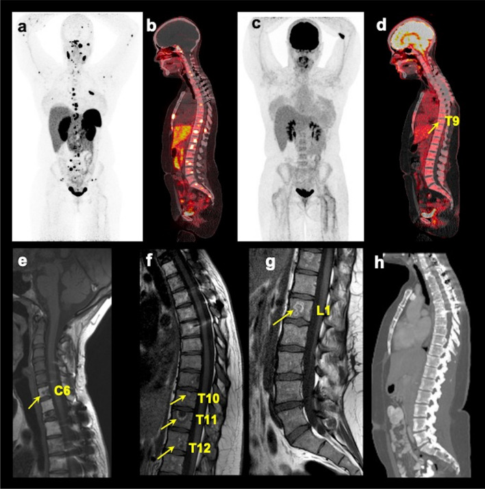
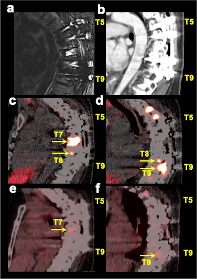
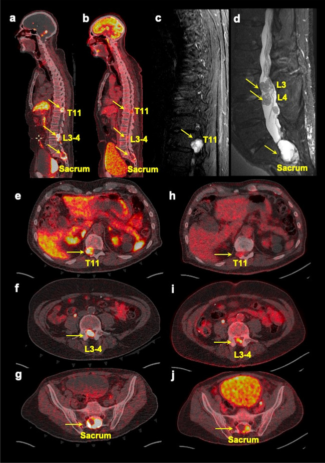
Materials and methods: Between 2014 and 2020, PPGL participants with spinal bone metastases prospectively underwent [68Ga]DOTATATE PET/CT, [18F]FDG PET/CT, MRI of the cervical-thoracolumbar spine (MRIspine), contrast-enhanced MRI of the neck and thoraco-abdominopelvic regions (MRIWB), and contrast-enhanced CT of the neck and thoraco-abdominopelvic regions (CTWB). Per-patient and per-lesion detection rates were calculated. Counting of spinal bone metastases was limited to a maximum of one lesion per vertebrae. A composite of all functional and anatomic imaging served as an imaging comparator. The McNemar test compared detection rates between the scans. Two-sided p values were reported.

Results: Forty-three consecutive participants (mean age, 41.7 ± 15.7 years; females, 22) with MRIspine were included who also underwent [68Ga]DOTATATE PET/CT (n = 43), [18F]FDG PET/CT (n = 43), MRIWB (n = 24), and CTWB (n = 33). Forty-one of 43 participants were positive for spinal bone metastases, with 382 lesions on the imaging comparator. [68Ga]DOTATATE PET/CT demonstrated a per-lesion detection rate of 377/382 (98.7%) which was superior compared to [18F]FDG (72.0%, 275/382, p < 0.001), MRIspine (80.6%, 308/382, p < 0.001), MRIWB (55.3%, 136/246, p < 0.001), and CTWB (44.8%, 132/295, p < 0.001). The per-patient detection rate of [68Ga]DOTATATE PET/CT was 41/41 (100%) which was higher compared to [18F]FDG PET/CT (90.2%, 37/41, p = 0.13), MRIspine (97.6%, 40/41, p = 1.00), MRIWB (95.7%, 22/23, p = 1.00), and CTWB (81.8%, 27/33, p = 0.03).

Conclusions: [68Ga]DOTATATE PET/CT should be the modality of choice in PPGL-related spinal bone metastases due to its superior detection rate.

Clinical relevance statement: In a prospective study of 43 pheochromocytoma/paraganglioma participants with spinal bone metastases, [68Ga]DOTATATE PET/CT had a superior per-lesion detection rate of 98.7% (377/382), compared to [18F]FDG PET/CT (p < 0.001), MRI of the spine (p < 0.001), whole-body CT (p < 0.001), and whole-body MRI (p < 0.001).

Key points: • Data regarding head-to-head comparison between functional and anatomic imaging modalities to detect spinal bone metastases in pheochromocytoma/paraganglioma are limited. • [68Ga]DOTATATE PET/CT had a superior per-lesion detection rate of 98.7% in the detection of spinal bone metastases associated with pheochromocytoma/paraganglioma compared to other imaging modalities: [18]F-FDG PET/CT, MRI of the spine, whole-body CT, and whole-body MRI. • [68Ga]DOTATATE PET/CT should be the modality of choice in the evaluation of spinal bone metastases associated with pheochromocytoma/paraganglioma.

Keywords: Fluorodeoxyglucose F18; Gallium GA 68 DOTATATE; Neuroendocrine tumors; Paraganglioma; Pheochromocytoma.


Conflict of interest statement

The authors of this manuscript declare no relationships with any companies, whose products or services may be related to the subject matter of the article.

Figures

Fig. 1
Fig. 1
Schematic diagram representing enrollment of participants in the study. The diagram of flow of participants’ inclusion and exclusion
Fig. 2
Fig. 2
Multimodality imaging of spinal bone metastases in a pheochromocytoma/paraganglioma participant. The images of whole-body 68Ga-DOTA(0)-Tyr(3)-octreotate ([68Ga]DOTATATE; a anterior maximum intensity projection (MIP) and (b) fused sagittal PET/CT), 18F-fluoro-2-deoxy-d-glucose ([18F]FDG; c MIP and (d) fused sagittal PET/CT), T1-weighted sagittal MRI of the cervical (e), thoracic (f), and lumbar (g) spine, and contrast-enhanced CT (h sagittal) of a 25-year-old woman with germline pathogenic variant in a gene encoding for succinate dehydrogenase B subunit are shown. This figure shows superiority of [68Ga]DOTATATE PET/CT in the detection of additional spinal bone metastases at C2, C5, C7, T1-2, T4-8, L2, and L5 compared to [18F]FDG PET/CT in detecting spinal bone metastases at T9 (arrow on fused sagittal image and not appreciated on MIP image) and L4 (not appreciated on MIP and fused sagittal images), and MRI spine at C6, T10-12, and L1 (arrows), respectively. The whole-body CT was read negative for any spinal bone metastases. This participant did not undergo whole-body MRI
Fig. 3
Fig. 3
Multimodality imaging in a spinal bone metastatic pheochromocytoma/paraganglioma participant with spinal hardware. The images of sagittal short tau inversion recovery (STIR, a) MRI, contrast-enhanced CT (b sagittal), fused sagittal 68Ga-DOTA(0)-Tyr(3)-octreotate ([68Ga]DOTATATE; c, d images at two different sagittal planes) PET/CT, and fused sagittal 18F-fluoro-2-deoxy-d-glucose ([18F]FDG PET/CT; e, f images at the same two sagittal planes as [68Ga]DOTATATE PET/CT) focused on thoracic spine of a 48-year-old woman with negative germline testing in pheochromocytoma and paraganglioma susceptibility genes are shown. This participant had spinal hardware placed at the T5–T9 vertebrae which therefore were excluded from the analysis due to the associated artifacts on MRI and CT. However, PET/CT imaging clearly is advantageous in the evaluation of patients with spinal bone hardware as demonstrated by the spinal bone metastases at T7–T9 on [68Ga]DOTATATE PET/CT (arrows) and at T7 and T9 on [18F]FDG PET/CT (arrows), where spinal hardware is placed. The uptake on [18F]FDG PET/CT is comparatively much fainter compared to [68Ga]DOTATATE PET/CT. This participant did not undergo whole-body MRI
Fig. 4
Fig. 4
Multimodality imaging in a spinal bone metastatic pheochromocytoma/paraganglioma participant with spinal canal involvement. The whole-body fused sagittal (a, b) images of 68Ga-DOTA(0)-Tyr(3)-octreotate ([68Ga]DOTATATE, a) PET/CT, 18F-fluoro-2-deoxy-d-glucose ([18F]FDG, b) PET/CT, and sagittal short tau inversion recovery (STIR, c, d) images of thoracic (c), lumbar (d), and axial (ej) fused PET/CT images of [68Ga]DOTATATE (eg) and [18F]FDG (hj) of a 71-year-old man with negative germline testing in pheochromocytoma and paraganglioma susceptibility genes. This figure shows spinal bone metastases (arrows, aj) in T11, L3–L4, and sacrum with spinal canal involvement. In such cases, a dedicated MRI of the spine (showing intradural tumors, here) to derive more anatomic information should be obtained

References

    1. Pacak K (2022) New biology of pheochromocytoma and paraganglioma. Endocr Pract 28:1253–1269 10.1016/j.eprac.2022.09.003 - DOI - PMC - PubMed
    1. Taïeb D, Wanna GB, Ahmad M et al (2023) Clinical consensus guideline on the management of phaeochromocytoma and paraganglioma in patients harbouring germline SDHD pathogenic variants. Lancet Diabetes Endocrinol 11:345–361 10.1016/S2213-8587(23)00038-4 - DOI - PMC - PubMed
    1. Taïeb D, Nölting S, Perrier ND et al (2024) Management of phaeochromocytoma and paraganglioma in patients with germline SDHB pathogenic variants: an international expert consensus statement. Nat Rev Endocrinol 20:168–184 10.1038/s41574-023-00926-0 - DOI - PubMed
    1. Ayala-Ramirez M, Palmer JL, Hofmann MC et al (2013) Bone metastases and skeletal-related events in patients with malignant pheochromocytoma and sympathetic paraganglioma. J Clin Endocrinol Metab 98:1492–1497 10.1210/jc.2012-4231 - DOI - PMC - PubMed
    1. Hamidi O, Young WF Jr, Iniguez-Ariza NM et al (2017) Malignant pheochromocytoma and paraganglioma: 272 patients over 55 years. J Clin Endocrinol Metab 102:3296–3305 10.1210/jc.2017-00992 - DOI - PMC - PubMed

Publication types

MeSH terms

LinkOut - more resources