. 2024 Apr 16;9(10):e168594.
doi: 10.1172/jci.insight.168594.

Altered lipid homeostasis is associated with cerebellar neurodegeneration in SNX14 deficiency

Affiliations

Altered lipid homeostasis is associated with cerebellar neurodegeneration in SNX14 deficiency

Yijing Zhou et al. JCI Insight. .

Abstract

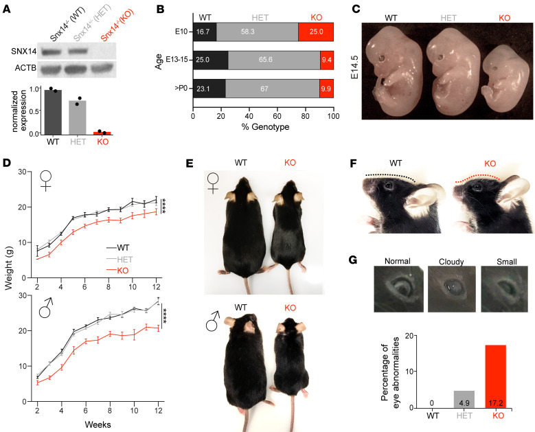
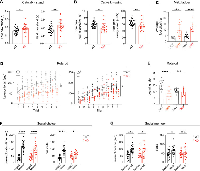
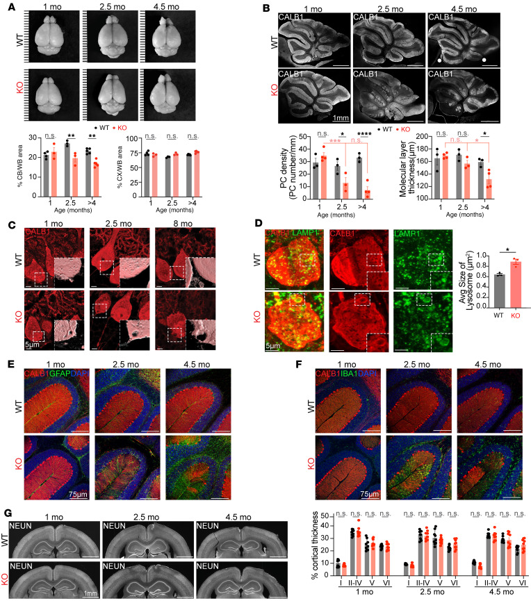
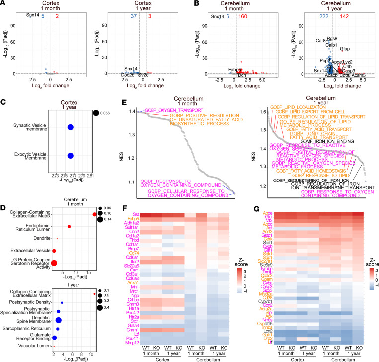
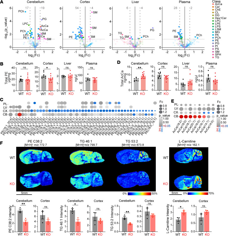
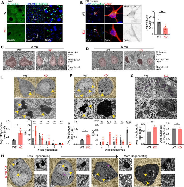
Dysregulated lipid homeostasis is emerging as a potential cause of neurodegenerative disorders. However, evidence of errors in lipid homeostasis as a pathogenic mechanism of neurodegeneration remains limited. Here, we show that cerebellar neurodegeneration caused by Sorting Nexin 14 (SNX14) deficiency is associated with lipid homeostasis defects. Recent studies indicate that SNX14 is an interorganelle lipid transfer protein that regulates lipid transport, lipid droplet (LD) biogenesis, and fatty acid desaturation, suggesting that human SNX14 deficiency belongs to an expanding class of cerebellar neurodegenerative disorders caused by altered cellular lipid homeostasis. To test this hypothesis, we generated a mouse model that recapitulates human SNX14 deficiency at a genetic and phenotypic level. We demonstrate that cerebellar Purkinje cells (PCs) are selectively vulnerable to SNX14 deficiency while forebrain regions preserve their neuronal content. Ultrastructure and lipidomic studies reveal widespread lipid storage and metabolism defects in SNX14-deficient mice. However, predegenerating SNX14-deficient cerebella show a unique accumulation of acylcarnitines and depletion of triglycerides. Furthermore, defects in LD content and telolysosome enlargement in predegenerating PCs suggest lipotoxicity as a pathogenic mechanism of SNX14 deficiency. Our work shows a selective cerebellar vulnerability to altered lipid homeostasis and provides a mouse model for future therapeutic studies.

Keywords: Lysosomes; Monogenic diseases; Neurodegeneration; Neuroscience.


Figures

Figure 1
Figure 1. SNX14-deficient mice show developmental delay and atypical facial features.
(A) Representative Western blot (WB) images show loss of SNX14 expression in Snx14-KO mice tissue. β-Actin (ACTB) was used as loading control. Bar graph shows WB band densitometry quantification of SNX14 relative to ACTB. n = 2 for each genotype. (B) Percentage of embryos/mice with the indicated genotypes obtained from heterozygous parent mattings. The χ2 test shows siginficant discrepancy between >P0 observed and expected values (P = 0.001) indicating embryonic lethality of KOs. E10, n = 12; E13–15, n = 32; >P0, n = 91. (C) Representative image of WT, HET, and KO E14.5 embryos showing smaller size of KOs. (D) Growth curves show consistently lower body weight of 2- to 12-week-old Snx14-KO males and females. Data represent mean ± SEM of n ≥ 3. Two-way ANOVA shows siginficant effect of genotype. ****P < 0.0001. (E) Representative images of 9-month-old WT and KO littermates of each sex. (F) Representative images showing the atypical face with forehead protrusion of 6-month-old KO mice (red line) compared with WT littermate. (G) Representative images of 8-month-old KO mice showing eye abnormalities, including cataracts (cloudy) and microphthalmia (small). Bar graph shows percentages of mice with eye abnormality for each genotype.
Figure 2
Figure 2. SNX14 deficiency in mice recapitulates motor and behavioral deficits of SCAR20.
(A and B) Catwalk analysis shows altered gait of KO mice with a longer stand (A) and shorter swing (B) than WT mice. Data are shown as mean ± SEM of n = 24 WT and n = 18 KO mice. Two-tailed Welch’s t test. (C) Metz ladder rung test shows altered limb placing and coordination of KO males and females. Data are shown as mean foot slip of 5 trials performed in consecutive days ± SEM of n = 10 WT males, n = 7 KO males, n = 12 WT females, and n = 12 KO females. Two-way ANOVA followed by Šidák’s test. (D) Accelerating rotarod reveals defects in motor performance of KO mice in the 9 trials performed over 3 consecutive days. Data are shown as mean latency to fall ± SEM of n = 11 WT males, n = 7 KO males, n = 13 WT females, and n = 11 KO. Two-way ANOVA shows siginficant effect of genotype. (E) KO females show impaired learning rate on accelerating rotarod performance over time (between trial 1 and 9). Data are shown as mean learning rate ± SEM of n = 9 WT males, n = 7 KO males, n = 13 WT females, and n = 10 KO females. Two-way ANOVA followed by Šidák’s test. (F and G) Three-chamber social interaction test showing similar preference for a mouse over an object between WT and KO mice (F) but impaired social novelty preference in KO mice (G). Data are shown as mean ± SEM of n = 24 WT and n = 17 KO. Two-way ANOVA followed by Tukey’s test. *P < 0.05, **P < 0.01, ***P < 0.001, ****P < 0.0001.
Figure 3
Figure 3. SNX14 deficiency causes selective cerebellar degeneration.
(A) Representative brain images from WT and KO mice at indicated age shows shrinkage of KO cerebellum (CB) over time. Ruler marks separated by 1 mm. Bar graphs show percentage area of CB or cerebral cortex (CX) relative to the whole brain (WB) in n = 3–5 mice. Two-way ANOVA followed by Šidák’s test. (B) Representative cerebellar sagittal sections immunostained with PC-specific anti-CALB1 antibody reveal progressive loss of PCs in KO mice. Bar graphs show PC linear density (right) and thickness of the molecular layer (left) in the cerebellar lobule III of n = 3–4 mice. Two-way ANOVA followed by Šidák’s test. (C) Representative immunostaining of PCs with anti-CALB1 antibody reveals progressive accumulation of vacuoles in KO mice. (D) Immunostaining of PCs with anti-CALB1 and lysosomes with anti-LAMP1 show enlarged lysosomes in KO mice. Bar graph shows average lysosome size per mouse. n = 3 mice (in WT, 29 PCs and 4,033 lysosomes were counted; in KO, 30 PCs and 3,247 lysosomes were counted). Two-tailed t test. (E and F) Representative immunostaining showing progressive accumulation of astrocytes labeled with anti-GFAP (E) and microglia with anti-IBA1 (F) in degenerating KO cerebella (base of lobules III and IV). (G) Coronal sections of cerebral cortices immunostained with anti-NeuN do not show differences between WT and KO mice. Bar graphs show percentage thickness occupied by each cortical layers (I–VI) in 4–5 cortical regions of 2 mice per genotype and age. Two-way ANOVA followed by Šidák’s test. In all graphs, data represent mean ± SEM. *P < 0.05, **P < 0.01, ****P < 0.0001. Scale bars: 1 mm, 5 μm, 75 μm.
Figure 4
Figure 4. Genes involved in lipid response are differentially expressed in SNX14-deficient cerebella.
(A and B) Volcano plots of differentially expressed genes (DEGs) in the cortex and cerebellum of WT versus KO mice at 1 month and 1 year. Dashed lines indicate statistical siginficance cut off (–log10[Padj] > 1.301 and log2[FC] = ± 0.5). Number of siginficantly down- and upregulated genes are displayed on the top of each plot in blue and red, respectively. (C and D) Dot plots of gene ontology (GO) enrichment analysis of the DEGs, with selected down- and upregulated GO categories marked in blue and red, respectively. Dot size indicates proportion of DEGs relative to the total number of genes in each category. (E) Waterfall plots of Gene Set Enrichment Analysis (GSEA) of cerebellum-specific siginficant gene ontology terms. Terms in orange, magenta, and black are related to lipid, oxygen, and iron, respectively. (F) Heatmap of the top 20 leading edge genes of each term displayed in the 1-month cerebellum GSEA shown in E. (G) Heatmap of the top 10 leading edge genes of each term displayed in the 1-year Cerebellum GSEA shown in E.
Figure 5
Figure 5. Unique deregulation of lipid metabolites in predegenerating KO cerebella.
(A) Volcano plots show deregulated lipids in 2-month-old Snx14-KO cerebellum (CB), cerebral cortex (CX), liver, and plasma. Horizontal gray lines indicate P < 0.05 cut-off. Data show increased concentrations of acylcarnitine (AcCa) species specifically in KO CB. (B) Bar graphs show total PE concentrations per tissue in n = 8 WT and n = 10 KO mice. Two-tailed t test. PEs are siginficantly reduced in Snx14-KO CX. (C) Dot plot depicting fold change (FC) (proportional to dot size) and P value (in gray intensity scale) of PE species detected in cerebral cortices for all analyzed tissues. Red dots represent siginficantly increased lipids, while blue dots represent siginficantly decreased lipids. (D) Bar graphs show total AcCa concentrations in n = 8 WT and n = 10 KO mice. Two-tailed t test. AcCa-s are siginficantly increased only in KO CB. (E) Dot plot depicting FC and P value of AcCa species detected in cerebellar samples for all analyzed tissues. Red dots represent siginficantly increased lipids, while blue dots represent siginficantly decreased lipids. (F) MALDI-MS imaging of brain cryosections show reduction of PE C38:2, TG 46:1, and TG 53:2 and cerebellar accumulation of L-carnitine in KO. The molecules were revealed in positive ion mode using DHB matrix, and the m/z (mass/charge ratio) of [M + H]+ are indicated. Heatmap colors depict the relative abundance of each metabolic species. Bar graphs show cerebellar or cortical intensity of each lipid species in n = 3 per genotype. In all panels, data are shown as mean ± SEM. *P < 0.05, **P < 0.01. Key of each lipid class can be found Supplemental Figure 6 legend. Scale bar: 5 mm.
Figure 6
Figure 6. Lipid storage organelles are affected in SNX14-deficient tissue.
(A) Representative BODIPY 493/503 (BD493) labeling shows less lipid droplets (LDs) in 2-month-old KO mice liver sections. (B) Representative BD493 and anti-CALB1 labeling shows less LDs in Snx14-KO primary cerebellar culture PCs. Bar graphs show average number of LDs per CALB1+ PC in n = 6 mice per genotype used for PC cultures. Total number of CALB1+ PC quantified: n = 69 WT and n = 50 KO. Two-tailed t test. (C and D) Representative TEM image of PC layer in WT and KO mice at 2 (C) and 6 (D) months of age. (E) Representative TEM images of PCs show less but larger telolysosomes in 2-month-old KO mice. Bottom graphs show the average area of telolysosomes (left) and the percentage of PCs with indicated number of telolysosomes (right) in n = 3 mice per genotype (6–10 PCs per mouse). Two-tailed t test (left) and 2-way ANOVA followed by Šidák’s test (right). (F) Representative TEM image of PCs showing less but larger telolysosomes in 6-month-old KO mice. Bottom graphs show the average area of telolysosomes (left) and percentage of PCs with indicated number of telolysosomes (right) in n = 3 mice per genotype (6–10 PCs per mouse). Two-tailed t test (left) and 2-way ANOVA followed by Šidák’s test (right). (G) Representative TEM image of PC mitochondria at 6 months of age. Bottom bar graphs show the average area (left) and roundness (right) of mitochondria in n = 3 mice per genotype (10 PCs per mouse). Two-tailed t tests. (H) Representative TEM images show a spectrum of less to more degenerating PCs from 6-month-old KO mice. Yellow arrowheads point to insets of enlarged telolysosomes in E and F, and mitochondria and enlarged telolysosomes in H. ER swelling highlighted in magenta and indicated with an asterisk. In all panels, data represent mean ± SEM. *P < 0.05, **P < 0.01, ***P < 0.001, ****P < 0.0001. Scale bars: 200 μm, 50 μm, 15 μm, 5 μm, 20 μm, 5 μm, 2 μm, 0.5 μm.

References

    1. Taylor JP, et al. Toxic proteins in neurodegenerative disease. Science. 2002;296(5575):1991–1995. doi: 10.1126/science.1067122. - DOI - PubMed
    1. Scrivo A, et al. Selective autophagy as a potential therapeutic target for neurodegenerative disorders. Lancet Neurol. 2018;17(9):802–815. doi: 10.1016/S1474-4422(18)30238-2. - DOI - PMC - PubMed
    1. Martini-Stoica H, et al. The autophagy-lysosomal pathway in neurodegeneration: a TFEB perspective. Trends Neurosci. 2016;39(4):221–234. doi: 10.1016/j.tins.2016.02.002. - DOI - PMC - PubMed
    1. Hernandez-Diaz S, Soukup SF. The role of lipids in autophagy and its implication in neurodegeneration. Cell Stress. 2020;4(7):167–186. doi: 10.15698/cst2020.07.225. - DOI - PMC - PubMed
    1. Welte MA. Expanding roles for lipid droplets. Curr Biol. 2015;25(11):R470–R481. doi: 10.1016/j.cub.2015.04.004. - DOI - PMC - PubMed