. 2024 Apr 16;19(4):e0301990.
doi: 10.1371/journal.pone.0301990. eCollection 2024.

SIRT3 regulates cardiolipin biosynthesis in pressure overload-induced cardiac remodeling by PPARγ-mediated mechanism

Affiliations

SIRT3 regulates cardiolipin biosynthesis in pressure overload-induced cardiac remodeling by PPARγ-mediated mechanism

Ling-Xin Liu et al. PLoS One. .

Abstract

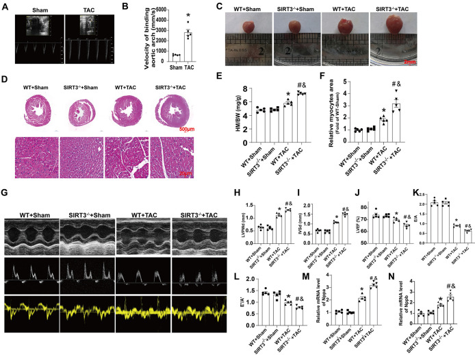
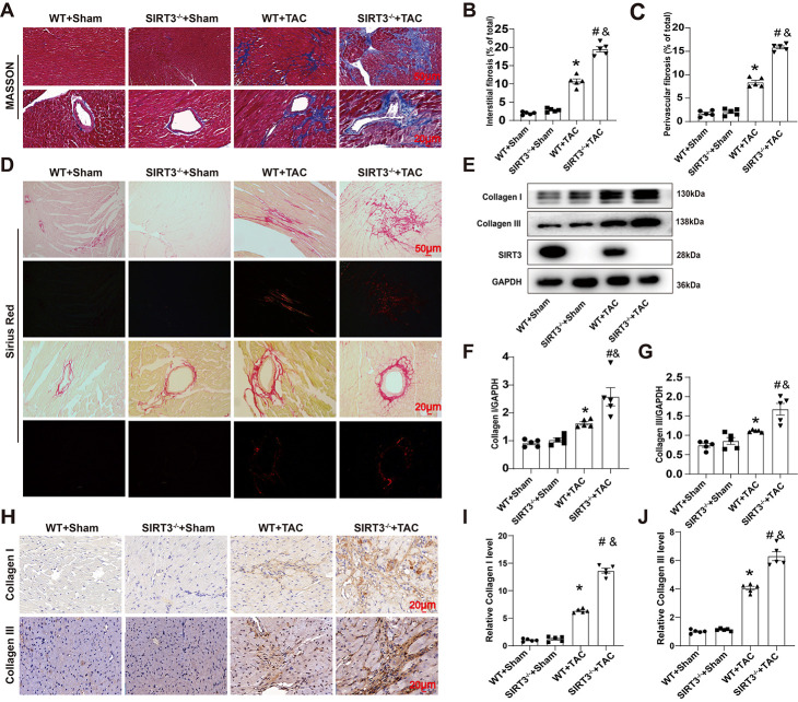
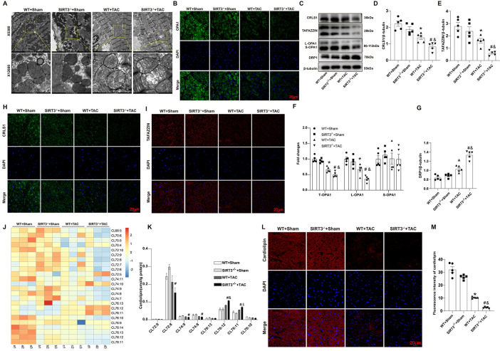
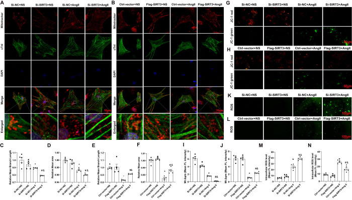
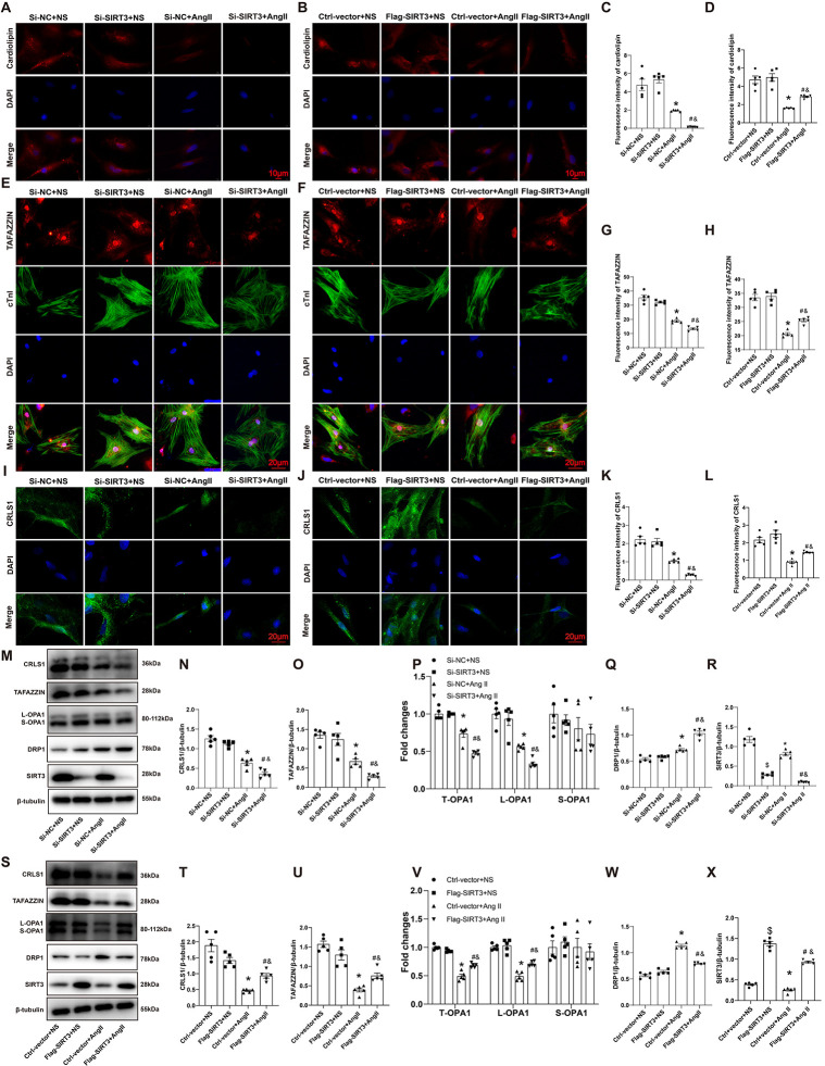
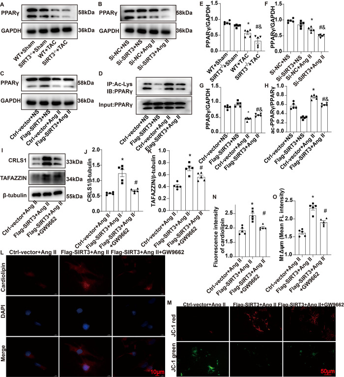
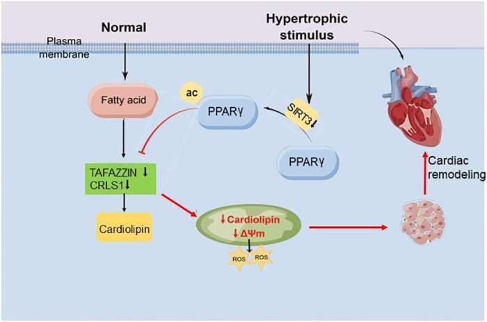
Cardiac remodeling is the primary pathological feature of chronic heart failure (HF). Exploring the characteristics of cardiac remodeling in the very early stages of HF and identifying targets for intervention are essential for discovering novel mechanisms and therapeutic strategies. Silent mating type information regulation 2 homolog 3 (SIRT3), as a major mitochondrial nicotinamide adenine dinucleotide (NAD)-dependent deacetylase, is required for mitochondrial metabolism. However, whether SIRT3 plays a role in cardiac remodeling by regulating the biosynthesis of mitochondrial cardiolipin (CL) is unknown. In this study, we induced pressure overload in wild-type (WT) and SIRT3 knockout (SIRT3-/-) mice via transverse aortic constriction (TAC). Compared with WT mouse hearts, the hearts of SIRT3-/- mice exhibited more-pronounced cardiac remodeling and fibrosis, greater reactive oxygen species (ROS) production, decreased mitochondrial-membrane potential (ΔΨm), and abnormal mitochondrial morphology after TAC. Furthermore, SIRT3 deletion aggravated TAC-induced decrease in total CL content, which might be associated with the downregulation of the CL synthesis related enzymes cardiolipin synthase 1 (CRLS1) and phospholipid-lysophospholipid transacylase (TAFAZZIN). In our in vitro experiments, SIRT3 overexpression prevented angiotensin II (AngII)- induced aberrant mitochondrial function, CL biosynthesis disorder, and peroxisome proliferator-activated receptor gamma (PPARγ) downregulation in cardiomyocytes; meanwhile, SIRT3 knockdown exacerbated these effects. Moreover, the addition of GW9662, a PPARγ antagonist, partially counteracted the beneficial effects of SIRT3 overexpression. In conclusion, SIRT3 regulated PPARγ-mediated CL biosynthesis, maintained the structure and function of mitochondria, and thereby protected the myocardium against cardiac remodeling.


Conflict of interest statement

The authors have declared that no competing interests exist.

Figures

Fig 1
Fig 1. SIRT3 deletion aggravated pathological development of cardiac remodeling induced by TAC.
(A) Velocity of binding aortic arch in Sham- or TAC-treated mice. (B) Statistical analysis of velocity of binding aortic arch of Sham- or TAC-treated mice (n = 5). (C) Representative gross appearance of Sham- or TAC-treated WT and SIRT3-/- mice heart. (D) HE-stained cross-sections of mouse hearts. (E) Heart mass (HM) normalized to body weight (BW). (F) Relative myocyte size by HE staining. (G) Representative echocardiographic images of M-mode, pulse-wave Doppler and tissue Doppler in 4 groups of mice. (H-L). Statistical analysis of LVPWd, IVSd, LVEF, E/A and E’/A’. M-N. Statistical analysis of relative mRNA level of Nppa and Nppb. n = 5 per group. *p<0.05 vs. WT+Sham group, &p<0.05 vs. SIRT3-/-+Sham group, #p<0.05 vs. WT+TAC group.
Fig 2
Fig 2. SIRT3 deletion aggravated TAC-induced myocardial fibrosis.
(A) Masson-stained heart sections. (B) Statistical analysis of interstitial fibrosis fraction. (C) Statistical analysis of perivascular fibrosis fraction. (D) Sirius red‐stained heart sections. (E) Representative Western blot images of Collagen I, Collagen III and SIRT3 protein expressions in the myocardium. (F-G). Quantifications of Collagen I, Collagen III protein expressions in the myocardium. (H-J) Representative immunohistochemical staining and quantifications of Collagen I, Collagen III and TUNEL in the myocardium. n = 5 per group. *p<0.05 vs. WT+Sham group, &p<0.05 vs. SIRT3-/-+Sham group, #p<0.05 vs. WT+TAC group.
Fig 3
Fig 3. SIRT3 deletion impaired mitochondrial structure and cardiolipin biosynthesis.
(A) Representative transmission electron microscope images. (B) Representative images of immunofluorescent staining of OPA1 in the myocardium. (C-G) Representative Western blot images and quantifications of CRLS1, TAFAZZIN, OPA1(L-OPA1 and S-OPA1) and DRP1 protein expressions in the myocardium. (H-I) Representative immunofluorescentimmunohistochemical staining of CRLS1 and TAFAZZIN expression in the myocardium. (J) Heatmap of cardiolipin content in four groups (n = 3 per group). (K) Statistical analysis of content of different cardiolipin species (n = 3 per group). (L-M) Representative immunofluorescent staining and quantifications of cardiolipin expression in the myocardium. n = 5 per group. *p<0.05 vs. WT+Sham group, &p<0.05 vs. SIRT3-/-+Sham group, #p<0.05 vs. WT+TAC group.
Fig 4
Fig 4. SIRT3 affected mitochondrial function in AngII-induced cardiomyocytes.
(A-B) Representative images of mitochondrial morphology in NRCMs. (C-F) Quantification of relative mean branch length and relative mean area of mitochondrion in NRCMs. (G-J) Representative images and quantification of Δψm in NRCMs. Δψm was stained with JC-1 followed by fluroscence analysis. (K-N) Representative images and quantification of intracellular ROS in NRCMs. n = 5 per group, *p<0.05 vs. Si-NC+NS or Ctrl-vector+NS group, &p<0.05 vs. Si-SIRT3+NS or Flag-SIRT3+NS group, #p<0.05 vs. Si-NC+AngII group or Ctrl-vector +AngII group.
Fig 5
Fig 5. SIRT3 affected CL content and CL synthesis related enzymes in AngII-treated cardiomyocytes.
(A-D) Representative immunofluorescence staining images and quantifications of cardiolipin in NRCMs. (E-H) Representative immunofluorescence staining images and quantifications of TAFAZZIN in NRCMs. (I-L) Representative immunofluorescence staining images and quantifications of CRLS1 in NRCMs. (M-X) Representative western blot images and quantifications of CRLS1, TAFAZZIN, OPA1(L-OPA1 and S-OPA1), DRP1 and SIRT3 protein expressions in NRCMs. n = 5 per group, *p<0.05 vs. Si-NC+NS or Ctrl-vector+NS group, &p<0.05 vs. Si-SIRT3+NS or Flag-SIRT3+NS group, #p<0.05 vs. Si-NC+AngII group or Ctrl-vector +AngII group.
Fig 6
Fig 6. SIRT3 regulated CL biosynthesis by modulating PPARγ activation.
(A and E) Representative Western blot images and quantifications of PPARγ protein expressions in the mouse myocardium. (B, C, F and G). Representative Western blot images and quantifications of PPARγ protein expressions in NRCMs. (D and H). Representative Western blot images and quantifications of acetylated PPARγ expressions in NRCMs. (I-K) Representative Western blot images and quantifications of CRLS1 and TAFAZZIN protein expressions in NRCMs. (L and N) Representative immunofluorescence staining images and quantifications of cardiolipin in NRCMs. (M and O) Representative images and quantification of Δψm in NRCMs. Δψm was stained with JC-1 followed by fluroscence analysis. n = 5 per group, *p<0.05 vs. WT+ Sham group (E), Si-NC+NS group (F), Ctrl-vector+NS (G, H), Ctrl-vector+ AngII (J, K, N, O); &p<0.05 vs. SIRT3-/-+Sham group (E), Si-SIRT3+NS group(F), Flag-SIRT3+NS (G, H); #p<0.05 vs. WT+TAC group (E), Si-NC+AngII group (F) or Ctrl-vector+AngII group (G, H), Flag-SIRT3+AngII group (J, K, N, O).
Fig 7
Fig 7. Diagram for the role of SIRT3 in CL biosynthesis regulation and cardiac remodeling.

Similar articles

References

    1. Bertero E, Maack C. Metabolic remodelling in heart failure. Nat Rev Cardiol. 2018;15(8):457–70. doi: 10.1038/s41569-018-0044-6 . - DOI - PubMed
    1. Bedi KC, Snyder NW, Brandimarto J, Aziz M, Mesaros C, Worth AJ, et al.. Evidence for Intramyocardial Disruption of Lipid Metabolism and Increased Myocardial Ketone Utilization in Advanced Human Heart Failure. Circulation. 2016;133(8):706–16. doi: 10.1161/CIRCULATIONAHA.115.017545 . - DOI - PMC - PubMed
    1. Keceli G, Gupta A, Sourdon J, Gabr R, Schär M, Dey S, et al.. Mitochondrial Creatine Kinase Attenuates Pathologic Remodeling in Heart Failure. Circ Res. 2022;130(5):741–59. doi: 10.1161/CIRCRESAHA.121.319648 . - DOI - PMC - PubMed
    1. Schwarzer M, Osterholt M, Lunkenbein A, Schrepper A, Amorim P, Doenst T. Mitochondrial reactive oxygen species production and respiratory complex activity in rats with pressure overload-induced heart failure. J Physiol. 2014;592(17):3767–82. doi: 10.1113/jphysiol.2014.274704 . - DOI - PMC - PubMed
    1. Wang D, Yin Y, Wang S, Zhao T, Gong F, Zhao Y, et al.. FGF1ΔHBS prevents diabetic cardiomyopathy by maintaining mitochondrial homeostasis and reducing oxidative stress via AMPK/Nur77 suppression. Signal Transduct Target Ther. 2021;6(1):133. doi: 10.1038/s41392-021-00542-2 . - DOI - PMC - PubMed