. 2024 Apr 23;121(17):e2315018121.
doi: 10.1073/pnas.2315018121. Epub 2024 Apr 16.

Structure and dynamics of a pentameric KCTD5/CUL3/Gβγ E3 ubiquitin ligase complex

Affiliations

Structure and dynamics of a pentameric KCTD5/CUL3/Gβγ E3 ubiquitin ligase complex

Duc Minh Nguyen et al. Proc Natl Acad Sci U S A. .

Abstract

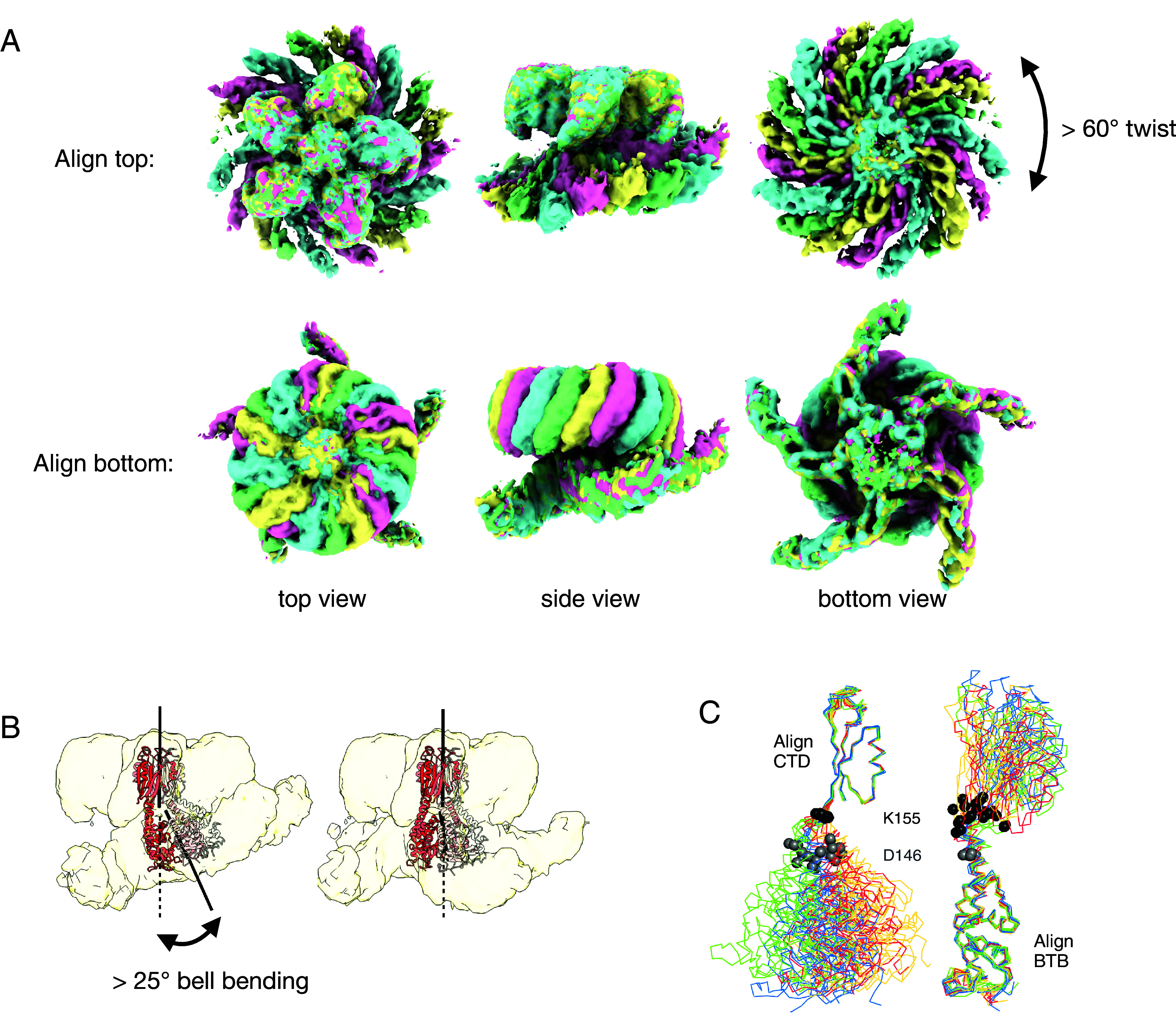
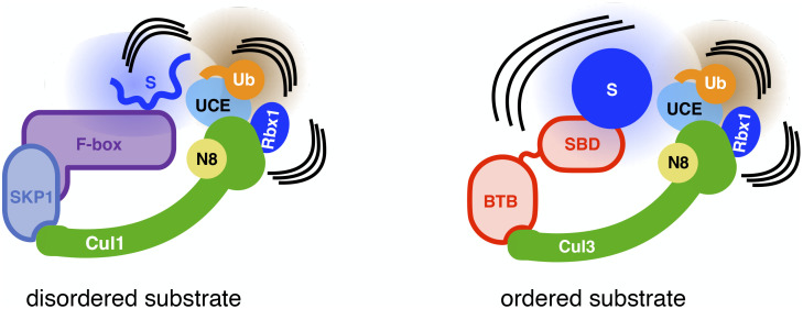
Heterotrimeric G proteins can be regulated by posttranslational modifications, including ubiquitylation. KCTD5, a pentameric substrate receptor protein consisting of an N-terminal BTB domain and a C-terminal domain, engages CUL3 to form the central scaffold of a cullin-RING E3 ligase complex (CRL3KCTD5) that ubiquitylates Gβγ and reduces Gβγ protein levels in cells. The cryo-EM structure of a 5:5:5 KCTD5/CUL3NTD/Gβ1γ2 assembly reveals a highly dynamic complex with rotations of over 60° between the KCTD5BTB/CUL3NTD and KCTD5CTD/Gβγ moieties of the structure. CRL3KCTD5 engages the E3 ligase ARIH1 to ubiquitylate Gβγ in an E3-E3 superassembly, and extension of the structure to include full-length CUL3 with RBX1 and an ARIH1~ubiquitin conjugate reveals that some conformational states position the ARIH1~ubiquitin thioester bond to within 10 Å of lysine-23 of Gβ and likely represent priming complexes. Most previously described CRL/substrate structures have consisted of monovalent complexes and have involved flexible peptide substrates. The structure of the KCTD5/CUL3NTD/Gβγ complex shows that the oligomerization of a substrate receptor can generate a polyvalent E3 ligase complex and that the internal dynamics of the substrate receptor can position a structured target for ubiquitylation in a CRL3 complex.

Keywords: BTB proteins; G proteins; cryo electron microscopy; ubiquitin ligase.


Conflict of interest statement

Competing interests statement:The authors declare no competing interest.

Figures

Fig. 1.
Fig. 1.
KCTD5 is a CRL3 substrate receptor for Gβγ. (A) CUL3NTD and Gβγ bind directly and independently to ALFA-tagged KCTD5 (ALFA-K5). Coomassie-stained SDS-PAGE. ALFA-resin fractions were eluted with SDS-sample buffer after washing the resin to remove unbound components. (B) BLI of sensor-anchored KCTD5 with CUL3NTD (Top) and Gβγ (Bottom). (CE) anti-Gβ1 western blots. (C) CUL3Nedd8/Rbx1/UBE2L3/ARIH1 ubiquitylates Gβ in a KCTD5-dependent manner (all reactions 2 min, 37 °C). (D) CRL3KCTD5/ARIH1/UBE2L3 catalyzes the ubiquitination of a single site on Gβ and has moderate ubiquitin-K48 chain extension activity. K5: KCTD5; K0: ubiquitin with all seven native lysines replaced with arginines; K48R ubiquitin with a single K48R substitution, K48 only: ubiquitin with six lysine to arginine substitutions, preserving the native lysine at position 48. (E) CRL3KCTD5 uses UBE2L3/ARIH1 but not UBE2D2 for the attachment of the initial ubiquitylation of Gβ. All experiments were repeated at least three times, and representative data are shown.
Fig. 2.
Fig. 2.
Structure of the pentameric KCTD5/CUL3NTD/Gβγ complex. (A) Cryo-EM map of the KCTD5CTD/Gβγ “top” part of the complex. Densities for KCTD5 are colored different shades of red. Gβ is blue and Gγ is purple. (B) Cryo-EM map of the KCTD5BTB/CUL3NTD “bottom” part of the complex. CUL3NTD is green. (C) Side view of the model fit to a map of the intact complex showing only one Gβγ and CUL3NTD chain for clarity. (D) Contact interfaces in the complex as per panel C in an “open book” format with buried surfaces in gray. The primary and distal interfaces from adjacent subunits in “c” are indicated with dark/light shades, respectively. (E) Schematic of the interfaces with key residues indicated. L209* is KCTD5 with a stop codon introduced at residue position 209. (F) Ubiquitylation activity of KCTD5 mutants. (G) Ubiquitylation activity of Gβ mutants. F and G are anti-Gβ western blots.
Fig. 3.
Fig. 3.
Structural variability in the complex. (A) Alignment of the consensus cryo-EM maps for states A, B, C, and D (green, cyan, pink, and yellow, respectively). Note that the two pseudo-C5 axes between the top and bottom of the complex are not colinear in these consensus maps. (B) Multi-body refinement reveals additional “bell-bending” variability between the two moieties for each state. (C) Structural alignments between the KCTD5 models built into the consensus cryo-EM maps show that the conformational variability in the complex is largely limited to a ~10 residue region between the BTB and CTD domains. Twenty models corresponding to the five chains of states A, B, C, and D are shown, colored according to the maps in A. The Cα atoms of KCTD5 residues 146 and 155 are shown in gray and black spheres, respectively. Bell-binding generates additional variability, but the changes remain localized to the same linker region.
Fig. 4.
Fig. 4.
Model of an active CRL3KCTD5 complex. (A) Side view of conformational state B of the KCTD5/CUL3NTD/Gβγ complex with one CUL3 arm modeled to include the C-terminal catalytic machinery based on the TS2 state of the CRL1FBXW7 complex (PDB ID 7B5M). Rbx1 is in gray, AriH1 (Ariadne and Rcat domains) is in white, and ubiquitin is in yellow. Only one CUL3 arm and one Gβγ unit are shown on the KCTD5 pentamer for clarity. The conformational state shown here places the NZ atom of Gβ K23 within 9 Å of the ubiquitin G75 CA atom with an unobstructed path to the thioester bond of the ubiquitin/AriH1~ubiquitin conjugate. The structure of the complex is consistent with membrane-anchored Gβγ. (B and C) Surface representation of one conformer of the extended complex. The small yellow circles indicate the positions of the ubiquitin G75 Cα in the ensemble of the conformations of the complex based on multi-body maps for states A–D. (D) Ubiquitylation of Gβ WT and point mutants (anti-Gβ western blot). (E) Kinetics of the ubiquitylation reaction for KCTD5 WT (black) and linker deletion mutants Δ2 (green), Δ4 (red), and Δ7 (blue). Error bars are SD with n = 3.
Fig. 5.
Fig. 5.
Reducing KCTD5 levels alters Gβγ expression and ubiquitination patterns. (A) The level of Gβγ is altered with KCTD5 knockdown or overexpression. BiFC was measured between the split YFP fusion proteins Gβ1-NYFP(1–158) and Gγ2-CYFP(159–238) in the presence or absence of overexpressed HA-KCTD5 or 100 nM siRNA. Fluorescence intensities are presented as mean ± SEM for four different experiments. Data were analyzed by two-way ANOVA followed by Tukey’s multiple comparisons test where **P < 0.01. (B) Pan-Gβ1-4 expression measured by western blot in parental and KCTD5 KO cell lines stably expressing either the WT, the C-terminal truncated (1 to 208*), F128A, or L161R KCTD5 mutants. For the sake of comparison, both parental and KCTD5 KO cell lines expressed a non-KCTD5 control protein (Venus, a YFP). Bars represent the average values from five independent experiments expressed as relative values normalized to expression of β-tubulin. Error bars are SEM. Statistical analysis was done by one-way ANOVA followed by a Dunnett post hoc test, *P < 0.05, **P < 0.01, and ns means nonsignificant. (C) KCTD5 levels alter ubiquitylation of Gβγ and their interacting proteins. Western blot analysis from parental and KCTD5 KO lines expressing FLAG-ubiquitin, HA-TAP-Gβ1, and HA-Gγ2 that were treated or not with 10 μM MG-132 for 8 h, lysed, and subjected to streptavidin purification. Blots are representative of three independent experiments.
Fig. 6.
Fig. 6.
Model for the role of CRL dynamics in positioning an activated ubiquitin C terminus near to a target substrate. S: substrate, SBD: substrate-binding domain, UCE: ubiquitin-carrying enzyme. See text for details.

References

    1. Rusnac D.-V., Zheng N., Structural biology of CRL ubiquitin ligases. Adv. Exp. Med. Biol. 1217, 9–31 (2020). - PubMed
    1. Harper J. W., Schulman B. A., Cullin-RING ubiquitin ligase regulatory circuits: A quarter century beyond the F-box hypothesis. Annu. Rev. Biochem. 90, 403–429 (2021). - PMC - PubMed
    1. Wang P., Song J., Ye D., CRL3s: The BTB-CUL3-RING E3 ubiquitin ligases. Adv. Exp. Med. Biol. 1217, 211–223 (2020). - PubMed
    1. Errington W. J., et al. , Adaptor protein self-assembly drives the control of a cullin-RING ubiquitin ligase. Structure 20, 1141–1153 (2012). - PubMed
    1. Canning P., et al. , Structural basis for Cul3 protein assembly with the BTB-Kelch family of E3 ubiquitin ligases. J. Biol. Chem. 288, 7803–7814 (2013). - PMC - PubMed