

Photobiomodulation therapy in improvement of harmful neural plasticity in sodium salicylate-induced tinnitus
- PMID: 38626075
- PMCID: PMC11020422
- DOI: 10.1371/journal.pone.0296607
Photobiomodulation therapy in improvement of harmful neural plasticity in sodium salicylate-induced tinnitus
Abstract
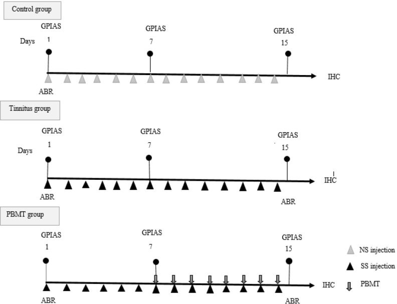
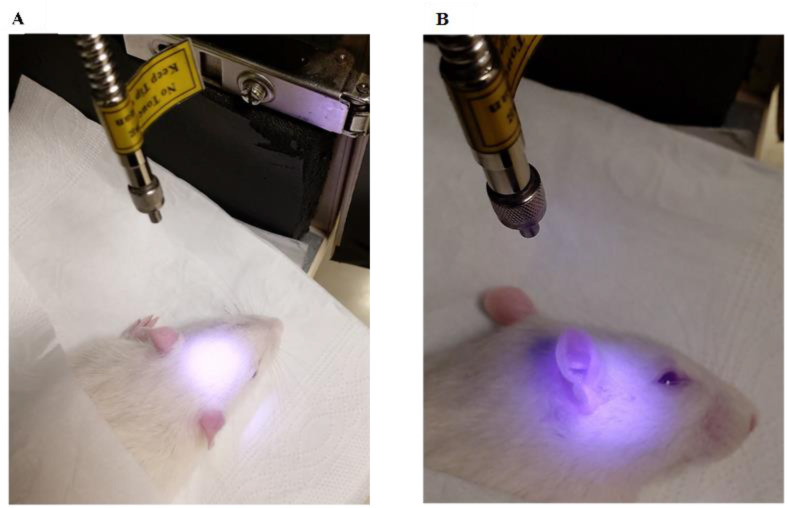
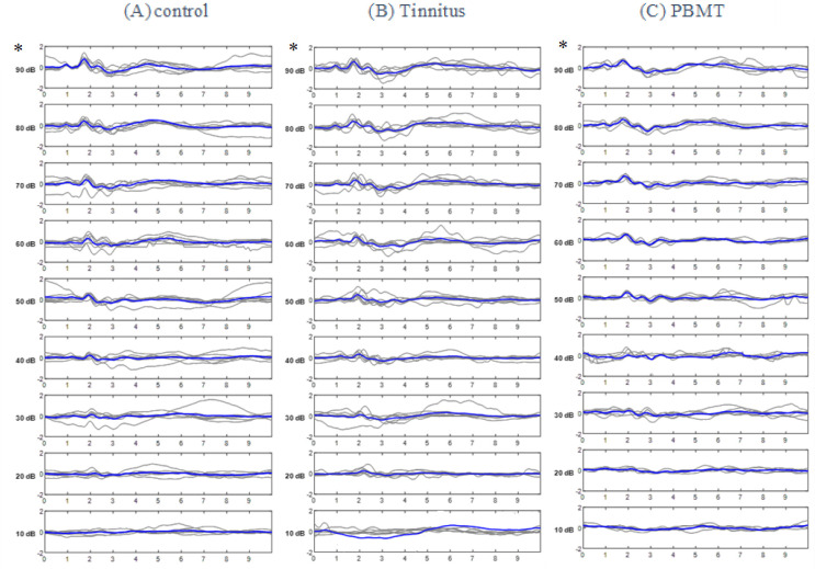
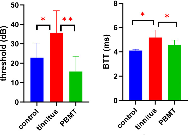
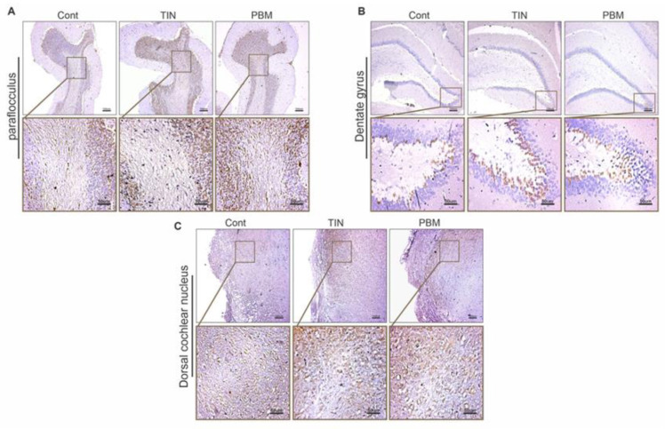
Tinnitus is a common annoying symptom without effective and accepted treatment. In this controlled experimental study, photobiomodulation therapy (PBMT), which uses light to modulate and repair target tissue, was used to treat sodium salicylate (SS)-induced tinnitus in a rat animal model. Here, PBMT was performed simultaneously on the peripheral and central regions involved in tinnitus. The results were evaluated using objective tests including gap pre-pulse inhibition of acoustic startle (GPIAS), auditory brainstem response (ABR) and immunohistochemistry (IHC). Harmful neural plasticity induced by tinnitus was detected by doublecortin (DCX) protein expression, a known marker of neural plasticity. PBMT parameters were 808 nm wavelength, 165 mW/cm2 power density, and 99 J/cm2 energy density. In the tinnitus group, the mean gap in noise (GIN) value of GPIAS test was significantly decreased indicated the occurrence of an additional perceived sound like tinnitus and also the mean ABR threshold and brainstem transmission time (BTT) were significantly increased. In addition, a significant increase in DCX expression in the dorsal cochlear nucleus (DCN), dentate gyrus (DG) and the parafloccular lobe (PFL) of cerebellum was observed in the tinnitus group. In PBMT group, a significant increase in the GIN value, a significant decrease in the ABR threshold and BTT, and also significant reduction of DCX expression in the DG were observed. Based on our findings, PBMT has the potential to be used in the management of SS-induced tinnitus.
Copyright: © 2024 Montazeri et al. This is an open access article distributed under the terms of the Creative Commons Attribution License, which permits unrestricted use, distribution, and reproduction in any medium, provided the original author and source are credited.
Conflict of interest statement
The authors have declared that no competing interests exist.
Figures


Similar articles
-
Transcriptional profile changes caused by noise-induced tinnitus in the cochlear nucleus and inferior colliculus of the rat.Ann Med. 2024 Dec;56(1):2402949. doi: 10.1080/07853890.2024.2402949. Epub 2024 Sep 13. Ann Med. 2024. PMID: 39268590 Free PMC article.
-
Dorsal Cochlear Nucleus Fusiform-cell Plasticity is Altered in Salicylate-induced Tinnitus.Neuroscience. 2019 May 21;407:170-181. doi: 10.1016/j.neuroscience.2018.08.035. Epub 2018 Sep 12. Neuroscience. 2019. PMID: 30217755 Free PMC article.
-
Exposure to sodium salicylate disrupts VGLUT3 expression in cochlear inner hair cells and contributes to tinnitus.Physiol Res. 2020 Feb 19;69(1):181-190. doi: 10.33549/physiolres.934180. Epub 2019 Dec 19. Physiol Res. 2020. PMID: 31852197 Free PMC article.
-
Understanding tinnitus: the dorsal cochlear nucleus, organization and plasticity.Brain Res. 2012 Nov 16;1485:40-53. doi: 10.1016/j.brainres.2012.03.044. Epub 2012 Mar 27. Brain Res. 2012. PMID: 22513100 Free PMC article. Review.
-
Tinnitus as a plastic phenomenon and its possible neural underpinnings in the dorsal cochlear nucleus.Hear Res. 2005 Aug;206(1-2):200-26. doi: 10.1016/j.heares.2005.02.013. Hear Res. 2005. PMID: 16081009 Review.
Cited by
-
The Role of Photobiomodulation to Modulate Ion Channels in the Nervous System: A Systematic Review.Cell Mol Neurobiol. 2024 Nov 23;44(1):79. doi: 10.1007/s10571-024-01513-1. Cell Mol Neurobiol. 2024. PMID: 39579175 Free PMC article.
References
-
- Jarach CM, Lugo A, Scala M, van den Brandt PA, Cederroth CR, Odone A, et al.. Global Prevalence and Incidence of Tinnitus: A Systematic Review and Meta-analysis. JAMA neurology. 2022;79(9):888–900. Epub 2022/08/09. doi: 10.1001/jamaneurol.2022.2189 ; PubMed Central PMCID: PMC9361184. - DOI - PMC - PubMed
MeSH terms
Substances
LinkOut - more resources
Full Text Sources
Medical
Miscellaneous
