. 2024 Jul 1;210(1):63-76.
doi: 10.1164/rccm.202308-1370OC.

Recessively Inherited Deficiency of Secreted WFDC2 (HE4) Causes Nasal Polyposis and Bronchiectasis

Affiliations

Recessively Inherited Deficiency of Secreted WFDC2 (HE4) Causes Nasal Polyposis and Bronchiectasis

Gerard W Dougherty et al. Am J Respir Crit Care Med. .

Abstract

Rationale: Bronchiectasis is a pathological dilatation of the bronchi in the respiratory airways associated with environmental or genetic causes (e.g., cystic fibrosis, primary ciliary dyskinesia, and primary immunodeficiency disorders), but most cases remain idiopathic. Objectives: To identify novel genetic defects in unsolved cases of bronchiectasis presenting with severe rhinosinusitis, nasal polyposis, and pulmonary Pseudomonas aeruginosa infection. Methods: DNA was analyzed by next-generation or targeted Sanger sequencing. RNA was analyzed by quantitative PCR and single-cell RNA sequencing. Patient-derived cells, cell cultures, and secretions (mucus, saliva, seminal fluid) were analyzed by Western blotting and immunofluorescence microscopy, and mucociliary activity was measured. Blood serum was analyzed by electrochemiluminescence immunoassay. Protein structure and proteomic analyses were used to assess the impact of a disease-causing founder variant. Measurements and Main Results: We identified biallelic pathogenic variants in WAP four-disulfide core domain 2 (WFDC2) in 11 individuals from 10 unrelated families originating from the United States, Europe, Asia, and Africa. Expression of WFDC2 was detected predominantly in secretory cells of control airway epithelium and also in submucosal glands. We demonstrate that WFDC2 is below the limit of detection in blood serum and hardly detectable in samples of saliva, seminal fluid, and airway surface liquid from WFDC2-deficient individuals. Computer simulations and deglycosylation assays indicate that the disease-causing founder variant p.Cys49Arg structurally hampers glycosylation and, thus, secretion of mature WFDC2. Conclusions: WFDC2 dysfunction defines a novel molecular etiology of bronchiectasis characterized by the deficiency of a secreted component of the airways. A commercially available blood test combined with genetic testing allows its diagnosis.

Keywords: Pseudomonas; chronic airway disease; infertility.


Figures
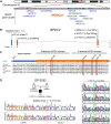
Figure 1.
Figure 1.
Identification of biallelic WFDC2 variants in 11 individuals from 10 global families. (A) WFDC2 localizes within the narrow WFDC gene cluster on human chromosome 20q13 with 14 of 18 members of the WFDC gene family. (B) The canonical transcript of WFDC2 (ENST00000372676) comprises four exons (Figure E3B); disease-causing WFDC2 variants identified in this study are indicated above respective exons. (C) WFDC2 encodes a secreted protein containing two WFDC domains, which are highly conserved throughout vertebrates as shown by multiple sequence alignments. Exon 1 encodes the signal peptide, which is cleaved before secretion. Exon 2 and exon 3 encode the N- and C-terminal WFDC domains, respectively. The WFDC domains comprise eight cysteine residues, whose disulfide bridges form a characteristic core motif. We identify seven distinct WFDC2 variants, including a start codon variant, two distinct large deletions, and four missense variants (indicated by red rectangles), affecting highly conserved residues within the WFDC domains. (D) OP-2032 II1 harbors WFDC2 variant c.145C>T, p.Cys49Arg, as well as a 1,570-bp deletion spanning exons 1 and 2. Segregation analysis verifies these variants are inherited from the father and mother, respectively. Additional pedigrees and Sanger sequencing are shown in the data supplement (Figure E1).
Figure 2.
Figure 2.
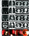
Individuals with biallelic WFDC2 variants present with bronchiectasis in all lung fields as well as nasal polyposis. (A–D, left panels) Paranasal sinus computed tomography (CT) images of individuals OP-2147 II1, OP-2032 II, OP-4281 II1, and CSU-150 II2 show chronic rhinosinusitis with pronounced nasal polyposis (marked by asterisks). (A–D, middle and right panels) In addition, thoracic CT images of these individuals indicate bronchiectasis and bronchial wall thickening present in all lung fields. (E) Nasal endoscopy of OP-398 II1 demonstrates several nasal polyps (dashed lines) and plugging of thick mucus (right image).
Figure 3.
Figure 3.
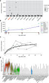
WFDC2 and SLPI are the only WFDC domain family genes robustly expressed in respiratory epithelial cells. (A) Comparison of WFDC domain gene family expression by RNA sequencing from human nasal epithelial cells (NECs) obtained by NB, air–liquid interface cultured NECs (ALI), Epstein–Barr virus (EBV)-transformed lymphocytes (EBV), and whole blood (blood) demonstrates high expression of WFDC2 and SLPI but no other WFDC family genes in respiratory cells. (B) Expression of WFDC2 and SLPI follow a comparable pattern during ciliogenesis in ALI-cultured NECs (time points 0, 1, 3, 15, and 30 days after airlift). Other WFDC genes, including WFDC1, EPPIN, WFDC8, WFDC9, WFDC12, and WFDC13, are not detected. (C) WFDC2 expression measured by quantitative PCR in bronchial epithelial cells cultured at the ALI from three healthy individuals (gray symbols; black line represents the data fitted to a mixed linear model). WFDC2 is increasingly expressed in a differentiation-dependent manner. (D) Single-cell RNA sequencing of different nasal, airway, and parenchymal samples from 107 healthy human donors (23, 42) demonstrates that WFDC2 is expressed primarily in secretory cells. Data shown here are the expression of WFDC2 per cell population in the merged dataset (biopsies, brushings, and dissections from all nasal, tracheal, bronchial, and parenchymal locations, from 107 healthy donors). NB = nasal brushing.
Figure 4.
Figure 4.
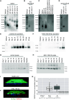
Secreted WFDC2 is detectable in healthy control but not WFDC2-deficient individuals. (A) Western blotting (WB) of saliva samples demonstrates the presence of WFDC2 (25 kD) in the healthy control and the healthy mother (OP-2032 I2) of individual OP-2032 II1. By contrast, this band is severely reduced or absent in saliva from individuals with pathogenic WFDC2 variants (OP-4281 II1, OP-2147 II1, and OP-2032 II1). In healthy saliva, a diffuse protein band of approximately 25 kD that represents glycosylated WFDC2 is detectable. (B) WB reveals that WFDC2 is absent from apical secretions of air–liquid interface (ALI)-cultured nasal epithelial cells (NECs) of WFDC2-mutant individual OP-2032 II1. (C) By WB, WFDC2 is detectable in saliva and the apical secretions of ALI-cultured NECs of healthy individuals but not in NECs, fibroblasts, or EBV-transformed lymphocytes as well as supernatants of fibroblasts and EBV-transformed lymphocytes (l = lysate; s = supernatant). (D) WB demonstrates that WFDC2 is detectable in seminal fluid from healthy control but not WFDC2-mutant individual OP-2147 II1. (E) WB demonstrates that WFDC2 is detectable in apical secretions of healthy control bronchial epithelial cells (BECs) cultured at the ALI by Day 5, preceding ciliation, whereas (F) WFDC2 is weakly detectable in apical secretions from individual UNC-186 II2. A total of 10 and 20 μg of recombinant WFDC2 (HEK293 expression) is loaded as positive control. (G) In contrast, lysates from control BECs show low levels of WFDC2, whereas (H) UNC-186 II2 shows accumulation of WFDC2 reactive material. A total of 10 and 20 μg of recombinant WFDC2 (HEK293 expression) is loaded as positive control. (I) Whole mount immunofluorescence images of ALI-cultured BECs from a healthy control and (J) UNC-186 II2 stained with anti-WFDC2 (red), anti-acetylated tubulin to label cilia (green), and Hoechst 33342 to label nuclei. WFDC2 accumulates intracellularly in cells from UNC-186 II2. (K) Measurement of WFDC2 in blood serum shows that WFDC2 concentration is below the limit of detection in samples of individuals with biallelic WFDC2 mutations UNC-186 II2, OP-2032 II1, OP-2147 II1, and OP-4281 II1 (OP-2032 II1 and OP-4281 II1 are tested twice, OP-2147 II1 is tested thrice with the same result). Median serum concentration of individuals with PCD (n = 44) and CF (n = 13) are elevated compared with published reference values (23) for healthy children (dotted line, 36.3 pmol/L, age 10–15 yr) and adults (dashed line, 33.8 pmol/L, age 23–38 yr). CF = cystic fibrosis; PCD = primary ciliary dyskinesia.
Figure 5.
Figure 5.
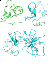
Disease-causing founder variant p.Cys49Arg impairs WFDC2 secretion through occlusion of the N-linked glycosylation site at Asn44. (A) The most populated conformation of the disulfide-free mature human WFDC2 with the Cys49Arg mutation showing the β carbon distances of seven disulfides (C36-C62, C45-C66, C55-C70, C80-C110, C93-C114, C97-C109, and C103-C119). (B) The most populated conformation of the wild-type disulfide-free mature human WFDC2 showing the β carbon distances of eight disulfides (C36-C62, C45-C66, C49-C61, C55-C70, C80-C110, C93-C114, C97-C109, and C103-C119). (C) Close-up view of the occluded glycosylation site of the Cys49Arg mutant. (D) Close-up view of the fully exposed glycosylation site of the wild type. Distances shown by dashed lines are in angstroms (Å). The most populated conformation of the mutant or wild type was derived from 220 distinct and independent simulations for each set, with an aggregated simulation time of 208.560 μs.
Figure 6.
Figure 6.
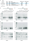
Proteomic content analysis of saliva among healthy control, WFDC2-mutant, and respiratory disease control groups. (A) Saliva from healthy control individuals (n = 3), individuals with WFDC2 mutations (OP-2032 II1, OP-2147 II1, and OP-4281 II1), as well as control individuals with respiratory disease (primary ciliary dyskinesia [PCD], n = 2; cystic fibrosis [CF], n = 2) are subjected to liquid chromatography with tandem mass spectrometry (LC-MS/MS) to determine differentially expressed proteins. The gene symbol, UniProt identifier, and gene name, as well as log2 protein ratios of WFDC2-mutant compared with healthy control group (WFDC2/control) and WFDC2-mutant compared with respiratory disease control group (WFDC2/disease) are shown. WFDC2-specific peptides are not identified in the WFDC2-mutant group. The respiratory disease control group represents individuals with PCD OP-3499 II2 (DNAH5, c.10616G>A, p.Arg3539His + c.5557A>T, p.Lys1853*) and OP-3180 II1 (SPAG1, c.1282_1294del13, p.Ala428Profs*17, homozygous) as well as individuals with CF, OS-MeHo II1 and OS-MaSc II1. (B and C) WFDC2 Western blotting (WB) demonstrates a diffuse band of approximately 25 kD (blue brackets) that is detectable in healthy control subjects as well as individuals with PCD and CF but not individuals with WFDC2 mutations OP-2147 II1 and OP-2032 II1. The immunoreactive band of approximately 26 kD in OP-2147 II1 and OP-2032 II1 is likely nonspecific, as this band does not shift in response to PNGase F (Figure E5), and LC-MS/MS does not identify WFDC2-specific peptides in individuals with WFDC2 mutations. (D and E) SLPI WB demonstrates variable expression of an approximately 14-kD protein (green arrow) among healthy control subjects, individuals with WFDC2 mutations, and control individuals with PCD and CF. (F and G) SPINK5 WB demonstrates significantly reduced expression of an approximately 26-kD protein (blue arrow) in individuals with WFDC2 mutations compared with healthy and disease control groups. Patients with PCD are represented by OP-3499 II2, OP-4168 II1 (RSPH1, c.85G>T, p.Glu29Ter, homozygous), and OP-1331 II1 (ODAD4, c.425_426insT, p.Lys142AsnfsTer12, homozygous); patients with CF are represented by OS-MeHo II1, OS-MeSc II1, and OS-MeHa II1, which all harbor pathogenic CFTR variant c.1521_1523del, p.Phe508del in the homozygous state. Orange circle indicates individual with PCD, OP-1331 II1, and individual with CF, OS-MeHa II1, with bronchiectasis but no history of Pseudomonas aeruginosa infection. 10 μg of protein per sample is analyzed by WB.

Comment in

Similar articles

Cited by

References

    1. Polverino E, Goeminne PC, McDonnell MJ, Aliberti S, Marshall SE, Loebinger MR, et al. European Respiratory Society guidelines for the management of adult bronchiectasis. Eur Respir J . 2017;50:1700629. - PubMed
    1. Flume PA, Chalmers JD, Olivier KN. Advances in bronchiectasis: endotyping, genetics, microbiome, and disease heterogeneity. Lancet . 2018;392:880–890. - PMC - PubMed
    1. Shoemark A, Ozerovitch L, Wilson R. Aetiology in adult patients with bronchiectasis. Respir Med . 2007;101:1163–1170. - PubMed
    1. Pembridge T, Chalmers JD. Precision medicine in bronchiectasis. Breathe (Sheff) . 2021;17:210119. - PMC - PubMed
    1. Oriano M, Amati F, Gramegna A, De Soyza A, Mantero M, Sibila O, et al. Protease-antiprotease imbalance in bronchiectasis. Int J Mol Sci . 2021;22:5996. - PMC - PubMed

Publication types

Substances

Grants and funding