. 2024 Apr 16;15(1):3271.
doi: 10.1038/s41467-024-47062-5.

The physiological interactome of TCR-like antibody therapeutics in human tissues

Affiliations

The physiological interactome of TCR-like antibody therapeutics in human tissues

Estelle Marrer-Berger et al. Nat Commun. .

Abstract

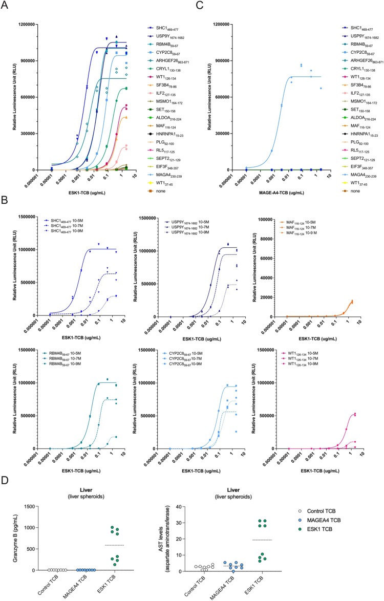
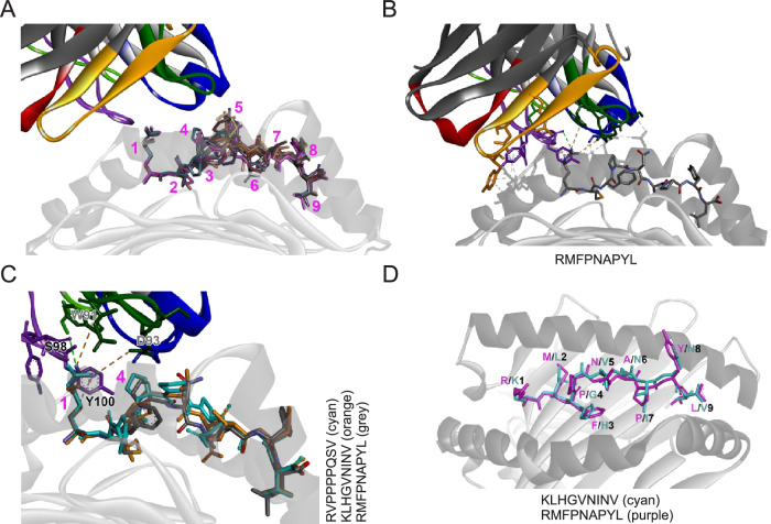
Selective binding of TCR-like antibodies that target a single tumour-specific peptide antigen presented by human leukocyte antigens (HLA) is the absolute prerequisite for their therapeutic suitability and patient safety. To date, selectivity assessment has been limited to peptide library screening and predictive modeling. We developed an experimental platform to de novo identify interactomes of TCR-like antibodies directly in human tissues using mass spectrometry. As proof of concept, we confirm the target epitope of a MAGE-A4-specific TCR-like antibody. We further determine cross-reactive peptide sequences for ESK1, a TCR-like antibody with known off-target activity, in human liver tissue. We confirm off-target-induced T cell activation and ESK1-mediated liver spheroid killing. Off-target sequences feature an amino acid motif that allows a structural groove-coordination mimicking that of the target peptide, therefore allowing the interaction with the engager molecule. We conclude that our strategy offers an accurate, scalable route for evaluating the non-clinical safety profile of TCR-like antibody therapeutics prior to first-in-human clinical application.


Conflict of interest statement

All authors listed with Roche Pharma affiliation are directly employed by Roche Pharma. T.W., P.U., and C.K. are inventors on related patents. P.U. and C.K. declare stock ownership. N.T. has been a paid consultant to Roche Pharma. The remaining authors declare no competing interests.

Figures

Fig. 1
Fig. 1. An experimental framework de novo identifies on-target binding of the functional MAGE-A4-specific TCB recognizing the peptide GVYDGREHTV presented by HLA-A*02:01.
A Schematic of the MAGE-A4 TCB and its mode of action. B Average ratio of endogenous GVYDGREHTV peptide area under the curve (AUC) by comparison to spiked-in isotopically labeled GVYDGREHTV peptide AUC (n = 4), measured in elution fractions from HLA-A2 immunoprecipitation in A375 tumor cells lines and MAGE-A4 KO (A375-KO) and equivalent lines in a xenograft model (XA375 and XA375-KO). Horizontal bars depict the mean of quadruplicate analyses. The estimated copy number of endogenous GVYDGREHTV per cell (shown on top of each bar graph) is calculated by comparison to a spiked-in isotopically labeled GVYDGREHTV peptide, assuming 100% recovery for both standard and endogenous peptides at the end of the workflow. C A375 wild-type cells and A375-KO cells were incubated with human PBMCs at an E:T ratio of 10:1. Depicted are dose-response curves of tumor cell lysis after 24 h of incubation with different concentrations of MAGE-A4 TCB as determined by quantification of lactate dehydrogenase release in the supernatant (plotted are the mean of triplicate repeat analyses and error bars depict the SD). D Schematic workflow using TCR-like antibodies to enrich interacting HLA-peptide complexes. Tissues were lysed, and solubilized HLA-peptide complexes were immunoprecipitated using the MAGE-A4 tool antibody as a bait. Peptides were enriched in acidic conditions through a 5 kDa molecular weight cut-off (MWKO) filter and analyzed by LC-MS2. E Peptide intensity as measured in three biological replicate analyses demonstrates significant enrichment (p = 0.0015, unpaired t-test, two-tailed) of the target peptide GVYDGREHTV in XA375, and not in MAGE-KO equivalent tissues (XA375-KO) nor with a control TCB (CTRL) (F) LC-MS spectrum leading to identification of the target peptide GVYDGREHTV in XA375 after enrichment using the MAGE-A4 antibody. The spectral identifier, the measured mass of the precursor peptide ion (m/z), charge state (z), and retention time (RT), at which the peptide ion was selected for fragmentation, are stated within the spectral panel. C-terminal fragment ions are indicated as y, N-terminal fragments are designated b, pre: unfragmented precursor peptide, +: singly charged ion, ++: doubly charged ion.
Fig. 2
Fig. 2. Off-target interaction of ESK1 with common HLAp sequences in primary liver tissue.
A Volcano plots showing significant (one-factor ANOVA, -log10(p)≥1.5) enrichment of peptides for ESK1 and MAGE-A4 in liver tissue (all peptides, left panel; peptides predicted to bind to HLA-A*02:01, right panel). B Sequence motif of the ESK1-enriched peptides that are predicted to bind to HLA-A*02:01. Shown is the frequency (probability) of each amino acid in each position across the peptide sequence from N- to C-terminus (left to right, positions 1-9). C Sequence motifs from Gejman et al. (left panel) and our study (right panel). D Venn diagram depicting the lack of overlap of peptide sequences shortlisted by Gejman et al. and our study as Esk1 binders.
Fig. 3
Fig. 3. Off-target binding activates effector cells and conveys killing of liver spheroids in the presence of ESK1.
A T2 cells were first pulsed with the 16 ESK1-enriched peptides and then co-incubated with JNFAT effector cells in the presence of different concentrations of ESK1 TCB. The target peptide (WT1126-134) was used as a positive control, while WT137-45, a non-relevant peptide, was used as a negative control. Effector cell activation was evaluated after 6 h by measuring luminescence signal using ONEGlo. B Titration curves for JNFAT assay with indicated peptides and ESK1 TCB concentrations. C Equivalent to (A) using MAGE-A4 TCB, and MAGE-A4 target peptide MAGA4230-239 as a control. D Liver spheroids were cocultured with peripheral blood mononuclear cells at a ratio of 5 effector to 1 target cell and target cell killing was evaluated by granzyme B secretion and aspartate aminotransferase expression. Each graph represents two independent experiments with two human donors in quadruplicates.
Fig. 4
Fig. 4. Structural mimicry is a feature of ESK1 off-target activity.
A Overlay of RMFPNAPYL (purple, from X-ray structure with PDB ID 4WUU) with models of the confirmed cross-reactive peptides in the presence of the Fab of ESK1. The HLA-A*02:01 peptide binding groove is indicated in gray. The numbers indicate the approximate position of the amino acid side chain at the respective peptide position. B RMFPNAPYL peptide within the HLA-A*02:01 binding groove and interactions with the ESK1 Fab (PDB ID 4WUU). C Non-bonded interactions between the Fab of ESK1 and RMFPNAPYL (gray) in the HLA-A*02:01 peptide-HLA complex (X-ray crystal structure with PDB ID 4WUU). Models of the cross-reactive peptides SHC1469-477 (cyan) and RBM4B59-67 (orange) are shown for comparison. D Overlay of RMFPNAPYL peptide (purple, from X-ray structure with PDB ID 4WUU) with modeled RBM4B59-67 peptide (cyan) in the peptide binding groove of HLA-A*02:01.
Fig. 5
Fig. 5. MAGE-A4 TCB does not show off-target reactivity in lung and intestinal tissue.
A Volcano plots showing significant enrichment (one-factor ANOVA, -log10(p)≥1.5) of peptides for anti-HLA-DQ (clone SPVL3), upper left quadrants) and MAGE-A4 (upper right quadrants) in lung and intestine tissue (all peptide, left panels; peptides predicted to bind to HLA-A*02:01, right panels). B Lung alveolus-on-chip and intestinal differentiated organoids were cocultured with peripheral blood mononuclear cells at a ratio of 10 effector to 1 target cell. Target cell killing was evaluated by granzyme B secretion and caspase 3/7 probe imaging (MFI median fluorescence intensity). Each graph represents two independent experiments with two human donors in triplicates.

References

    1. Dolgin E. First soluble TCR therapy opens ‘new universe’ of cancer targets. Nat. Biotechnol. 2022;40:441–444. doi: 10.1038/s41587-022-01282-6. - DOI - PubMed
    1. Liddy N, et al. Monoclonal TCR-redirected tumor cell killing. Nat. Med. 2012;18:980–987. doi: 10.1038/nm.2764. - DOI - PubMed
    1. Middleton MR, et al. Tebentafusp, A TCR/Anti-CD3 bispecific fusion protein targeting gp100, potently activated antitumor immune responses in patients with metastatic melanoma. Clin. Cancer Res. 2020;26:5869–5878. doi: 10.1158/1078-0432.CCR-20-1247. - DOI - PMC - PubMed
    1. Nathan P, et al. Overall survival benefit with tebentafusp in metastatic uveal melanoma. N. Engl. J. Med. 2021;385:1196–1206. doi: 10.1056/NEJMoa2103485. - DOI - PubMed
    1. Linette GP, et al. Cardiovascular toxicity and titin cross-reactivity of affinity-enhanced T cells in myeloma and melanoma. Blood. 2013;122:863–871. doi: 10.1182/blood-2013-03-490565. - DOI - PMC - PubMed