. 2024 Aug;45(8):1632-1643.
doi: 10.1038/s41401-024-01282-z. Epub 2024 Apr 16.

Ripretinib inhibits HIV-1 transcription through modulation of PI3K-AKT-mTOR

Affiliations

Ripretinib inhibits HIV-1 transcription through modulation of PI3K-AKT-mTOR

Jin-Feng Cai et al. Acta Pharmacol Sin. 2024 Aug.

Abstract

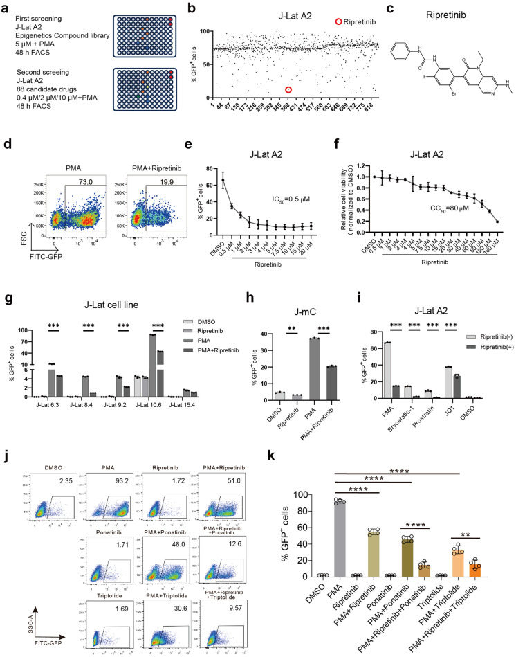
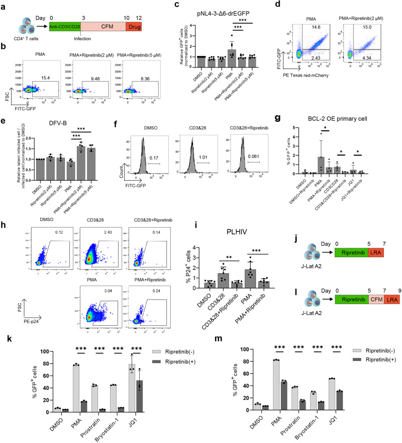
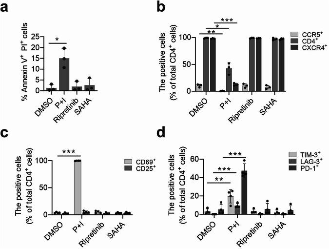
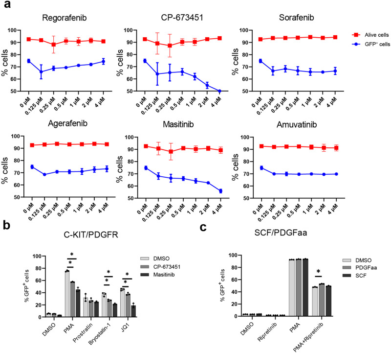
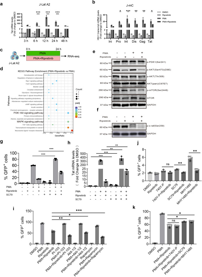
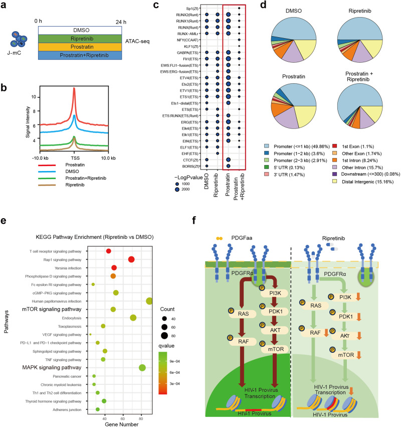
Despite the effectiveness of antiretroviral therapy (ART) in prolonging the lifespan of individuals infected with HIV-1, it does not offer a cure for acquired immunodeficiency syndrome (AIDS). The "block and lock" approach aims to maintain the provirus in a state of extended transcriptional arrest. By employing the "block and lock" strategy, researchers endeavor to impede disease progression by preventing viral rebound for an extended duration following patient stops receiving ART. The crux of this strategy lies in the utilization of latency-promoting agents (LPAs) that are suitable for impeding HIV-1 provirus transcription. However, previously documented LPAs exhibited limited efficacy in primary cells or samples obtained from patients, underscoring the significance of identifying novel LPAs that yield substantial outcomes. In this study, we performed high-throughput screening of FDA-approved compound library in the J-Lat A2 cell line to discover more efficacious LPAs. We discovered ripretinib being an LPA candidate, which was validated and observed to hinder proviral activation in cell models harboring latent infections, as well as CD4+ T cells derived from infected patients. We demonstrated that ripretinib effectively impeded proviral activation through inhibition of the PI3K-AKT-mTOR signaling pathway in the HIV-1 latent cells, thereby suppressing the opening states of cellular chromatin. The results of this research offer a promising drug candidate for the implementation of the "block and lock" strategy in the pursuit of an HIV-1 cure.

Keywords: HIV-1; PI3K-AKT-mTOR; block and lock; latency-promoting agents; ripretinib.


Conflict of interest statement

The authors declare no competing interests.

Figures

Fig. 1
Fig. 1. Screening of an FDA-approved compound library identified ripretinib as an LPA candidate.
a The process of high-throughput screening in the J-Lat A2 cell line. The J-Lat A2 cells were treated with 5 μM drugs plus PMA for 48 h. The blocking efficiency was then determined by measuring the percentage of GFP+ cells. b In the summary of screening results, reactivation efficiency was measured using flow cytometry, specifically by measuring the percentage of GFP+ cells. Each bar represents the average value of triplicates. c Chemical structure of ripretinib. d Representative flow cytometer plots of the suppression effect of ripretinib to PMA. J-Lat A2 cells were cotreated with indicated concentrations of ripretinib in combination with PMA (50 ng/mL). Percentage of GFP+ cells (e) and cell viability (f) are plotted. g Effects of ripretinib on J-Lat cell lines. h Effects of ripretinib on J-mC cells. i Ripretinib can inhibit HIV-1 reactivation induced by different LRAs in J-Lat A2 cells. Ripretinib exerts synergistic effect with triptolide (5 nM) or ponatinib (1 μM) in inhibiting the reactivation of PMA in J-Lat A2 cells. Representative flow cytometry plots (j) and histogram plots (k) are shown. Data are presented as means ± SD for three technical replicates. P values were calculated using an unpaired t-test, with significance levels denoted as follows: **P < 0.01, ***P < 0.001, ****P < 0.0001.
Fig. 2
Fig. 2. Ripretinib broadly inhibits HIV-1 reactivation in vitro and ex vivo.
a Primary CD4+ T cells from healthy individuals were isolated and activated by αCD3/αCD28 antibodies for 3 d. Pseudovirus was added for infection, and drugs were added 7 d after infection. Cell reactivation levels were detected 9 d after infection. Ripretinib suppresses provirus activation in the pNL4-3-Δ6-drEGFP infected primary CD4+ T cells, as shown by the representative flow cytometer plots (b) and histogram plots (c) of pNL4-3-Δ6-drEGFP infected CD4+ T cells. Ripretinib suppresses the activation of the provirus in the DFV-B infected primary CD4+ T cells, as shown by the representative flow cytometer plots (d) and histogram plots (e) of DFV-B infected CD4+ cells. Ripretinib can inhibit HIV-1 reactivation induced by different LRAs in Bcl-2-transduced latent infected primary CD4+ T cells, as depicted in the representative flow cytometry plots (f) and histogram plots (g). Ripretinib can decrease HIV-1 p24 levels in activated CD4+ T cells from ART-suppressed individuals, as depicted in the representative flow cytometry plots (h) and histogram plots (i). j, k Ripretinib pretreatment inhibited LRA-induced reactivation of J-Lat A2. J-Lat A2 cells were treated with ripretinib for 5 d and then were treated with LRAs (j) and histogram plots (k). Inhibitory effect of ripretinib can be maintained for some time. J-Lat A2 cells were treated with ripretinib for 5 d, cultured in a component-free medium (CFM) for 2 d, and then treated with LRAs (l) and histogram plots (m). Data are presented as means ± SD for three technical replicates. P values were calculated using an unpaired t-test. Significance levels are denoted with *P < 0.05, **P < 0.01, ***P < 0.001.
Fig. 3
Fig. 3. Ripretinib does not cause apparent cytotoxicity, cell activation, or cell dysfunction.
CD4+ T cells were isolated from healthy donors and then treated with DMSO, 1 μM ripretinib, 1 μM SAHA or PMA plus ionomycin. a Ripretinib did not cause apparent cytotoxicity, as revealed using annexin V/propidium iodide (PI) staining. b Ripretinib had no significant effect on the expression of CD4, CCR5, or CXCR4. c Ripretinib did not significantly upregulate CD25 expression or CD69 expression on CD4+ T cells. d Ripretinib did not significantly upregulate the expression of PD-1, TIM-3, and LAG-3 on CD4+ T cells. Data are presented as means ± SD for three technical replicates. P values were calculated using an unpaired t test, with significance levels indicated as follows: *P < 0.05, **P < 0.01, ***P < 0.001.
Fig. 4
Fig. 4. Multiple drugs targeting C-kit or PDGFRα inhibit the reactivation of HIV-1 provirus.
a Inhibitory effect of Regorafenib, CP-673451, sorafenib, agerafenib, masitinib, and amuvatinib on provirus activation and the corresponding cell activity. b Inhibitory effects of CP-673451 and masitinib on different LRAs. c Proportion of activation of provirus in J-Lat A2 cells treated with SCF (2 ng/μL) and PDGFaa (2 ng/μL). Each point represents the average of three data points. The count is expressed as mean ± standard deviation. P values were calculated using an unpaired t test, with significance level indicated as follows: *P < 0.05.
Fig. 5
Fig. 5. Ripretinib inhibits HIV-1 provirus activation by inhibiting the PI3K-AKT and MAPK signaling pathways.
Ripretinib significantly inhibits HIV-1 transcription. HIV-1 Tat transcription level with indicated treatment in J-Lat A2 cells (a). HIV-1 gene transcription level with indicated treatment in J-mC cells (b). c J-Lat A2 cells were treated with PMA (10 ng/mL), PMA plus ripretinib (1 μM) for 24 h, representatively. The cells were collected for RNA-seq. d PI3K-AKT and MAPK signaling pathways were significantly downregulated in the J-Lat 10.6 cells. Enrichment plots depict KEGG pathways downregulated by ripretinib plus PMA versus PMA. e Phosphorylation and basal expression levels of PDK1, AKT, mTOR protein in the J-Lat A2 cells. f Phosphorylation levels of c-raf in J-Lat A2 cells. g Percentage of GFP+ cells when treated with ripretinib or SC79 (1 μM). h SC79 increased the Tat mRNA levels of HIV-1-infected J-Lat 10.6 cells which were treated with ripretinib. i Ripretinib displayed a synergistic effect with PI-103 (5 μM), Akti-1/2 (10 μM), and rapamycin (1 μM). j Ripretinib effectively inhibited the reactivation of MHY1485 (5 μM) and SC79 (1 μM). k MHY1485 and SC79 offset the inhibitory effects of ripretinib on PMA. Data are presented as means ± SD for three technical replicates. P values were calculated using an unpaired t test, with *P < 0.05, **P < 0.01, ***P < 0.001 and ns indicating non-significant differences.
Fig. 6
Fig. 6. Ripretinib inhibits HIV-1 proviral reactivation by suppressing cellular chromatin opening.
a J-Lat A2 cells were treated with DMSO, ripretinib, prostratin, and prostratin plus ripretinib for 24 h. The cells were collected for ATAC-seq analysis. b ATAC-seq signaling intensity in the 10-kb region upstream and downstream of the transcription initiation site (TSS). c Enrichment of transcription factor motif on ATAC-seq data. d Distribution of peaks in functional regions on ATAC-seq data. e Enrichment plots depict KEGG pathways downregulated by ripretinib versus DMSO. f Ripretinib treatment decreased phosphorylation of PDK1 (Ser 241), AKT (Ser473 and Thr308) and p-c-raf (Ser259). Decreased AKT activity reduces mTOR (ser2448) phosphorylation levels, while ripretinib inhibits chromatin opening, which ultimately inhibiting HIV-1 provirus transcription.

Similar articles

Cited by

References

    1. Hütter G, Nowak D, Mossner M, Ganepola S, Müssig A, Allers K, et al. Long-term control of HIV by CCR5Δ32/Δ32 stem-cell transplantation. N Engl J Med. 2009;360:692–8. 10.1056/NEJMoa0802905 - DOI - PubMed
    1. Gupta RK, Abdul-Jawad S, McCoy LE, Mok HP, Peppa D, Salgado M, et al. HIV-1 remission following CCR5Δ32/Δ32 haematopoietic stem-cell transplantation. Nature. 2019;568:244–8. 10.1038/s41586-019-1027-4 - DOI - PMC - PubMed
    1. Deeks SG, Archin N, Cannon P, Collins S, Jones RB, de Jong MAWP, et al. Research priorities for an HIV cure: international AIDS society global scientific strategy 2021. Nat Med. 2021;27:2085–98. 10.1038/s41591-021-01590-5 - DOI - PubMed
    1. Chen J, Zhou T, Zhang Y, Luo S, Chen H, Chen D, et al. The reservoir of latent HIV. Front Cell Infect Microbiol. 2022;12:945–6. - PMC - PubMed
    1. Simonetti FR, Sobolewski MD, Fyne E, Shao W, Spindler J, Hattori J, et al. Clonally expanded CD4+ T cells can produce infectious HIV-1 in vivo. Proc Natl Acad Sci USA. 2016;113:1883–8. 10.1073/pnas.1522675113 - DOI - PMC - PubMed

MeSH terms

LinkOut - more resources