. 2024 Aug;31(8):1243-1250.
doi: 10.1038/s41594-024-01283-w. Epub 2024 Apr 16.

Molecular basis of Gabija anti-phage supramolecular assemblies

Affiliations

Molecular basis of Gabija anti-phage supramolecular assemblies

Xiao-Yuan Yang et al. Nat Struct Mol Biol. 2024 Aug.

Abstract

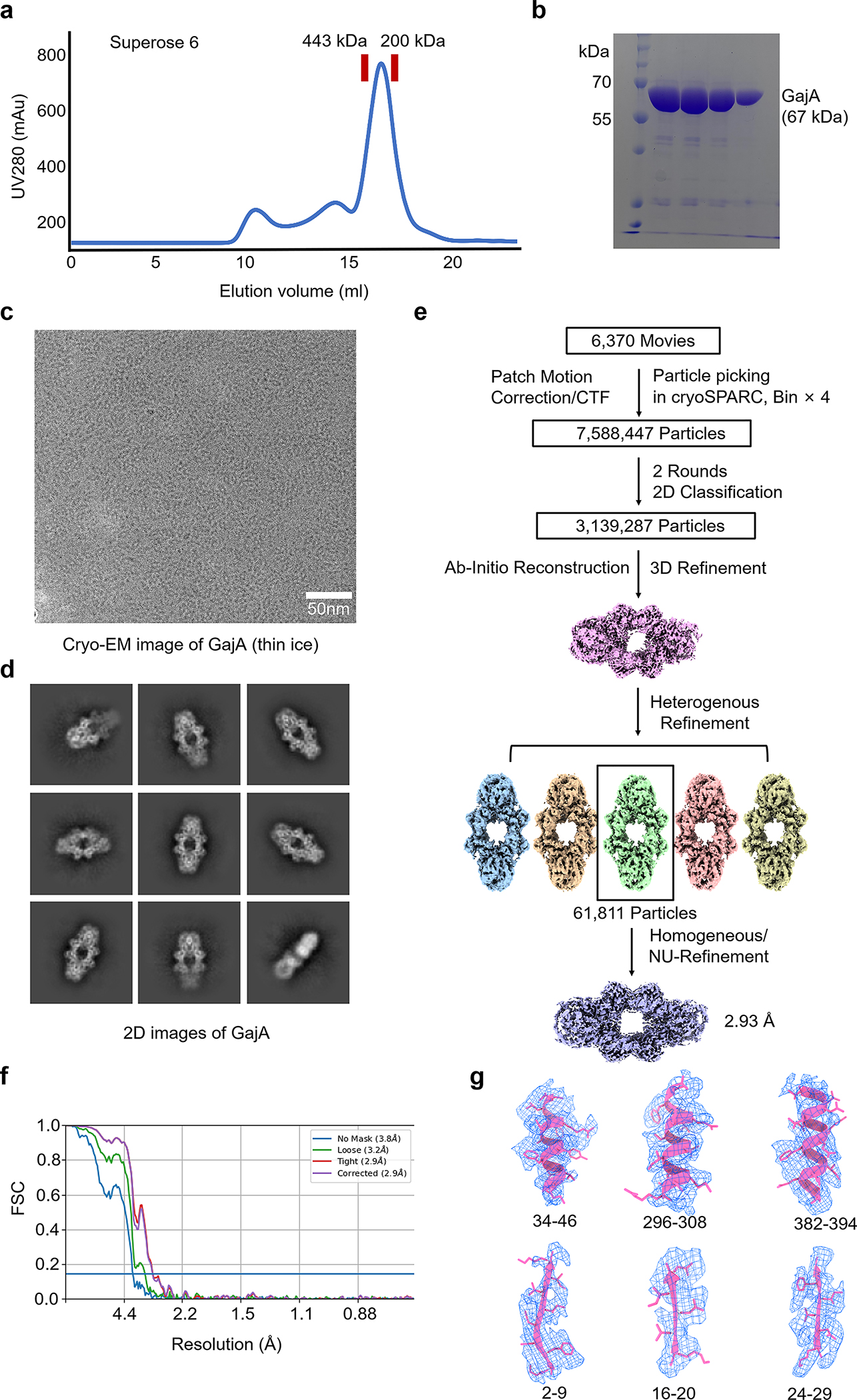
As one of the most prevalent anti-phage defense systems in prokaryotes, Gabija consists of a Gabija protein A (GajA) and a Gabija protein B (GajB). The assembly and function of the Gabija system remain unclear. Here we present cryo-EM structures of Bacillus cereus GajA and GajAB complex, revealing tetrameric and octameric assemblies, respectively. In the center of the complex, GajA assembles into a tetramer, which recruits two sets of GajB dimer at opposite sides of the complex, resulting in a 4:4 GajAB supramolecular complex for anti-phage defense. Further biochemical analysis showed that GajA alone is sufficient to cut double-stranded DNA and plasmid DNA, which can be inhibited by ATP. Unexpectedly, the GajAB displays enhanced activity for plasmid DNA, suggesting a role of substrate selection by GajB. Together, our study defines a framework for understanding anti-phage immune defense by the GajAB complex.


Conflict of interest statement

Competing interests

All authors declare they have no competing interests.

Figures

Extended data Fig.1 |
Extended data Fig.1 |. Cryo-EM reconstruction of GajA in thin ice.
a, b, Gel filtration profile (a) and SDS-PAGE gel (b) of GajA purification. c, Cryo-EM image of GajA in thin ice. d, Representative 2D class averages of GajA calculated from thin-ice cryo-EM images. e, Data processing workflow for 3D reconstruction of GajA tetramer from thin-ice cryo-EM images. f, FSC curve of reconstructed GajA tetramer from thin-ice cryo-EM images. g, Representative cryo-EM density of GajA tetramer fit with α-helixes and β-strands. The density map is shown at contour levels of 0.03.
Extended data Fig.2 |
Extended data Fig.2 |. Cryo-EM reconstruction of GajA in thicker ice.
a, Cryo-EM image of GajA in thick ice. b, 2D class averages of GajA calculated from thick-ice cryo-EM images. c, Data processing workflow for 3D reconstruction of GajA tetramer from thick-ice cryo-EM images. d, FSC curve of reconstructed GajA tetramer from thick-ice cryo-images. e, Local resolution of reconstructed GajA tetramer from thick-ice cryo-images. f, Cryo-EM density of GajA tetramer fit with α-helixes and β-strands. The density map is shown at contour levels of 0.03.
Extended data Fig.3 |
Extended data Fig.3 |. Architecture of GajA.
a, Ribbon diagram of GajA N-terminal ATPase domain with secondary structures indicated. b, Overlaid structures of GajA N-terminal ATPase domain (green) and Rad50 ATPase domain (PDB ID 5DNY, magenta). c, Ribbon diagram of GajA C-terminal Toprim domain with secondary structures indicated. d, The dimerization domain of GajA in complex with a phage protein Gad1 (magenata).
Extended data Fig.4 |
Extended data Fig.4 |. Interfaces in GajA tetramer.
a-c, Enlarged views of interface I (a), interface II (b), and interface III (c) in GajA tetramer. Key residues on the interfaces were highlighted in sticks. d, Superimposed structures of the active sites from GajA (green) and BpOLD (PDB ID 6NK8, grey). e, Structure of GajA in complex with dsDNA (Yellow) that was predicted by RoseTTAFoldNA. f, Electrostatic surface representation of GajA with dsDNA. The catalytic center of GajA is highlighted by a red circle. Negatively charged residues surrounding the catalytic center of GajA coordinate dsDNA. g, Key residues involved in coordinating dsDNA are highlighted in sticks.
Extended data Fig.5 |
Extended data Fig.5 |. Oligomerization state of GajB and GajAB.
a, Gel filtration profile of GajB indicates that GajB alone assembles as a monomer. b, Gel filtration profile of GajAB indicates that GajAB assembles as a tetramer of heterodimer. c, Native mass spectrometry analysis revealed that there are four copies of GajA and four copies of GajB in the GajAB complex.
Extended data Fig.6 |
Extended data Fig.6 |. Cryo-EM reconstruction of GajAB.
a, Cryo-EM image of GajAB complex. b, 2D class averages of GajAB complex. c, Data processing workflow for 3D reconstruction of GajAB complex. d, e, Local resolution (d) and FSC curve (e) of reconstructed GajAB complex without symmetry setting. f, g, Local resolution (f) and FSC curve (g) of reconstructed GajAB complex with D2 symmetry setting.
Extended data Fig.7 |
Extended data Fig.7 |. Structural comparison of GajB and UvrD.
a, Overlaid structures of GajB (magenta, pink, yellow, and orange, AlphaFold predicted structure) and UvrD (PDB ID 2IS2, blue). b, Sequence alignment of ATP binding motifs between GajB and UvrD. c, Overlaid structures of GajB (magenta) and UvrD (blue) showed that domain 2A of GajB is not well positioned to coordinate ATP. d, Expanded view of key residues involved in coordinating ssDNA from GajB (magenta, AlphaFold predicted structure) and UvrD (blue). e, Overlaid structures of GajB (magenta, AlphaFold predicted structure) and UvrD (blue) revealed that domain 2B in GajB lacks key motifs for coordinating dsDNA.
Extended data Fig.8 |
Extended data Fig.8 |. Interfaces in GajAB.
a, Key residues mediating interactions between GajB domain 1B (magenta) and GajA ATPase domain (green). b, Key residues mediating cis-interactions between GajB domain 1A (pink) and GajA ATPase domain (green). c, Key residues mediating trans-interactions between GajB 1A (pink) and GajA ATPase (blue). d, Key residues mediating interactions of two neighboring GajB protomers.
Fig. 1 |
Fig. 1 |. Cryo-EM structure of GajA.
a, b, Cryo-EM density map (a) and ribbon diagrams (b) of GajA tetramer. c, Domain architecture of GajA. The ABC ATPase domain is indicated in green, dimerization domain in orange, and Toprim in blue. d, e, Ribbon diagram of a GajA protomer determined by cryo-EM reconstruction (d) or AlphaFold prediction (e) with domains colored as in c.
Fig. 2 |
Fig. 2 |. Assembly of GajA.
a, Assembly of tetrameric GajA with three key interfaces indicated, which are denoted as Interface I, II, and III. b, Details of interface I mediated by the first haves of ABC ATPase domains with secondary structures indicated. c, Details of interface II mediated by the second halves of ABC ATPase domains with secondary structure indicated. d, Details of interface III mediated by the toprim domains with secondary structure indicated. e, Catalytic center of the toprim domains. Distance between the active sites of dimeric toprim domains are highlighted. f, Key residues in the catalytic center of the toprim domain that are highlighted in sticks.
Fig. 3 |
Fig. 3 |. Structure of the GajAB complex.
a, b, Cryo-EM density map (a) and ribbon diagrams (b) of the GajAB complex with GajA in cold colors and GajB in warm colors. c, Domain architecture of GajB. The 1A domain is indicated in pink, 1B domain in magenta, 2A in yellow, and 2B in orange. d, e, Ribbon diagram of a GajB protomer predicted by AlphaFold (d) or determined by cryo-EM reconstruction (e) with domains colored as in c.
Fig. 4 |
Fig. 4 |. Assembly of GajAB.
a, Assembly of GajAB with a dimeric GajA (green and blue) engaged with two GajB protomers (pink and magenta). b, Cis-interactions mediated by GajA ATPase domain and GajB. c, Trans-interactions mediated by GajA ATPase domain and GajB. d, Interactions between two neighboring GajB protomers, which are mediated by the 1B domain of GajB.
Fig. 5 |
Fig. 5 |. Anti-phage defense of GajAB.
a, GajA can process dsDNA in the presence of magnesium, which can be inhibited by ATP. b, dsDNA cannot be processed by GajB. c, GajAB cleaves dsDNA in the presence of magnesium, which can be inhibited by ATP. d, e, pUC19 plasmids were processed by GajA, GajB, and GajAB for 5 minute (d) and 10 minutes (e) at room temperature, respectively. GajAB displayed higher activities than GajA, underscoring the importance of GajB in promoting the catalytic activity of GajA. f, GajB alone or the GajAB complex displays ATPase activity in the presence of ssDNA. Histograms correspond to the mean of three independent experiments; error bars represent the s.d. g, Anti-phage defense of GajA, GajB, GajAB, GajA E379A mutant, and the complex of GajA E379A and GajB.
Fig. 6 |
Fig. 6 |. Mechanisms of GajAB assembly and function.
A schematic diagram to illustrate mechanisms of GajAB assembly and function.

References

    1. Tal N & Sorek R SnapShot: Bacterial immunity. Cell 185, 578–578 e571, doi:10.1016/j.cell.2021.12.029 (2022). - DOI - PubMed
    1. Koonin EV, Makarova KS & Zhang F Diversity, classification and evolution of CRISPR-Cas systems. Curr Opin Microbiol 37, 67–78, doi:10.1016/j.mib.2017.05.008 (2017). - DOI - PMC - PubMed
    1. Duncan-Lowey B & Kranzusch PJ CBASS phage defense and evolution of antiviral nucleotide signaling. Curr Opin Immunol 74, 156–163, doi:10.1016/j.coi.2022.01.002 (2022). - DOI - PubMed
    1. Doron S et al. Systematic discovery of antiphage defense systems in the microbial pangenome. Science 359, doi:10.1126/science.aar4120 (2018). - DOI - PMC - PubMed
    1. Gao L et al. Diverse enzymatic activities mediate antiviral immunity in prokaryotes. Science 369, 1077–1084, doi:10.1126/science.aba0372 (2020). - DOI - PMC - PubMed

MeSH terms

LinkOut - more resources