. 2024 May;57(5):256-261.
doi: 10.5483/BMBRep.2024-0011.

Cisd2 deficiency impairs neutrophil function by regulating calcium homeostasis via Calnexin and SERCA

Affiliations

Cisd2 deficiency impairs neutrophil function by regulating calcium homeostasis via Calnexin and SERCA

Un Yung Choi et al. BMB Rep. 2024 May.

Abstract

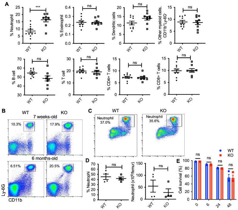
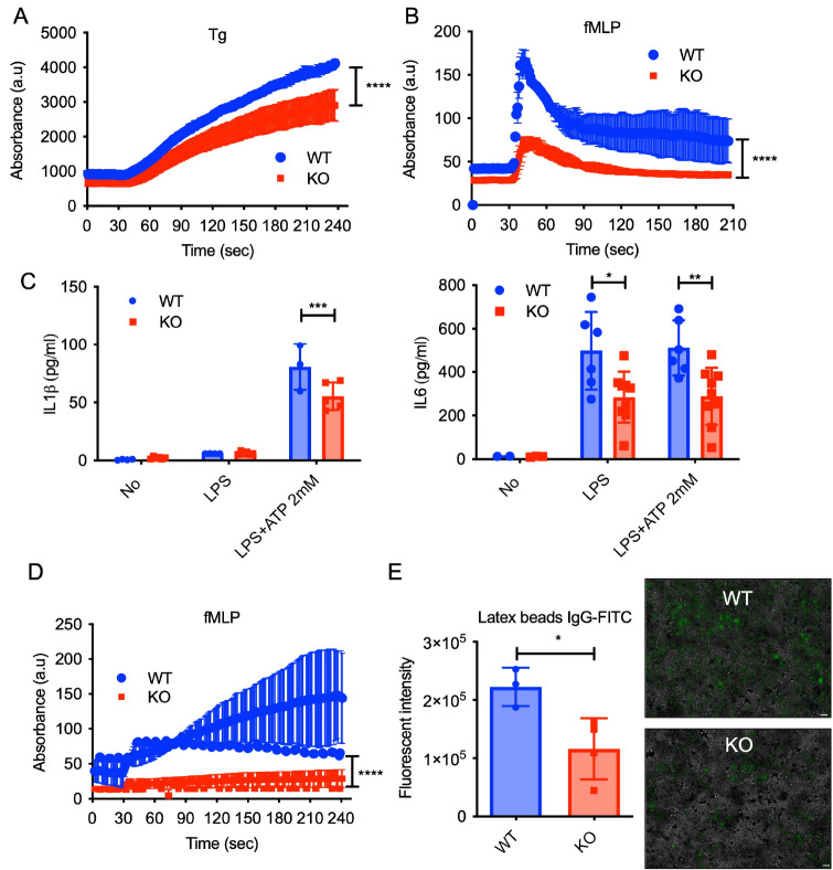
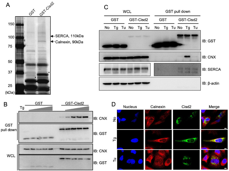
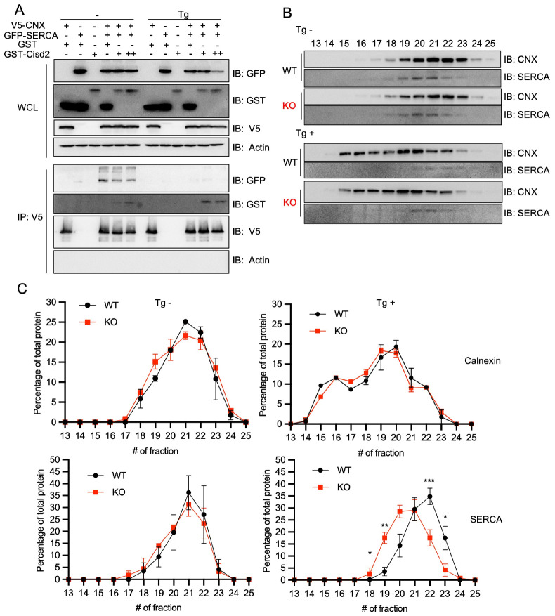
In the context of aging, the susceptibility to infectious diseases increases, leading to heightened morbidity and mortality. This phenomenon, termed immunosenescence, is characterized by dysregulation in the aging immune system, including abnormal alterations in lymphocyte composition, elevated basal inflammation, and the accumulation of senescent T cells. Such changes contribute to increased autoimmune diseases, enhanced infection severity, and reduced responsiveness to vaccines. Utilizing aging animal models becomes imperative for a comprehensive understanding of immunosenescence, given the complexity of aging as a physiological process in living organisms. Our investigation focuses on Cisd2, a causative gene for Wolfram syndrome, to elucidate on immunosenescence. Cisd2 knockout (KO) mice, serving as a model for premature aging, exhibit a shortened lifespan with early onset of aging-related features, such as decreased bone density, hair loss, depigmentation, and optic nerve degeneration. Intriguingly, we found that the Cisd2 KO mice present a higher number of neutrophils in the blood; however, isolated neutrophils from these mice display functional defects. Through mass spectrometry analysis, we identified an interaction between Cisd2 and Calnexin, a protein known for its role in protein quality control. Beyond this function, Calnexin also regulates calcium homeostasis through interaction with sarcoendoplasmic reticulum calcium transport ATPase (SERCA). Our study proposes that Cisd2 modulates calcium homeostasis via its interaction with Calnexin and SERCA, consequently influencing neutrophil functions. [BMB Reports 2024; 57(5): 256-261].


Conflict of interest statement

CONFLICTS OF INTEREST

The authors have no conflicting interests.

Figures

Fig. 1
Fig. 1
Immunophenotyping of blood cells in Cisd2 knockout and WT mice. (A) Quantitative flow cytometric analysis of various myeloid cells and lymphocytes in blood of WT and Cisd2 KO mice (n = 9-10 each group). Each dot plot shows the percentage of immune cell population in CD45+ cell populations. (B) Representative flow cytometric analysis of neutrophils (CD11+ and Ly6G+ cells) neutrophils in WT and Cisd2 KO mice at indicated age. (C) Representative flow cytometric analysis of neutrophils in 6-8 months-old WT and Cisd2 KO bone marrow (n = 4 each group). Percentage of neutrophil in CD45+ cell populations is indicated. (D) Graph showing frequency (left) and absolute (right) number of bone marrow neutrophil in mice of the indicated genotype. (E) The percentage of live neutrophils isolated form bone marrow. Data are presented as the mean ± SEM. ***P = 0.0007. ns, not significant, by Student’s t test.
Fig. 2
Fig. 2
Neutrophil dysfunction of Cisd2 KO mice. Calcium influx in blood neutrophils from WT and Cisd2 KO mice measured by flow cytometry over time after (A) thapsigargin (TG; 1 μM) or (B) fMLP (1 μM) treatment. Baseline calcium level was established for 30 sec prior to addition of stimuli. (C) Levels of IL-1β or IL-6 in the cell supernatants of blood neutrophils from WT and Cisd2 KO mice primed with LPS (500 ng/ml) alone for 4 h or further stimulated with ATP (2 mM) for 30 min. Each point represents data from single mouse. (D) ROS in blood neutrophils from WT and Cisd2 KO mice measured by flow cytometry over time after fMLP (1 μM) treatment. Baseline ROS level was established for 30 sec prior to addition of stimuli. (E) Phagocytic activity of blood neutrophils from WT and Cisd2 KO mice using IgG-coated latex beads. Each point represents data from single mouse (left). The fluorescence intensity was read in a fluorescence plate reader at an excitation of 485 nm and an emission of 535 nm. Scale bar, 40 μm. Data are presented as the mean ± SEM. *P = 0.0286 by Student’s t test (right). Representative image of three independent experiments.
Fig. 3
Fig. 3
Cisd2 interacts with Calnexin and SERCA. (A) Silver-stained SDS-PAGE gel showing proteins recovered from GST pull down assay. HEK293T cells were transfected GST-only or GST-Cisd2 constructs, lysed, and subjected to GST pull down. Bands corresponding to 110-kDa and 90-kDa proteins (indicated by arrows) were excised and identified by mass-spectrometry. Asterisks indicate GST and GST-Cisd2. (B) Western blot analysis of whole cell lysates (WCL) and GST pull down products from HEK293T cells transfected with GST-only or GST-Cisd2 constructs and treated with thapsigargin (Tg) at varying doses (0, 0.01, 0.05, 0.1, and 0.3 μM) for 12 h before cell harvest. (C) Western blot analysis of whole cell lysates (WCL) and GST pull down products from HEK293T cells transfected with GST-only or GST-Cisd2 constructs and treated with 0.1 μM of thapsigargin (Tg) or 1 μg/ml of Tunicamycin (Tu) for 12 h before cell harvest. (D) Confocal image representing HeLa cell cultures transfected with GFP-Cisd2. The cells were treated with Tg (2 μM) or Tu (2 μg/ml) for 4 h. Cells were immunostained for Calnexin (red) and nuclei were stained by DAPI (blue). Scale bar, 10 μm.
Fig. 4
Fig. 4
Cisd2 interrupts Calnexin and SERCA complex formation. (A) HEK293T cells were transfected with V5-Calnexin (CNX), GFP-SERCA, GST, GST-Cisd2 constructs individually or in combinations. The expression level of GST-Cisd2 was varied between low (+) and high (++) levels. Two days post-transfection, whole cell lysates (WCL) were prepared and immunoprecipitated (IP) with anti-V5 antibodies. Co-immunoprecipitated proteins were detected by Western blotting by using anti-GFP, anti-GST, anti-V5, and anti-β-actin. (B) Representative Western blots of size-exclusion chromatography (SEC) fractions (13-25) from MEF cell lysates, prepared with or without Tg (0.3 μM for 6 h) treatment. Western blotting with anti-Calnexin (CNX), and anti-SERCA. (C) SEC fraction profiles represented as the percentage of Calnexin and SERCA in each fraction, indicating the amount per fraction relative to the total protein amount across all fractions. *P < 0.05, **P = 0.0094, ***P = 0.0009, by two-way ANOVA.

References

    1. Oh SJ, Lee JK, Shin OS. Aging and the immune system: the impact of immunosenescence on viral infection, immunity and vaccine immunogenicity. Immune Netw. 2019;19:e37. doi: 10.4110/in.2019.19.e37. - DOI - PMC - PubMed
    1. Liu Z, Liang Q, Ren Y, et al. Immunosenescence: molecular mechanisms and diseases. Signal Transduct Target Ther. 2023;8:200. doi: 10.1038/s41392-023-01451-2.29fd65001ed549ba89b568718601163c - DOI - PMC - PubMed
    1. Shaw AC, Goldstein DR, Montgomery RR. Age-dependent dysregulation of innate immunity. Nat Rev Immunol. 2013;13:875–887. doi: 10.1038/nri3547. - DOI - PMC - PubMed
    1. Chen YF, Kao CH, Kirby R, Tsai TF. Cisd2 mediates mitochondrial integrity and life span in mammals. Autophagy. 2009;5:1043–1045. doi: 10.4161/auto.5.7.9351. - DOI - PubMed
    1. Chen YF, Kao CH, Chen YT, et al. Cisd2 deficiency drives premature aging and causes mitochondria-mediated defects in mice. Genes Dev. 2009;23:1183–1194. doi: 10.1101/gad.1779509. - DOI - PMC - PubMed

MeSH terms