. 2024 Apr 4:14:e2024485.
doi: 10.4322/acr.2024.485. eCollection 2024.

Giant hepatic hemangioma in a patient with cirrhosis: challenging to manage

Affiliations

Giant hepatic hemangioma in a patient with cirrhosis: challenging to manage

Marlone Cunha-Silva et al. Autops Case Rep. .

Abstract

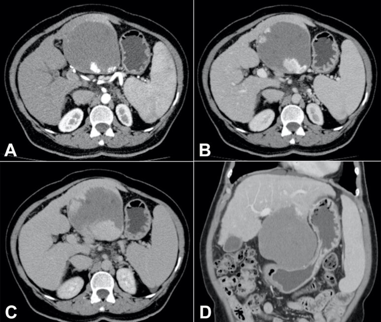
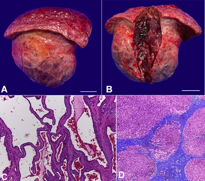
Giant hepatic hemangiomas are occasional in patients with cirrhosis. It remains a challenge to decide on the need for treatment and choose the most appropriate intervention. A 62-year-old woman was recently diagnosed with cirrhosis and complained of upper abdominal fullness, reduction in oral food intake, and weight loss of 6 kg over the last three years. Upper digestive endoscopy evidenced thin-caliber esophageal varices and significant extrinsic compression of the lesser gastric curvature. Abdominal computed tomography revealed an exophytic tumor in the left hepatic lobe, measuring 11.5 cm, which had progressive centripetal contrast enhancement from the arterial phase, compatible with hepatic hemangioma. Serum tumor markers were negative, and her liver function was unimpaired. The patient underwent surgical resection (non-anatomical hepatectomy of segments II and III) which had no immediate complications, and the histopathological evaluation confirmed cavernous hepatic hemangioma. Two weeks later, she was admitted to the emergency room with jaundice, signs of hepatic encephalopathy, and moderate ascites, and was further diagnosed with secondary bacterial peritonitis. As no perforations, abscesses, or fistulas were observed on subsequent imaging tests, clinical management was successfully carried out. This case highlights that giant hepatic hemangiomas may be symptomatic and warrant treatment. In the setting of cirrhosis and portal hypertension, physicians should be aware of the risk of hepatic decompensation following surgical resection, even in patients with Child-Pugh class A.

Keywords: Case Reports; Hemangioma; Liver Cirrhosis.


Conflict of interest statement

Conflict of interest: None.

Figures

Figure 1
Figure 1. Contrast-enhanced abdominal computed tomography: axial section, in the (A) arterial, (B) venous, and (C) late phases: exophytic tumor in the left hepatic lobe (segments II and III), measuring up to 11.5 cm, with centripetal contrast enhancement, compatible with hepatic hemangioma; (D) coronal section, in the venous phase: hepatic hemangioma with significant compression of the stomach; liver with rounded borders and irregular surface, splenomegaly.
Figure 2
Figure 2. Product of hepatectomy: macroscopic evaluation: (A) external surface of the giant hemangioma and part of the left hepatic lobe, with a nodular surface (scale bar = 4 cm); (B) internal tissue of the tumor after sectioning (scale bar = 4 cm); microscopic analysis: (C) tumor: proliferations of blood-filled vascular channels of varying sizes, lined by single layers of flattened endothelial cells and separated by fibrous septa of different thickness, compatible with cavernous hepatic hemangioma [H&E, 20x]; (D) non-tumoral liver parenchyma: fibrotic septa that delimit regenerating nodules of hepatocytes, characterizing cirrhosis (Laennec stage 4C) [Masson's Trichrome stain, 4x].

References

    1. Dong W, Qiu B, Xu H, He L. Invasive management of symptomatic hepatic hemangioma. Eur J Gastroenterol Hepatol. 2019;31(9):1079–1084. doi: 10.1097/MEG.0000000000001413. - DOI - PubMed
    1. Di Carlo I, Koshy R, Al Mudares S, Ardiri A, Bertino G, Toro A. Giant cavernous liver hemangiomas: is it the time to change the size categories? Hepatobiliary Pancreat Dis Int. 2016;15(1):21–29. doi: 10.1016/S1499-3872(15)60035-2. - DOI - PubMed
    1. Mastropasqua M, Kanematsu M, Leonardou P, Braga L, Woosley JT, Semelka RC. Cavernous hemangiomas in patients with chronic liver disease: MR imaging findings. Magn Reson Imaging. 2004;22(1):15–18. doi: 10.1016/j.mri.2003.02.001. - DOI - PubMed
    1. Ribeiro MA, Jr, Papaiordanou F, Gonçalves JM, Chaib E. Spontaneous rupture of hepatic hemangiomas: a review of the literature. World J Hepatol. 2010;2(12):428–433. doi: 10.4254/wjh.v2.i12.428. - DOI - PMC - PubMed
    1. Yu JS, Kim KW, Park MS, Yoon SW. Hepatic cavernous hemangioma in cirrhotic liver: imaging findings. Korean J Radiol. 2000;1(4):185–190. doi: 10.3348/kjr.2000.1.4.185. - DOI - PMC - PubMed