. 2024 Mar 7;4(3):100306.
doi: 10.1016/j.bpsgos.2024.100306. eCollection 2024 May.

Complement C5a Receptor Signaling Alters Stress Responsiveness and Modulates Microglia Following Chronic Stress Exposure

Affiliations

Complement C5a Receptor Signaling Alters Stress Responsiveness and Modulates Microglia Following Chronic Stress Exposure

Hsiao-Jou Cortina Chen et al. Biol Psychiatry Glob Open Sci. .

Abstract

Background: Accumulating evidence underscores the pivotal role of heightened inflammation in the pathophysiology of stress-related diseases, but the underlying mechanisms remain elusive. The complement system, a key effector of the innate immune system, produces the C5-cleaved activation product C5a upon activation, initiating inflammatory responses through the canonical C5a receptor 1 (C5aR1). While C5aR1 is expressed in stress-responsive brain regions, its role in stress responsiveness remains unknown.

Methods: To investigate C5a-C5aR1 signaling in stress responses, mice underwent acute and chronic stress paradigms. Circulating C5a levels and messenger RNA expression of C5aR1 in the hippocampus and adrenal gland were measured. C5aR1-deficient mice were used to elucidate the effects of disrupted C5a-C5aR1 signaling across behavioral, hormonal, metabolic, and inflammation parameters.

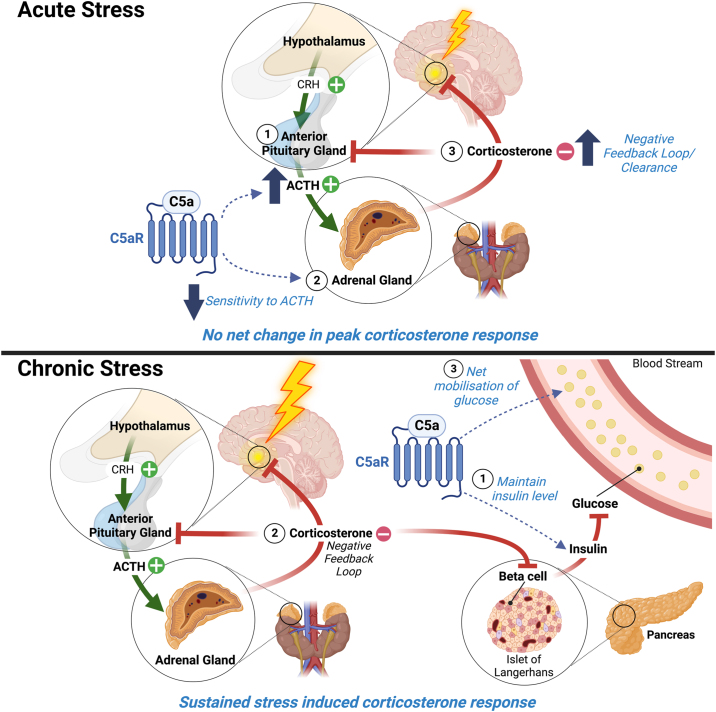
Results: Chronic restraint stress elevated circulating C5a levels while reducing C5aR1 messenger RNA expression in the hippocampus and adrenal gland. Notably, the absence of C5aR1 signaling enhanced adrenal sensitivity to adrenocorticotropic hormone, concurrently reducing pituitary adrenocorticotropic hormone production and enhancing the response to acute stress. C5aR1-deficient mice exhibited attenuated reductions in locomotor activity and body weight under chronic stress. Additionally, these mice displayed increased glucocorticoid receptor sensitivity and disrupted glucose and insulin homeostasis. Chronic stress induced an increase in C5aR1-expressing microglia in the hippocampus, a response mitigated in C5aR1-deficient mice.

Conclusions: C5a-C5aR1 signaling emerges as a key metabolic regulator during stress, suggesting that complement activation and dysfunctional C5aR1 signaling may contribute to neuroinflammatory phenotypes in stress-related disorders. The results advocate for further exploration of complement C5aR1 as a potential therapeutic target for stress-related conditions.

Keywords: C5a receptor; Complement system; Corticosterone; Glia; Hippocampus; Neuroinflammation.

Plain language summary

How the immune system, particularly the complement system, influences responses to stress has not been fully clear. In this study, we focus on C5a-C5aR1 signaling, a part of the immune system, and found that it significantly affects stress-related reactions in mice. In chronic stress, we observed increased inflammation, altered hormonal responses, and disrupted metabolic regulation. Mice lacking C5aR1 showed reduced stress-induced behavioral changes, indicating that this receptor may play a vital role in modulating the stress response. Understanding these immune mechanisms sheds light on stress-related disorders and may open avenues for therapeutic interventions.


Figures

Figure 1
Figure 1
(A) Plasma C5a levels and (B) hippocampal and (C) adrenal gland C5ar1 mRNA expression from wild-type mice following 7, 14, and 21 days of 2-hour restraint stress compared with nonstressed (day 0) control mice (n = 6 per group). Data are expressed as mean ± SEM, ∗p < .05, ∗∗p < .01, and ∗∗∗p < .001. C5ar1, C5a receptor 1; mRNA, messenger RNA.
Figure 2
Figure 2
(A) Plasma corticosterone concentrations and (B) blood glucose levels from WT and C5aR1−/− mice following a single episode of 30-minute restraint stress (n = 9–10 per group). Plasma corticosterone and blood glucose concentrations were determined from serial blood samples collected at 0, 30, 60, 90, and 120 minutes. (C, D) Blood glucose concentrations following (C) glucose tolerance test and (D) insulin tolerance test from WT and C5aR1−/− mice (n = 5–6 per group). (E) Plasma corticosterone concentrations following synacthen stimulation from WT and C5aR1−/− mice (n = 5–6 per group). (F) Plasma ACTH concentrations from WT and C5aR1−/− mice following a single episode of 30-minute restraint stress (n = 5 per group). Data are expressed as mean ± SEM. ∗p < .05, ∗∗p < .01, and ∗∗∗p < .001. ACTH, adrenocorticotropic hormone; C5aR1, C5a receptor 1; WT, wild-type.
Figure 3
Figure 3
(A) Schematic recording timeline for the PhenoMaster experiment, (B) line graph demonstrating 24-hour continuous recording of total locomotor activity under basal conditions, and (C) total locomotor activity from baseline recording (“B”) and following daily restraint stress for 7 days from WT and C5aR1−/− mice (n = 7–8 per group). (D) Body weight gain, (E) blood glucose, (F) plasma insulin, and (G) plasma corticosterone concentrations from WT and C5aR1−/− mice following 7, 14, and 21 days of 2-hour restraint stress compared with nonstressed control mice (n = 5–10 per group). (H) Hippocampal glucocorticoid receptor Nr3c1 and (I)Creb3 mRNA expression from WT and C5aR1−/− mice following 7, 14, and 21 days of 2-hour restraint stress compared with nonstressed control mice (n = 6 per group). Data are expressed as mean ± SEM with ∗p < .05, ∗∗p < .01, and ∗∗∗p < .001 when comparing between strains and †/‡p < .05, ††/‡‡p < .01, and †††/‡‡‡p < .001 when comparing with respective nonstressed (day 0) values. C5aR1, C5a receptor 1; mRNA, messenger RNA; WT, wild-type.
Figure 4
Figure 4
(A) Double immunolabeling of C5aR1 (red) with astrocytes (GFAP; green) and quantification in the CA3 region of the hippocampus of WT mice following 21 days of 2-hour restraint stress compared with nonstressed (day 0) control mice (n = 3 per group). (B) Double immunolabeling of C5aR1 (red) with microglia/macrophages (Iba-1; green) and quantification in the CA3 region of the hippocampus of WT mice following 21 days of 2-hour restraint stress compared with nonstressed (day 0) control mice (n = 3–4 per group). (C) GFAP-positive astrocytes and Iba-1 positive microglia/macrophages in the CA3 region of the hippocampus of WT and C5aR1−/− mice at 21 days of restraint stress compared with nonstressed control mice (n = 3–4 per group). Data are expressed as mean ± SEM. ∗∗p < .01, and ∗∗∗p < .001. Scale bars for all panels = 25 μm. C5aR1, C5a receptor 1; WT, wild-type.
Figure 5
Figure 5
Schematic summary of C5a-C5aR1 signaling on hypothalamic-pituitary-adrenal axis. (Top panel) Under acute stress, C5a-C5aR1 signaling enhances pituitary gland (1) output of ACTH while simultaneously reducing adrenal gland (2) sensitivity to ACTH. This results in no net change in the peak response of corticosterone (3) to an acute stressor. However, C5a-C5aR1 signaling alters the rate of corticosterone recovery to baseline levels by influencing increased negative feedback/clearance mechanisms. (Bottom panel) In conditions of chronic stress, C5a-C5aR1 signaling helps to maintain basal insulin levels (1) and sustains the corticosterone response (2) to stress by delaying habituation. This results in a net mobilization of blood glucose (3). ACTH, adrenocorticotropic hormone; C5aR1, C5a receptor 1; CRH, corticotropin-releasing hormone.

References

    1. Edmiston E., Ashwood P., Van de Water J. Autoimmunity, autoantibodies, and autism spectrum disorder. Biol Psychiatry. 2017;81:383–390. - PMC - PubMed
    1. Horowitz M.A., Zunszain P.A. Neuroimmune and neuroendocrine abnormalities in depression: Two sides of the same coin. Ann N Y Acad Sci. 2015;1351:68–79. - PubMed
    1. Miller A.H., Raison C.L. The role of inflammation in depression: From evolutionary imperative to modern treatment target. Nat Rev Immunol. 2016;16:22–34. - PMC - PubMed
    1. Novellino F., Saccà V., Donato A., Zaffino P., Spadea M.F., Vismara M., et al. Innate immunity: A common denominator between neurodegenerative and neuropsychiatric diseases. Int J Mol Sci. 2020;21 - PMC - PubMed
    1. Pape K., Tamouza R., Leboyer M., Zipp F. Immunoneuropsychiatry - Novel perspectives on brain disorders. Nat Rev Neurol. 2019;15:317–328. - PubMed

LinkOut - more resources