Case Reports
. 2024 Apr 6:35:100499.
doi: 10.1016/j.ensci.2024.100499. eCollection 2024 Jun.

CASE REPORT: Fulminant acute hemorrhagic Leukoencephalitis (AHLE): A rare and ruinous outcome with cerebral herniation (COVID-19)

Affiliations
Case Reports

CASE REPORT: Fulminant acute hemorrhagic Leukoencephalitis (AHLE): A rare and ruinous outcome with cerebral herniation (COVID-19)

Abeer Sabry Safan et al. eNeurologicalSci. .

Abstract

Background: Acute hemorrhagic leukoencephalitis (AHLE) is a very rare demyelinating disease with rapid fulminant inflammation of the white matter. Although the exact etiology is unknown, AHLE usually manifests post a viral or bacterial infection and less often seen post vaccination for measles or rabies. AHLE has a very poor prognosis and a high mortality rate. Owing to the rarity of this entity there is not clear consensus on the proper line of management. In this report, we present a case of AHLE as a para-infectious sequel to COVID-19 in a young patient.

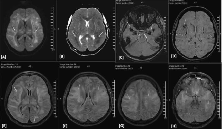
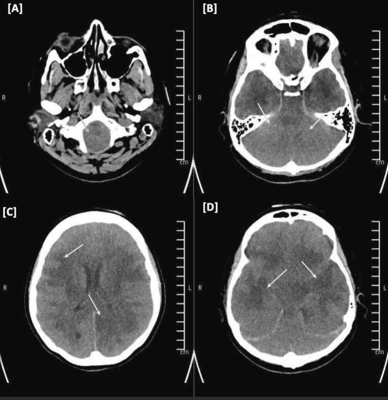
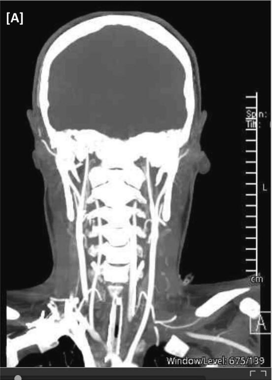
Clinical presentation: We report a 30-year-old turkish patient with an initial presentation of upper respiratory tract infection due to COVID-19. Initially, she was admitted to the hospital with generalized tonic-clonic seizure (GTCS) and deterioration in her level of consciousness lapsing into a coma. An initial CT scan showed diffuse brain edema and an MRI head confirmed the suspicion of Acute hemorrhagic leukoencephalitis (AHLE). Despite prompt and diligent osmotic therapy and pulsed intravenous (IV) methylprednisolone, her condition rapidly depreciated and progressed into cerebral edema with gravid sequela of brainstem herniation.

Conclusions: AHLE is a very rare entity and perhaps its fulminant debilitating course and high mortality should warrant further studies on disease pathophysiology and its optimal treatment parameters. Life-saving decompressive hemicraniectomy should be considered in the multidisciplinary approach of the management with tailored osmotic and immunotherapy.

Keywords: Acute; Disease; Hemorrhagic; Hurst; Leukoencephalitis.


Conflict of interest statement

The authors have no conflicts of interest to disclose.

Figures

Fig. 1
Fig. 1
CT head none contrast enhanced. [A] initial CT head: Presence of generalized cerebral oedema and diffuse white matter hypodensity (orange arrows). [B—D] 12-h follow CT head: Significant progression of generalized cerebral oedema (orange arrows). Diffuse white matter hypodensity involving cerebrum and cerebellum. Bilateral thalamic hypodensities (yellow arrow). (For interpretation of the references to colour in this figure legend, the reader is referred to the web version of this article.)
Fig. 2
Fig. 2
MRI head contrast enhanced. [A-D] DWI/ADC sequences shows radiologic evidence of increased intracranial pressure, scattered bilateral white matter, thalamus and brainstem diffusion restriction (yellow arrows) along with bilateral microhemorrhages (blue arrows). [E-H] FLAIR images showed evidence of bilateral leptomeningeal enhancement noted on postcontrast images (green arrows) and diffused cerebral edema (orange arrows). Intracranial angiography is unremarkable (not shown). Overall impression in favor of acute disseminated hemorrhagic encephalitis (For interpretation of the references to colour in this figure legend, the reader is referred to the web version of this article.)
Fig. 3
Fig. 3
CT head none contrast enhanced. [A-D] Significant interval increase of the diffuse cerebral brain oedema which extended to involve the brainstem, cervico-medullary junction and extending to the visualized part of the cervical spinal cord. The ventricular system,the basal cisterns are completely effaced with diffuse blurring of the gray-white matter junction with loss of differentiation. (yellow arrows). Crowding with almost complete obliteration of the foramen magnum signifying tonsillar herniation (blue arrows) (For interpretation of the references to colour in this figure legend, the reader is referred to the web version of this article.)
Fig. 4
Fig. 4
CTA Intracrnaial angiography. [A] Faint flow of bilateral intracranial ICAs and MCAs with paucity of the distal branches. Absent flow of the vestibulobasilar system and bilateral ACAs. Cervical arteries opacified normally with contrast with no evidence of stenosis or occlusion.

References

    1. Hurst E.W. Acute hemorrhagic leukoencephalitis: a previously undefined entity. Med. J. Aust. 1941;2:1–6.
    1. Donnet A., Dufour H., Gambarelli D., Bruder N., Pellissier J.F., Grisoli F. Acute Weston Hurst necrotizing hemorrhagic leukoencephalitis. Rev. Neurol. 1996;152(12) https://pubmed.ncbi.nlm.nih.gov/9205699/ - PubMed
    1. Yildiz O., Pul R., Raab P., Hartmann C., Skripuletz T., Stangel M. Acute hemorrhagic leukoencephalitis (Weston-Hurst syndrome) in a patient with relapse-remitting multiple sclerosis. J. Neuroinflammation. 2015;12:175. - PMC - PubMed
    1. Grzonka P., Scholz M.C., De Marchis G.M., Tisljar K., Rüegg S., Marsch S., Fladt J., Sutter R. Acute hemorrhagic leukoencephalitis: a case and systematic review of the literature. Front. Neurol. 2020;11 doi: 10.3389/fneur.2020.00899. - DOI - PMC - PubMed
    1. Nabi S., Badshah M., Ahmed S., Nomani A.Z. Weston-Hurst syndrome: a rare fulminant form of acute disseminated encephalomyelitis (ADEM) BMJ Case Rep. 2016;2016 doi: 10.1136/bcr-2016-217215. bcr2016217215. - DOI - PMC - PubMed

Publication types

LinkOut - more resources