

Primary extranodal marginal zone mucosa-associated lymphoid tissue-type B-cell lymphoma involving the dura: A case report
- PMID: 38628522
- PMCID: PMC11021089
- DOI: 10.25259/SNI_792_2023
Primary extranodal marginal zone mucosa-associated lymphoid tissue-type B-cell lymphoma involving the dura: A case report
Abstract
Background: Primary extranodal marginal zone mucosa-associated lymphoid tissue-type B-cell lymphoma (EMZMBCL), which presents as a dural mass, is a rare intracranial tumor that mimics a subdural hematoma or meningioma.
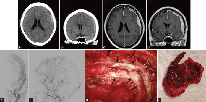
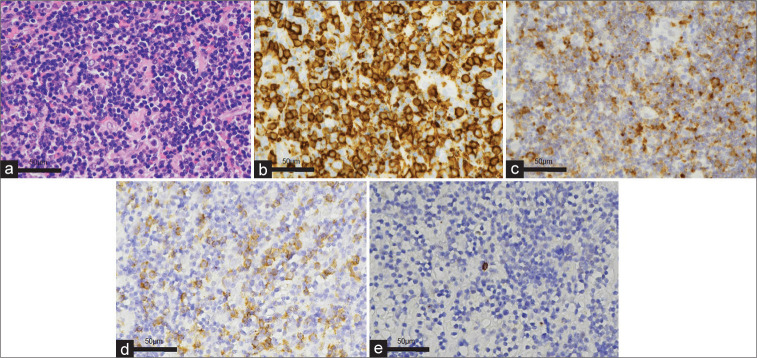
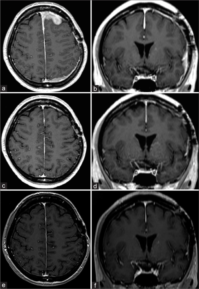
Case description: A 49-year-old woman presented to our hospital with transient right upper limb paresis, dysarthria for 10 min, and ongoing right upper-limb numbness. Computed tomography (CT) of the head revealed extra-axial lesions in the left frontal and parietal lobes. Based on the initial CT findings in the emergency room, an acute subdural hematoma was suspected. However, meningiomas and other intracranial tumors were also listed as differential diagnoses because there was no history of head trauma or coagulation abnormalities on blood examination, and further imaging studies were performed. Imaging findings suggested a subdural neoplastic lesion. A partial resection was performed for the lesion. Based on histopathological and immunohistochemical examinations, the patient was diagnosed with EMZMBCL. Whole-brain and intensity-modulated radiation therapies were administered as adjuvant therapies. The patient was discharged without neurological deficits.
Conclusion: EMZMBCL is a rare disease that should be considered in the differential diagnosis of subdural lesions, especially when there is no history of trauma or abnormalities in the coagulation system. The patient had a favorable outcome after selecting radiotherapy as the adjuvant therapy.
Keywords: Intracranial tumor; Meningioma; Primary extranodal marginal zone mucosa-associated lymphoid tissue-type B-cell lymphoma; Radiotherapy; Subdural hematoma.
Copyright: © 2024 Surgical Neurology International.
Figures
Similar articles
-
Primary extranodal marginal zone B-cell lymphoma of the mucosa-associated lymphoid tissue type in the central nervous system (MZL CNS) presented as traumatic subdural hematoma and subarachnoid bleeding - case report.Clin Neuropathol. 2013 Sep-Oct;32(5):384-92. doi: 10.5414/NP300579. Clin Neuropathol. 2013. PMID: 23557903
-
Tumoral Mimics of Subdural Hematomas: Case Report and Review of Diagnostic and Management Strategies in Primary B-Cell Lymphoma of the Subdural Space.World Neurosurg. 2020 Jan;133:49-54. doi: 10.1016/j.wneu.2019.09.091. Epub 2019 Sep 25. World Neurosurg. 2020. PMID: 31562973 Review.
-
Primary dural lymphoma mimicking a subdural hematoma.J Clin Neurosci. 2010 Mar;17(3):380-2. doi: 10.1016/j.jocn.2009.02.014. Epub 2010 Jan 15. J Clin Neurosci. 2010. PMID: 20079653
-
Case report: Primary intracranial mucosa-associated lymphoid tissue lymphoma presenting as two primary tumors involving the cavernous sinus and extra-axial dura, respectively.Front Oncol. 2023 Jan 4;12:927086. doi: 10.3389/fonc.2022.927086. eCollection 2022. Front Oncol. 2023. PMID: 36686768 Free PMC article.
-
Extranodal marginal zone B-cell lymphoma of malt type involving the cavernous sinus.Leuk Lymphoma. 2001 Sep-Oct;42(5):1133-7. doi: 10.3109/10428190109097736. Leuk Lymphoma. 2001. PMID: 11697633 Review.
Cited by
-
Primary subdural lymphoma mimicking chronic subdural hematoma: a case report and a narrative review of some recent similar cases in the literature.Int J Emerg Med. 2025 Feb 25;18(1):35. doi: 10.1186/s12245-025-00836-0. Int J Emerg Med. 2025. PMID: 40000935 Free PMC article.
References
Publication types
LinkOut - more resources
Full Text Sources