

Non-invasive suppression of the human nucleus accumbens (NAc) with transcranial focused ultrasound (tFUS) modulates the reward network: a pilot study
- PMID: 38628972
- PMCID: PMC11018963
- DOI: 10.3389/fnhum.2024.1359396
Non-invasive suppression of the human nucleus accumbens (NAc) with transcranial focused ultrasound (tFUS) modulates the reward network: a pilot study
Abstract
Background: The nucleus accumbens (NAc) is a key node of the brain reward circuit driving reward-related behavior. Dysregulation of NAc has been demonstrated to contribute to pathological markers of addiction in substance use disorder (SUD) making it a potential therapeutic target for brain stimulation. Transcranial focused ultrasound (tFUS) is an emerging non-invasive brain stimulation approach that can modulate deep brain regions with a high spatial resolution. However, there is currently no evidence showing how the brain activity of NAc and brain functional connectivity within the reward network neuromodulated by tFUS on the NAc.
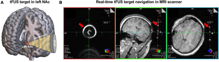
Methods: In this pilot study, we carried out a single-blind, sham-controlled clinical trial using functional magnetic resonance imaging (fMRI) to investigate the underlying mechanism of tFUS neuromodulating the reward network through NAc in ten healthy adults. Specifically, the experiment consists of a 20-min concurrent tFUS/fMRI scan and two 24-min resting-state fMRI before and after the tFUS session.
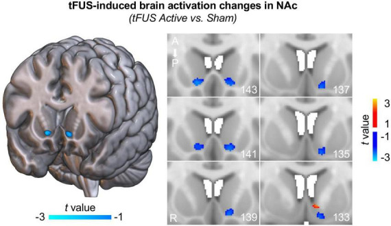
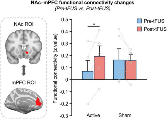
Results: Firstly, our results demonstrated the feasibility and safety of 20-min tFUS on NAc. Additionally, our findings demonstrated that bilateral NAc was inhibited during tFUS on the left NAc compared to sham. Lastly, increased functional connectivity between the NAc and medial prefrontal cortex (mPFC) was observed after tFUS on the left NAc, but no changes for the sham group.
Conclusion: Delivering tFUS to the NAc can modulate brain activations and functional connectivity within the reward network. These preliminary findings suggest that tFUS could be potentially a promising neuromodulation tool for the direct and non-invasive management of the NAc and shed new light on the treatment for SUD and other brain diseases that involve reward processing.
Keywords: fMRI; focused ultrasound; nucleus accumbens; reward network; tFUS.
Copyright © 2024 Peng, Connolly, Sutton, Robinson, Baker-Vogel, Short and Badran.
Conflict of interest statement
The authors declare that the research was conducted in the absence of any commercial or financial relationships that could be construed as a potential conflict of interest.
Figures
Similar articles
-
Transcranial focused ultrasound to the posterior cingulate cortex modulates default mode network and subjective experience: an fMRI pilot study.Front Hum Neurosci. 2024 Jun 4;18:1392199. doi: 10.3389/fnhum.2024.1392199. eCollection 2024. Front Hum Neurosci. 2024. PMID: 38895168 Free PMC article.
-
Transcranial Focused Ultrasound to the Right Prefrontal Cortex Improves Mood and Alters Functional Connectivity in Humans.Front Hum Neurosci. 2020 Feb 28;14:52. doi: 10.3389/fnhum.2020.00052. eCollection 2020. Front Hum Neurosci. 2020. PMID: 32184714 Free PMC article.
-
Transcranial focused ultrasound of the amygdala modulates fear network activation and connectivity.Brain Stimul. 2024 Mar-Apr;17(2):312-320. doi: 10.1016/j.brs.2024.03.004. Epub 2024 Mar 4. Brain Stimul. 2024. PMID: 38447773 Clinical Trial.
-
Neuroimaging and neuromodulation approaches to study eating behavior and prevent and treat eating disorders and obesity.Neuroimage Clin. 2015 Mar 24;8:1-31. doi: 10.1016/j.nicl.2015.03.016. eCollection 2015. Neuroimage Clin. 2015. PMID: 26110109 Free PMC article. Review.
-
Transcranial Focused Ultrasound (tFUS) and Transcranial Unfocused Ultrasound (tUS) Neuromodulation: From Theoretical Principles to Stimulation Practices.Front Neurol. 2019 Jun 11;10:549. doi: 10.3389/fneur.2019.00549. eCollection 2019. Front Neurol. 2019. PMID: 31244747 Free PMC article. Review.
Cited by
-
The therapeutic potential of low-intensity focused ultrasound for treating substance use disorder.Front Psychiatry. 2024 Nov 19;15:1466506. doi: 10.3389/fpsyt.2024.1466506. eCollection 2024. Front Psychiatry. 2024. PMID: 39628494 Free PMC article. Review.
-
Cross-species striatal hubs: Linking anatomy to resting-state connectivity.Neuroimage. 2024 Nov 1;301:120866. doi: 10.1016/j.neuroimage.2024.120866. Epub 2024 Sep 24. Neuroimage. 2024. PMID: 39322095 Free PMC article.
-
Investigating low intensity focused ultrasound pulsation in anhedonic depression-A randomized controlled trial.Front Hum Neurosci. 2025 Mar 24;19:1478534. doi: 10.3389/fnhum.2025.1478534. eCollection 2025. Front Hum Neurosci. 2025. PMID: 40196448 Free PMC article.
-
Low-intensity transcranial focused ultrasound amygdala neuromodulation: a double-blind sham-controlled target engagement study and unblinded single-arm clinical trial.Mol Psychiatry. 2025 Apr 24. doi: 10.1038/s41380-025-03033-w. Online ahead of print. Mol Psychiatry. 2025. PMID: 40275098
-
Research hotspots and frontiers of neuromodulation technology in the last decade: a visualization analysis based on the Web of Science database.Front Hum Neurosci. 2025 Apr 11;19:1574721. doi: 10.3389/fnhum.2025.1574721. eCollection 2025. Front Hum Neurosci. 2025. PMID: 40292332 Free PMC article. Review.
References
-
- Badran B. W., Caulfield K. A., Stomberg-Firestein S., Summers P. M., Dowdle L. T., Savoca M., et al. (2020). Sonication of the anterior thalamus with MRI-guided transcranial focused ultrasound (tFUS) alters pain thresholds in healthy adults: A double-blind, sham-controlled study. Brain Stimul. 13 1805–1812. 10.1016/j.brs.2020.10.007 - DOI - PMC - PubMed
LinkOut - more resources
Full Text Sources