. 2024 Jun 3;221(6):e20222171.
doi: 10.1084/jem.20222171. Epub 2024 Apr 17.

Dominant negative OTULIN-related autoinflammatory syndrome

Affiliations

Dominant negative OTULIN-related autoinflammatory syndrome

Sophia Davidson et al. J Exp Med. .

Abstract

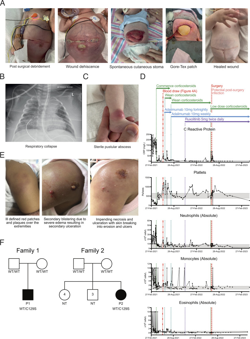
OTU deubiquitinase with linear linkage specificity (OTULIN) regulates inflammation and cell death by deubiquitinating linear ubiquitin chains generated by the linear ubiquitin chain assembly complex (LUBAC). Biallelic loss-of-function mutations causes OTULIN-related autoinflammatory syndrome (ORAS), while OTULIN haploinsuffiency has not been associated with spontaneous inflammation. However, herein, we identify two patients with the heterozygous mutation p.Cys129Ser in OTULIN. Consistent with ORAS, we observed accumulation of linear ubiquitin chains, increased sensitivity to TNF-induced death, and dysregulation of inflammatory signaling in patient cells. While the C129S mutation did not affect OTULIN protein stability or binding capacity to LUBAC and linear ubiquitin chains, it did ablate OTULIN deubiquitinase activity. Loss of activity facilitated the accumulation of autoubiquitin chains on LUBAC. Altered ubiquitination of LUBAC inhibits its recruitment to the TNF receptor signaling complex, promoting TNF-induced cell death and disease pathology. By reporting the first dominant negative mutation driving ORAS, this study expands our clinical understanding of OTULIN-associated pathology.


Conflict of interest statement

Disclosures: D. Komander is a founder, shareholder, and SAB member of Entact Bio. S.L. Masters reported personal fees from Odyssey Therapeutics and NRG Therapeutics outside the submitted work. No other disclosures were reported.

Figures

None
Graphical abstract
Figure 1.
Figure 1.
Patients with the heterozygous C129S variant in OTULIN exhibit clinical features consistent with ORAS. (A) Photographs detailing surgical wound progression of P1. (B) Acute respiratory deterioration in P1. (C) Sterile pustular abscesses at sites of previous trauma, e.g., injection site on ankle from P1. (D) Concentration of CRP and frequencies of platelets, neutrophils, monocytes, and eosinophils in patient blood measured weekly. Gray sections indicate normal range, and therapeutic interventions and key events in P1’s treatment are denoted by dotted lines. (E) Photographs of P2’s tender erythematous plaques and patches over the trunk and bilateral lower limbs and ulcerating wounds with necrotizing fasciitis. (F) Pedigrees of the kindreds presenting ORAS-like disease and carrying C129S heterozygous mutations of OTULIN, P1, P2, and NT (indicates not tested).
Figure 2.
Figure 2.
C129S OTULIN mutant protein associates with increased M1-Ub and cell death in patient cells. (A) C129S OTULIN protein thermostability is comparable to WT OTULIN protein, as assessed by Tycho. (B) Transcription of OTULIN, SHARPIN, HOIL-1, and HOIP in HD1, HD2, and patient fibroblasts was assessed by qPCR. (C) Protein levels of OTULIN, SHARPIN, HOIL-1, HOIP, and actin were assessed in HDs (HD1, HD2) and patient fibroblasts by immunoblot. (D) Fibroblast lysates from HD1, HD2, and patient lines were treated with the OTULIN ABP at stated concentrations to assess OTULIN activity. OTULIN and Tubulin (loading control) were assessed by immunoblot. Arrows indicate unlabeled (OTULIN) and labeled (OTULIN+ABP) OTULIN. * indicates non-specific band. Densitometry analysis of labeled and unlabeled OTULIN bands was performed and graphed as a percentage of total signal. (E and F) TUBE reagent was used to pull-down polyubiquitin in cell lysates. Linear Ub chains (M1-Ub) were then specifically probed for in TUBE pull-down and WCL samples. Equal loading was confirmed by blotting for actin in the WCL. This was performed in (E) lysates from HD1, HD2, and P1 fibroblasts and in (F) lysates from HEK 293T cells transiently transfected with EV, WT, C129S, or Y244C OTULIN. (A and B) Data were pooled from at least three experiments, where each dot is representative of a single experimental result and statistical significance was assessed by unpaired t test, means ± SEMs. (C–F) Data representative of at least three experiments; indicated molecular weight values in kilodaltons (kD). Source data are available for this figure: SourceData F2.
Figure S1.
Figure S1.
The C129S mutation ablates OTULIN deubiquitinase activty, but not OTULIN protein stability or substrate binding. (A) Fibroblast lysates from HD1, HD2, and P1 lines assessed for protein levels of OTULIN, HOIP, HOIL-1, SHARPIN, and actin (loading control) were assessed by immunoblot. Densitometry analysis of protein expression was performed using Fiji and graphed as a function of actin loading control. Data were pooled from four experiments, where each dot indicates an individual experimental result, means ± SEM, and statistical significance was assessed by one-way ANOVA. (B) Recombinant WT OTULIN and C129A and C129S mutant OTULIN proteins were incubated at stated concentrations with M1-linked di-Ub chains. Cleavage capacity was assessed from 0 to 60 min. Arrows indicate uncleaved M1–di-Ub and cleaved Ub; indicated molecular weight values in kD. Data is representative of three repeats. (C) HD1 and P1 fibroblasts were stimulated with TNF-FLAG for indicated time points; the TNFR1 signaling complex was then assessed by Flag pull-down. Immunoblotting was performed for RIPK1, M1-Ub, HOIP, SHARPIN, OTULIN, and actin as a loading control; indicated molecular weight values in kD. Graphs show densitometry quantification of proteins immunoprecipitated at 5 min with TNF-FLAG relative to the signal observed in WCL. Data pooled from three experiments where HD indicates HD1 or HD4 and lines connect values from the same experiment. Significance assessed by ratio paired t test and ** indicates P < 0.01. (D) C129S and C129A binding capacity to M1-Ub was assessed by SPR; data representative of three repeats and KD values summarized in column graph; significance assessed by paired Student’s t test where ** indicates P < 0.01. (E) Densitometry quantification of OTULIN pulled down in four repeats of endogenous SHARPIN IP (Fig. 5 C); significance assessed by Student’s t test, where * indicates P < 0.05. Source data are available for this figure: SourceData FS1.
Figure 3.
Figure 3.
Heterozygous C129S OTULIN fibroblasts exhibit increased sensitivity to TNF-induced cell death. (A and B) NFκB activity was assessed by luciferase reporter assay, where HEK 293T cells were transfected with NFκB luciferase reporter, Renilla control, and (A) stated concentrations of EV, WT, C129S, Y244C, and N341D OTULIN or (B) as a ratio between WT and C129S OTULIN. (C) HD4 and P1 fibroblasts were stimulated with TNF-FLAG for indicated time points; the TNFR1 signaling complex was then assessed by Flag pull-down. Immunoblotting was performed for RIPK1, M1-Ub, HOIP, SHARPIN, and actin as a loading control. (D) HD1, HD2, and P1 fibroblast lines were treated with TNF (100 ng/ml) for indicated time points. Immunoblot was then performed to assess phosphorylated (p) and total levels of p65, p38, JNK as well as total levels IκBa and OTULIN. Actin was used as a loading control. (E–G) Fibroblasts from up to four HDs, P1, two ORAS patients (OTULINL272P/L272P and OTULING174DfsX2/G174DfsX2) and three OTULIN-haploinsufficent patients (OTULIN+/E95X, OTULIN+/R263Q, and OTULIN+/del(5p-)) were used. (E and F) Indicated fibroblast lines were treated with 10 ng/ml TNF for stated time points. Supernatants were then assessed for secretion of (E) IL-8 and (F) IL-6. (G) Stated fibroblasts were treated with TNF (100 ng/ml), CHX (50 μg/ml), QVD-OPh (10 μM), and Nec-1 (10 μM) or a combination thereof, as indicated. Cell death was measured over a period of 24 h by cellular inclusion of propidium iodide dye. (A–G) Data is pooled from at least three experiments, where each dot indicates an individual experimental result, means ± SEM, and statistical significance was assessed by (A, E, and F) two-way ANOVA or (B) one-way ANOVA, where ** indicates P < 0.01 and *** denotes P < 0.001. (C and D) Data representative of at least three experiments, indicated molecular weight values in kD. Source data are available for this figure: SourceData F3.
Figure 4.
Figure 4.
C129S OTULIN heterozygosity drives elevated IFNαβ signaling in patient cells. (A–C) Transcription of inflammatory genes including IFNB1, IRF7, IL6, and TNF was assessed by qPCR in (A) patient and HD PBMCs, in (B) THP-1 cells stably expressing WT or C129S OTULIN or non-transduced controls, and in (C) fibroblasts from up to four HDs, P1, two ORAS patients (OTULINL272P/L272P and OTULING174DfsX2/G174DfsX2), and three OTULIN-haploinsufficent patients (OTULIN+/E95X, OTULIN+/R263Q and OTULIN+/del(5p-)). Inflammatory genes of interest are expressed as fold change relative to the (A and C) average of the healthy donors or (B) non-transduced THP-1s. (D) Activation of IFNαβ signaling was assessed in fibroblasts from indicated donors by ISG Score. ISG Score was generated from the assessment of six known ISGs (IRF7, ISG15, MX1, SIGLEC1, IFIT1, and RSAD2). (A) Data representative of one experiment, technical replicates graphed. (B–D) Data are pooled from at least three experiments, where each dot indicates an individual experimental result, means ± SEM, and statistical significance was assessed by one-way ANOVA. * indicates P < 0.05. For C and D, the following comparisons to P1 were performed: HDs, ORAS, and OTULIN-haploinsufficent.
Figure 5.
Figure 5.
C129S OTULIN dominantly promotes LUBAC autoubiquitination. (A) Cleavage of M1-linked di-Ub was assessed in competitive settings. Increasing concentrations of C129S OTULIN were incubated with WT OTULIN M1-linked di-Ub for 0–180 min. (B) FLAG-tagged WT and C129S OTULIN were stably expressed in HEK 293T cells. Flag IP and WCL lysates from WT OTULIN, C129S OTULIN, and parental HEK 293T cells were immunoblotted for M1-Ub, OTULIN, FLAG, HOIP, SHARPIN, HOIL-1, and actin (loading control). (C and D) Endogenous SHARPIN IP was performed on two HDs and P1 fibroblasts. (C) SHARPIN IP and WCL lysates were immunoblotted for OTULIN, HOIP, SHAPRIN, and actin (loading control). (D) SHARPIN-IP and WCL lysates were treated with the OTULIN ABP probe to assess OTULIN activity when bound to LUBAC. OTULIN, SHARPIN, and actin (loading control) were assessed by immunoblot; arrows indicate unlabeled (OTULIN) and labeled (OTULIN+ABP) OTULIN, and this is quantified by densitometry. (A–D) Data is representative of three repeats and indicated molecular weight values in kD. Source data are available for this figure: SourceData F5.

References

    1. Boisson, B., Laplantine E., Dobbs K., Cobat A., Tarantino N., Hazen M., Lidov H.G., Hopkins G., Du L., Belkadi A., et al. . 2015. Human HOIP and LUBAC deficiency underlies autoinflammation, immunodeficiency, amylopectinosis, and lymphangiectasia. J. Exp. Med. 212:939–951. 10.1084/jem.20141130 - DOI - PMC - PubMed
    1. Boisson, B., Laplantine E., Prando C., Giliani S., Israelsson E., Xu Z., Abhyankar A., Israël L., Trevejo-Nunez G., Bogunovic D., et al. . 2012. Immunodeficiency, autoinflammation and amylopectinosis in humans with inherited HOIL-1 and LUBAC deficiency. Nat. Immunol. 13:1178–1186. 10.1038/ni.2457 - DOI - PMC - PubMed
    1. Damgaard, R.B., Elliott P.R., Swatek K.N., Maher E.R., Stepensky P., Elpeleg O., Komander D., and Berkun Y.. 2019. OTULIN deficiency in ORAS causes cell type-specific LUBAC degradation, dysregulated TNF signalling and cell death. EMBO Mol. Med. 11:e9324. 10.15252/emmm.201809324 - DOI - PMC - PubMed
    1. Damgaard, R.B., Jolin H.E., Allison M.E.D., Davies S.E., Titheradge H.L., McKenzie A.N.J., and Komander D.. 2020. OTULIN protects the liver against cell death, inflammation, fibrosis, and cancer. Cell Death Differ. 27:1457–1474. 10.1038/s41418-020-0532-1 - DOI - PMC - PubMed
    1. Damgaard, R.B., Walker J.A., Marco-Casanova P., Morgan N.V., Titheradge H.L., Elliott P.R., McHale D., Maher E.R., McKenzie A.N.J., and Komander D.. 2016. The deubiquitinase OTULIN is an essential negative regulator of inflammation and autoimmunity. Cell. 166:1215–1230.e20. 10.1016/j.cell.2016.07.019 - DOI - PMC - PubMed