NASH triggers cardiometabolic HFpEF in aging mice
- PMID: 38630423
- PMCID: PMC11336017
- DOI: 10.1007/s11357-024-01153-9
NASH triggers cardiometabolic HFpEF in aging mice
Abstract
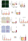
Both heart failure with preserved ejection fraction (HFpEF) and non-alcoholic fatty liver disease (NAFLD) develop due to metabolic dysregulation, has similar risk factors (e.g., insulin resistance, systemic inflammation) and are unresolved clinical challenges. Therefore, the potential link between the two disease is important to study. We aimed to evaluate whether NASH is an independent factor of cardiac dysfunction and to investigate the age dependent effects of NASH on cardiac function. C57Bl/6 J middle aged (10 months old) and aged mice (24 months old) were fed either control or choline deficient (CDAA) diet for 8 weeks. Before termination, echocardiography was performed. Upon termination, organ samples were isolated for histological and molecular analysis. CDAA diet led to the development of NASH in both age groups, without inducing weight gain, allowing to study the direct effect of NASH on cardiac function. Mice with NASH developed hepatomegaly, fibrosis, and inflammation. Aged animals had increased heart weight. Conventional echocardiography revealed normal systolic function in all cohorts, while increased left ventricular volumes in aged mice. Two-dimensional speckle tracking echocardiography showed subtle systolic and diastolic deterioration in aged mice with NASH. Histologic analyses of cardiac samples showed increased cross-sectional area, pronounced fibrosis and Col1a1 gene expression, and elevated intracardiac CD68+ macrophage count with increased Il1b expression. Conventional echocardiography failed to reveal subtle change in myocardial function; however, 2D speckle tracking echocardiography was able to identify diastolic deterioration. NASH had greater impact on aged animals resulting in cardiac hypertrophy, fibrosis, and inflammation.
Keywords: Fatty liver; Inflammation; Liver fibrosis; Metabolic dysfunction; Strain rate analysis.
© 2024. The Author(s).
Conflict of interest statement
P.F. is the founder and CEO of Pharmahungary, a group of R&D companies. A.F. and A.K. report personal fees from Argus Cognitive, Inc., outside the submitted work. All other authors declare no competing interests.
Figures


Similar articles
-
IL-1β neutralization prevents diastolic dysfunction development, but lacks hepatoprotective effect in an aged mouse model of NASH.Sci Rep. 2023 Jan 7;13(1):356. doi: 10.1038/s41598-022-26896-3. Sci Rep. 2023. PMID: 36611037 Free PMC article.
-
Comparison of murine steatohepatitis models identifies a dietary intervention with robust fibrosis, ductular reaction, and rapid progression to cirrhosis and cancer.Am J Physiol Gastrointest Liver Physiol. 2020 Jan 1;318(1):G174-G188. doi: 10.1152/ajpgi.00041.2019. Epub 2019 Oct 21. Am J Physiol Gastrointest Liver Physiol. 2020. PMID: 31630534 Free PMC article.
-
A trans fatty acid substitute enhanced development of liver proliferative lesions induced in mice by feeding a choline-deficient, methionine-lowered, L-amino acid-defined, high-fat diet.Lipids Health Dis. 2020 Dec 14;19(1):251. doi: 10.1186/s12944-020-01423-3. Lipids Health Dis. 2020. PMID: 33317575 Free PMC article.
-
A Comparison of the Gene Expression Profiles of Non-Alcoholic Fatty Liver Disease between Animal Models of a High-Fat Diet and Methionine-Choline-Deficient Diet.Molecules. 2022 Jan 27;27(3):858. doi: 10.3390/molecules27030858. Molecules. 2022. PMID: 35164140 Free PMC article. Review.
-
Non-alcoholic fatty liver disease and heart failure with preserved ejection fraction: from pathophysiology to practical issues.ESC Heart Fail. 2021 Apr;8(2):789-798. doi: 10.1002/ehf2.13222. Epub 2021 Feb 3. ESC Heart Fail. 2021. PMID: 33534958 Free PMC article. Review.
Cited by
-
Comparison of mouse models of heart failure with reduced ejection fraction.ESC Heart Fail. 2025 Feb;12(1):87-100. doi: 10.1002/ehf2.15031. Epub 2024 Sep 7. ESC Heart Fail. 2025. PMID: 39243187 Free PMC article.
-
Chronic alcohol consumption accelerates cardiovascular aging and decreases cardiovascular reserve capacity.Geroscience. 2025 Aug;47(4):5881-5901. doi: 10.1007/s11357-025-01613-w. Epub 2025 Mar 20. Geroscience. 2025. PMID: 40111699 Free PMC article.
-
The role of glucagon-like peptide-1 receptor (GLP-1R) agonists in enhancing endothelial function: a potential avenue for improving heart failure with preserved ejection fraction (HFpEF).Cardiovasc Diabetol. 2025 Feb 7;24(1):70. doi: 10.1186/s12933-025-02607-w. Cardiovasc Diabetol. 2025. PMID: 39920668 Free PMC article. Review.
-
Effects of sex and obesity on immune checkpoint inhibition-related cardiac systolic dysfunction in aged mice.Basic Res Cardiol. 2025 Feb;120(1):207-223. doi: 10.1007/s00395-024-01088-4. Epub 2024 Nov 8. Basic Res Cardiol. 2025. PMID: 39516409 Free PMC article.
-
CardiLect: A combined cross-species lectin histochemistry protocol for the automated analysis of cardiac remodelling.ESC Heart Fail. 2025 Apr;12(2):1398-1415. doi: 10.1002/ehf2.15155. Epub 2024 Nov 13. ESC Heart Fail. 2025. PMID: 39535377 Free PMC article.
References
-
- Peery AF, Crockett SD, Murphy CC, Jensen ET, Kim HP, Egberg MD, Lund JL, Moon AM, Pate V, Barnes EL, Schlusser CL, Baron TH, Shaheen NJ, Sandler RS. Burden and cost of gastrointestinal, liver, and pancreatic diseases in the United States: update 2021. Gastroenterology. 2022;162(2):621–44. 10.1053/j.gastro.2021.10.017. 10.1053/j.gastro.2021.10.017 - DOI - PMC - PubMed
-
- Fotbolcu H, Yakar T, Duman D, Karaahmet T, Tigen K, Cevik C, Kurtoglu U, Dindar I. Impairment of the left ventricular systolic and diastolic function in patients with non-alcoholic fatty liver disease. Cardiol J. 2010;17(5):457–63. - PubMed
-
- Karabay CY, Kocabay G, Kalayci A, Colak Y, Oduncu V, Akgun T, Kalkan S, Guler A, Kirma C. Impaired left ventricular mechanics in nonalcoholic fatty liver disease: a speckle-tracking echocardiography study. Eur J Gastroenterol Hepatol. 2014;26(3):325–31. 10.1097/meg.0000000000000008. 10.1097/meg.0000000000000008 - DOI - PubMed
-
- VanWagner LB, Wilcox JE, Colangelo LA, Lloyd-Jones DM, Carr JJ, Lima JA, Lewis CE, Rinella ME, Shah SJ. Association of nonalcoholic fatty liver disease with subclinical myocardial remodeling and dysfunction: a population-based study. Hepatology. 2015;62(3):773–83. 10.1002/hep.27869. 10.1002/hep.27869 - DOI - PMC - PubMed
MeSH terms
Grants and funding
LinkOut - more resources
Full Text Sources
Medical
Miscellaneous

