. 2024 Apr 23;121(17):e2403206121.
doi: 10.1073/pnas.2403206121. Epub 2024 Apr 17.

Lipoarabinomannan modification as a source of phenotypic heterogeneity in host-adapted Mycobacterium abscessus isolates

Affiliations

Lipoarabinomannan modification as a source of phenotypic heterogeneity in host-adapted Mycobacterium abscessus isolates

Kavita De et al. Proc Natl Acad Sci U S A. .

Abstract

Mycobacterium abscessus is increasingly recognized as the causative agent of chronic pulmonary infections in humans. One of the genes found to be under strong evolutionary pressure during adaptation of M. abscessus to the human lung is embC which encodes an arabinosyltransferase required for the biosynthesis of the cell envelope lipoglycan, lipoarabinomannan (LAM). To assess the impact of patient-derived embC mutations on the physiology and virulence of M. abscessus, mutations were introduced in the isogenic background of M. abscessus ATCC 19977 and the resulting strains probed for phenotypic changes in a variety of in vitro and host cell-based assays relevant to infection. We show that patient-derived mutational variations in EmbC result in an unexpectedly large number of changes in the physiology of M. abscessus, and its interactions with innate immune cells. Not only did the mutants produce previously unknown forms of LAM with a truncated arabinan domain and 3-linked oligomannoside chains, they also displayed significantly altered cording, sliding motility, and biofilm-forming capacities. The mutants further differed from wild-type M. abscessus in their ability to replicate and induce inflammatory responses in human monocyte-derived macrophages and epithelial cells. The fact that different embC mutations were associated with distinct physiologic and pathogenic outcomes indicates that structural alterations in LAM caused by nonsynonymous nucleotide polymorphisms in embC may be a rapid, one-step, way for M. abscessus to generate broad-spectrum diversity beneficial to survival within the heterogeneous and constantly evolving environment of the infected human airway.

Keywords: Mycobacterium abscessus; biofilm; immunomodulation; lipoarabinomannan; nontuberculous mycobacteria.


Conflict of interest statement

Competing interests statement:The authors declare no competing interest.

Figures

Fig. 1.
Fig. 1.
Schematic representation of the structure of Mabs LAM and analysis of EmbC mutations identified in host-adapted Mabs isolates. (A) Proposed structure of Mabs LAM showing acetyl and succinyl substituents on the Ara4 and Ara6 arabinan termini and the tentative location and structure of 3-linked mannoside chains on the mannan domain. Ara5 motifs, not shown in this figure, are thought to be extended linear Ara4 motifs harboring one additional Araf residue (31). (B) Mapping of the structural locations of the wild-type residues corresponding to the 15 patient-derived Mabs EmbC mutations. The predicted AlphaFold model is shown as cartoon. Individual domains corresponding to the N-terminal periplasmic domain (PN), transmembrane domain (TM) and C-terminal periplasmic domain (PC) are colored red, yellow, and cyan, respectively. The location of ethambutol inferred from the structural alignment of the crystal structure from M. smegmatis (PDB 7VBE) is shown as ball and stick representation along with the transparent surface representation. The regions predicted to be disordered are shown in magenta. (C) SDM and FoldX mutant stability prediction. SDM scores are unitless whereas the FoldX scores are given in kcal/mol. Positive and negative values correspond to stabilizing effects in SDM and FoldX, respectively, and vice versa. (D) Same predicted AlphaFold model as in panel (B) but the cartoon representation is differentially colored based on the allosteric coupling intensity values calculated using Ohm. Its corresponding color key is shown at the Bottom. Higher positive values of ACI (shown in red) correspond to potential allosteric hotspots linked to the ligand binding site.
Fig. 2.
Fig. 2.
Electrophoretic mobility of LAM from Mabs ATCC 19977, the embC null mutant, and the recombinant Mabs ATCC 19977 strains expressing different mutated variants of embC. (A) Purified LAMs from the different strains were run on a 10 to 20% Tricine gel followed by periodic acid-silver staining. The results presented are representative of three separate lipoglycan extractions and SDS-PAGE analyses. MWM, molecular weight marker. (B) Araf/Manp ratio of the purified LAMs from the different strains derived from the glycosyl linkage analysis presented in Table 2. Shown on the graph are the means ± SD of three analytical replicates. Asterisks denote statistically significant differences between the control strain, MabsΔembC::pMV306H-embCWT, and the embC mutants (****P < 0.0001; ***P < 0.0005; **P < 0.005; *P < 0.05; ordinary one-way ANOVA, Dunnett’s multiple comparison). (C) Gas chromatograms showing the presence of 3-linked mannose residues in the Mabs ATCC 19977 parent strain, the control WT strain MabsΔembC::pMV306H-embCWT (WT), the EmbCM310T mutant and the embC knockout mutant, comigrating with the authentic standard, 2,4,6-tri-O-methylD-mannitol acetate.
Fig. 3.
Fig. 3.
Impact of patient-derived embC mutations on the morphology, sliding motility, and ability of Mabs ATCC 19977 to form biofilms and serpentine cords. (A) Colony morphology of the control and embC mutant strains on 7H11-OADC agar plates after 5 d of incubation at 37 °C. Mabs ATCC 19977 R is the rough morphotype parent strain used throughout this study (and in all other figures). Mabs ATCC 19977 S is the reference smooth morphotype strain shown here for comparison. (B) Sliding motility of the control and embC mutant strains on agar medium. Mabs strains were drop-inoculated from liquid cultures diluted to 106 CFU/mL onto 7H9-ADC medium containing 0.34% agar and incubated at 37 °C for 5 d at which point the diameters of spread of the different strains were compared. (C) Biofilm formation in SCFM by the different strains was analyzed after 5 d of incubation at 37 °C by crystal violet staining as described previously (43). Shown are the means ± SD of absorbances measured at 562 nm for six biological replicates. Asterisks denote statistically significant differences between the control strain, MabsΔembC::pMV306H-embCWT, and the embC mutants (****P < 0.0001; ***P < 0.001; ordinary one-way ANOVA; Dunnett’s multiple comparison). (D) The control and mutant strains were inoculated in Tryptic Soy broth at a concentration of 104 CFU/mL and their ability to form serpentine cords was compared after 3 d of incubation at 37 °C. All assays were performed as described under Materials and Methods and the results presented are representative of two to three independent experiments.
Fig. 4.
Fig. 4.
Phenotypic properties of an Mabs ATCC 19977 rough sucT knockout mutant. (A) Allelic replacement at the sucT locus of Mabs ATCC 19977 (rough). Genomic DNA was extracted from the WT Mabs ATCC 19977 rough strain and knock-out (ΔsucT) mutant and allelic replacement in the mutant was confirmed by PCR as detailed in Materials and Methods. The expected sizes of the PCR products are 2,474 bp and 3,415 bp in the WT strain and knockout mutant, respectively. The WT and mutant strains were compared for their ability to form biofilms in SCFM (B), colony morphology (C), ability to form serpentine cords in Tryptic Soy broth (D), and sliding motility on 7H9-ADC agar plates (E) as described in Fig. 3. The M310T and I402V EmbC mutants were included in the biofilm assay for comparison. Shown on the graph are the averages ± SD of crystal violet absorbances measured at 562 nm for four biological replicates. Asterisks denote statistically significant differences between WT strain and mutant strains (****P < 0.0001; **P < 0.001; *P < 0.01; ordinary one-way ANOVA; Dunnett’s multiple comparison).
Fig. 5.
Fig. 5.
NF-κB activation in HEK-TLR2 cells by the Mabs ATCC 19977 control and mutant embC strains and surface extracts derived thereof. (A) The NF-κB activity of HEK-TLR2 cells stimulated with the control and a subset of embC mutant strains (MOI of 1) at 37 °C for 16 h was determined by reading the absorbance of the medium at 650 nm. Shown are the means ± SD of absorbances measured at 650 nm for three biological replicates. (B) NF-κB activity of HEK-TLR2 cells stimulated with TSE, either untreated or treated with papain, prepared from the MabsΔembC::pMV306H-embCWT control strain and three patient-derived EmbC mutants. Shown are the means ± SD of absorbances measured at 650 nm for three biological replicates. Asterisks denote statistically significant differences between the control strain, MabsΔembC::pMV306H-embCWT (or TSE derived thereof), and the embC mutants (or TSE derived thereof) (****P < 0.0001; **P < 0.01; *P < 0.05; ordinary one-way ANOVA; Dunnett’s multiple comparison). The results presented in (A) and (B) are representative of two to three independent experiments.
Fig. 6.
Fig. 6.
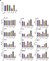
Intracellular replication, immune activation, and chemokine/cytokine secretion induced by the control and embC mutant strains in monocyte-derived THP-1 macrophages. THP-1 cells were infected at an MOI of 1. (A) Two and 48 h post infection, the macrophages were lysed, and intracellular bacteria enumerated by CFU plating. Shown is the fold-change in CFUs for each strain between 48 h and 2 h. (B) Levels of expression of activation markers for HLADR, CD80, CD86, and CD40 48 h postinfection was determined by flow cytometry. (C) Culture supernatants were analyzed 48 h postinfection for chemokine/cytokine secretion by multiplex immunoassay using Luminex. The results presented are the means ± SD of triplicate wells from one experiment and are representative of three independent experiments. Asterisks denote statistically significant differences between the control strain, MabsΔembC::pMV306H-embCWT, and the embC mutants (****P < 0.0001; ***P < 0.0005; **P < 0.005; ordinary one-way ANOVA; Dunnett’s multiple comparison).
Fig. 7.
Fig. 7.
Intracellular replication of embC mutants in human A549 epithelial cells. A549 lung alveolar type II epithelial cells were infected at an MOI of 1. Two and 48 h post infection, the cells were lysed and intracellular bacteria enumerated by CFU plating. The results presented are the means ± SD of triplicate wells from one experiment and are representative of two independent experiments. Asterisks denote statistically significant differences between the control strain, MabsΔembC::pMV306H-embCWT, and the embC mutants (****P < 0.0001; ***P < 0.0006; **P < 0.005; ordinary one-way ANOVA; Dunnett’s multiple comparison).

References

    1. Park I. K., Olivier K. N., Nontuberculous mycobacteria in cystic fibrosis and non-cystic fibrosis bronchiectasis. Semin. Respir. Crit. Care Med. 36, 217–224 (2015). - PMC - PubMed
    1. Prevots D. R., Marras T. K., Epidemiology of human pulmonary infection with nontuberculous mycobacteria: A review. Clin. Chest Med. 36, 13–34 (2015). - PMC - PubMed
    1. Martiniano S. L., Nick J. A., Daley C. L., Nontuberculous mycobacterial infections in cystic fibrosis. Clin. Chest Med. 43, 697–716 (2022). - PubMed
    1. Bryant J. M., et al. , Stepwise pathogenic evolution of Mycobacterium abscessus. Science 372, eabb8699 (2021). - PMC - PubMed
    1. Bryant J. M., et al. , Emergence and spread of a human-transmissible multidrug-resistant nontuberculous mycobacterium. Science 354, 751–757 (2016). - PMC - PubMed

LinkOut - more resources