

Structural insights into the unexpected agonism of tetracyclic antidepressants at serotonin receptors 5-HT1eR and 5-HT1FR
- PMID: 38630816
- PMCID: PMC11023502
- DOI: 10.1126/sciadv.adk4855
Structural insights into the unexpected agonism of tetracyclic antidepressants at serotonin receptors 5-HT1eR and 5-HT1FR
Abstract
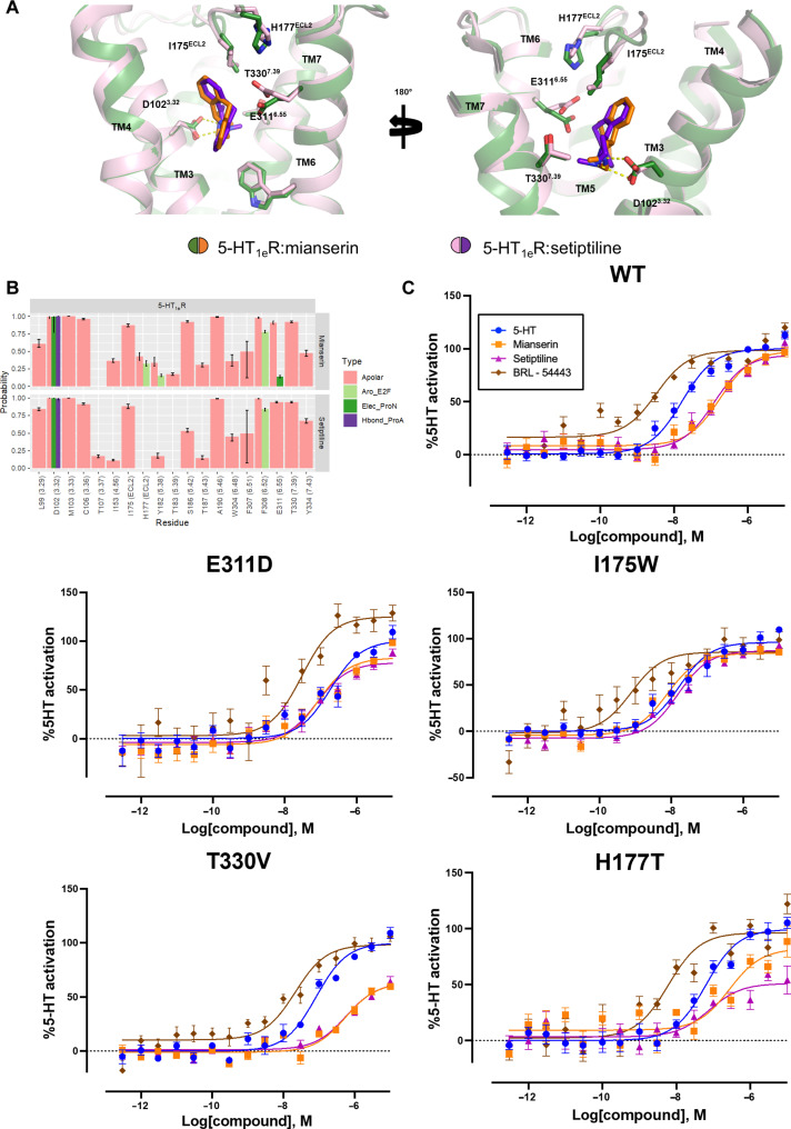
Serotonin [5-hydroxytryptamine (5-HT)] acts via 13 different receptors in humans. Of these receptor subtypes, all but 5-HT1eR have confirmed roles in native tissue and are validated drug targets. Despite 5-HT1eR's therapeutic potential and plausible druggability, the mechanisms of its activation remain elusive. To illuminate 5-HT1eR's pharmacology in relation to the highly homologous 5-HT1FR, we screened a library of aminergic receptor ligands at both receptors and observe 5-HT1eR/5-HT1FR agonism by multicyclic drugs described as pan-antagonists at 5-HT receptors. Potent agonism by tetracyclic antidepressants mianserin, setiptiline, and mirtazapine suggests a mechanism for their clinically observed antimigraine properties. Using cryo-EM and mutagenesis studies, we uncover and characterize unique agonist-like binding poses of mianserin and setiptiline at 5-HT1eR distinct from similar drug scaffolds in inactive-state 5-HTR structures. Together with computational studies, our data suggest that these binding poses alongside receptor-specific allosteric coupling in 5-HT1eR and 5-HT1FR contribute to the agonist activity of these antidepressants.
Figures


Update of
-
Structural Insights into the Unexpected Agonism of Tetracyclic Antidepressants at Serotonin Receptors 5-HT1eR and 5-HT1FR.bioRxiv [Preprint]. 2023 Oct 7:2023.10.05.561100. doi: 10.1101/2023.10.05.561100. bioRxiv. 2023. Update in: Sci Adv. 2024 Apr 19;10(16):eadk4855. doi: 10.1126/sciadv.adk4855. PMID: 37986777 Free PMC article. Updated. Preprint.
Similar articles
-
Structural Insights into the Unexpected Agonism of Tetracyclic Antidepressants at Serotonin Receptors 5-HT1eR and 5-HT1FR.bioRxiv [Preprint]. 2023 Oct 7:2023.10.05.561100. doi: 10.1101/2023.10.05.561100. bioRxiv. 2023. Update in: Sci Adv. 2024 Apr 19;10(16):eadk4855. doi: 10.1126/sciadv.adk4855. PMID: 37986777 Free PMC article. Updated. Preprint.
-
Assessing the comparative effects of interventions in COPD: a tutorial on network meta-analysis for clinicians.Respir Res. 2024 Dec 21;25(1):438. doi: 10.1186/s12931-024-03056-x. Respir Res. 2024. PMID: 39709425 Free PMC article. Review.
-
Surveillance for Violent Deaths - National Violent Death Reporting System, 50 States, the District of Columbia, and Puerto Rico, 2022.MMWR Surveill Summ. 2025 Jun 12;74(5):1-42. doi: 10.15585/mmwr.ss7405a1. MMWR Surveill Summ. 2025. PMID: 40493548 Free PMC article.
-
Defining disease severity in atopic dermatitis and psoriasis for the application to biomarker research: an interdisciplinary perspective.Br J Dermatol. 2024 Jun 20;191(1):14-23. doi: 10.1093/bjd/ljae080. Br J Dermatol. 2024. PMID: 38419411 Free PMC article. Review.
-
Use of β-adrenoreceptor drugs and Parkinson's disease incidence in women from the French E3N cohort study.J Parkinsons Dis. 2025 Jun;15(4):789-804. doi: 10.1177/1877718X251330993. Epub 2025 Apr 29. J Parkinsons Dis. 2025. PMID: 40302366
Cited by
-
The ABCs of psychedelics: a preclinical roadmap for drug discovery.Trends Pharmacol Sci. 2025 Aug 27:S0165-6147(25)00160-9. doi: 10.1016/j.tips.2025.07.017. Online ahead of print. Trends Pharmacol Sci. 2025. PMID: 40877079 Review.
References
-
- Barnes N. M., Ahern G. P., Becamel C., Bockaert J., Camilleri M., Chaumont-Dubel S., Claeysen S., Cunningham K. A., Fone K. C., Gershon M., Giovanni G. D., Goodfellow N. M., Halberstadt A. L., Hartley R. M., Hassaine G., Herrick-Davis K., Hovius R., Lacivita E., Lambe E. K., Leopoldo M., Levy F. O., Lummis S. C. R., Marin P., Maroteaux L., McCreary A. C., Nelson D. L., Neumaier J. F., Newman-Tancredi A., Nury H., Roberts A., Roth B. L., Roumier A., Sanger G. J., Teitler M., Sharp T., Villalón C. M., Vogel H., Watts S. W., Hoyer D., International Union of Basic and Clinical Pharmacology. CX. Classification of receptors for 5-hydroxytryptamine; pharmacology and function. Pharmacol. Rev. 73, 310–520 (2021). - PMC - PubMed
-
- Tepper S. J., Rapoport A. M., Sheftell F. D., Mechanisms of action of the 5-HT1B/1D receptor agonists. Arch. Neurol. 59, 1084–1088 (2002). - PubMed
MeSH terms
Substances
Grants and funding
LinkOut - more resources
Full Text Sources
