. 2024 Apr 19;10(16):eadi1782.
doi: 10.1126/sciadv.adi1782. Epub 2024 Apr 17.

RIPK3 deficiency blocks R-2-hydroxyglutarate-induced necroptosis in IDH-mutated AML cells

Affiliations

RIPK3 deficiency blocks R-2-hydroxyglutarate-induced necroptosis in IDH-mutated AML cells

Shuanghong Zhu et al. Sci Adv. .

Abstract

Mutant isocitrate dehydrogenases (IDHs) produce R-2-hydroxyglutarate (R-2HG), which inhibits the growth of most acute myeloid leukemia (AML) cells. Here, we showed that necroptosis, a form of programmed cell death, contributed to the antileukemia activity of R-2HG. Mechanistically, R-2HG competitively inhibited the activity of lysine demethylase 2B (KDM2B), an α-ketoglutarate-dependent dioxygenase. KDM2B inhibition increased histone 3 lysine 4 trimethylation levels and promoted the expression of receptor-interacting protein kinase 1 (RIPK1), which consequently caused necroptosis in AML cells. The expression of RIPK3 was silenced because of DNA methylation in IDH-mutant (mIDH) AML cells, resulting in R-2HG resistance. Decitabine up-regulated RIPK3 expression and repaired endogenous R-2HG-induced necroptosis pathway in mIDH AML cells. Together, R-2HG induced RIPK1-dependent necroptosis via KDM2B inhibition in AML cells. The loss of RIPK3 protected mIDH AML cells from necroptosis. Restoring RIPK3 expression to exert R-2HG's intrinsic antileukemia effect will be a potential therapeutic strategy in patients with AML.


Figures
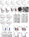
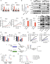
Fig. 1.
Fig. 1.. R-2HG caused necroptosis of wtIDH AML cells.
(A) Viability curves of AML cells treated with R-2HG at indicated time points. Viability rate were normalized to the vehicle-treated cells at the same time point. (B) Viability of Bone Marrow Mononuclear Cells (BMMNCs) from patients without mIDH treated with 300 μM R-2HG. (C) Viability of AML cells cotreated with 300 μM R-2HG and indicated cell death inhibitors for 72 hours: Nec-1, 25 μM; Z-VAD, 25 μM; Fer-1, 1 μM, 3-MA, 2 mM; VX-765, 25 μM. (D) Viability of AML cells from patients with wtIDH cotreated with 300 μM R-2HG and Nec-1 (25 μM) for 72 hours. (E) Transmission electron microscopy images of SKM-1 cells treated with vehicle or 300 μM R-2HG for 8 hours. Arrows indicate swollen mitochondria. (F) Percentage of SYTOX Green+ SKM-1 or THP-1 cells cotreated with R-2HG and Nec-1 (25 μM). (G) Percentage of SYTOX Green+ cells in primary AML cells from patients with wtIDH cotreated with R-2HG and Nec-1 (25 μM). (H) Expression of necroptosis-related proteins in SKM-1 and THP-1 cells treated with R-2HG for 8 hours. p-RIPK1, phosphorylation of RIPK1; p-MLKL, phosphorylation of MLKL. (I) Expression of necroptosis-related proteins in primary AML cells from patients with wtIDH treated with R-2HG for 8 hours. (J) R-2HG production in SKM-1 and THP-1 cells overexpressing wtIDH2 or mIDH2. (K) Viability of wtIDH2 or IDH2R172K overexpressed SKM-1 and THP-1 cells treated with Nec-1 (25 μM). (L) Phosphorylation of MLKL and total MLKL in wtIDH2 or IDH2R172K overexpressed SKM-1 and THP-1 cells treated with Nec-1 (25 μM). (M) Percentage of SYTOX Green+ wtIDH2 or IDH2R172K overexpressed SKM-1 or THP-1 cells treated with Nec-1 (25 μM). *P < 0.05, **P < 0.01, ***P < 0.001, and ****P < 0.0001.
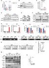
Fig. 2.
Fig. 2.. R-2HG–induced cell necroptosis depends on the high expression of RIPK1 in AML cells.
(A) mRNA levels of RIPK1, RIPK3, and MLKL in SKM-1 and THP-1 cells treated with 300 μM R-2HG for 8 hours. (B) mRNA level of RIPK1 in mIDH and wtIDH primary AML cells. (C) Western blot showing the indicated proteins in SKM-1 and THP-1 cells expressing Dox-inducible wtIDH2 and mIDH2 after 72 hours of Dox treatment. (D) Cell proliferation curve of SKM-1 and THP-1 cells infected with an empty vector or overexpressing (OE) RIPK1. (E) Percentage of SYTOX Green+ SKM-1 or THP-1 cells infected with an empty vector or overexpressing RIPK1. (F) Western blot showing the indicated proteins in SKM-1 and THP-1 cells infected with an empty vector or overexpressing RIPK1. (G) Viability of shRIPK1-transduced cells treated with 300 μM R-2HG for 72 hours. (H) Percentage of SYTOX Green+ cells in shRIPK1-transduced cells treated with 300 μM R-2HG. (I) Schematic illustration of AML mouse models treated with R-2HG. BM, bone marrow. (J) Engraftment of SKM-1 cells in the bone marrow of recipients. (K) Survival of SKM-1 cell–transplanted mice after the indicated treatment. KD, knockdown; ns, not significant (P ≥ 0.05). *P < 0.05, **P < 0.01, and ***P < 0.001.
Fig. 3.
Fig. 3.. Inhibition of KDM2B increases RIPK1 expression and induces necroptosis in AML cells.
(A) α-KG–dependent dioxygenases coexpressed with RIPK1 in AML cohorts (data from the GSE14468 and GSE10358 datasets). (B) Coexpression analysis of RIPK1 and KDM2B in our own AML cohort. (C) mRNA level of RIPK1 in SKM-1 and THP-1 cells expressing Dox-inducible scramble and shKDM2B after 5 days of Dox treatment. (D) Western blot showing the indicated proteins in SKM-1 and THP-1 cells expressing Dox-inducible scramble and shKDM2B after 5 days of Dox treatment. (E) Percentage of SYTOX Green+ SKM-1 or THP-1 cells expressing Dox-inducible scramble and shKDM2B after 5 days of Dox treatment. (F) Proliferation curves of SKM-1 or THP-1 cells expressing Dox-inducible scramble and shKDM2B at indicated time. (G) Western blot showing the indicated proteins in SKM-1 or THP-1 cells expressing Dox-inducible scramble and shKDM2B after 5 days of Dox treatment. (H) RIPK1 DNA enriched with anti-H3K4me3 antibody from R-2HG–treated cells. Rabbit immunoglobulin G (IgG) was included as the negative control. (I) RIPK1 DNA enriched with anti-H3K4me3 antibody from KDM2B-knockdown cells. Rabbit IgG was included as the negative control. (J) Percentage of SYTOX Green+ SKM-1 or THP-1 cells transduced with the indicated shRNAs. (K) Viability curves of SKM-1 and THP-1 cells transduced with the indicated shRNAs. *P < 0.05, **P < 0.01, ***P < 0.001, and ****P < 0.0001.
Fig. 4.
Fig. 4.. Methylation-dependent loss of RIPK3 expression represses necroptosis in response to R-2HG.
(A) mRNA levels of RIPK3 and MLKL in primary AML cells from patients without mIDH. (B) Expression of RIPK3 in primary AML cells from patients without mIDH. (C) RIPK3 mRNA levels in R-2HG–sensitive and R-2HG–resistant AML cell lines (data from dataset GSE87187). (D) Expression of RIPK3 in R-2HG–sensitive and R-2HG–resistant AML cell lines. (E) Expression of RIPK3 in vector control and RIPK3-overexpressing cells. (F) Expression of the indicated proteins in K562 or NB4 transduced with empty vector or overexpressing RIPK3 after treatment with vehicle or 300 μM R-2HG for 72 hours. (G) Percentage of SYTOX Green+ K562 or NB4 transduced with empty vector or overexpressing RIPK3 after treatment with vehicle or 300 μM R-2HG for 72 hours. (H) Viability of K562 or NB4 transduced with empty vector or overexpressing RIPK3 after treatment with vehicle or 300 μM R-2HG for 72 hours. (I) Methylation-specific PCR (MSP) of the RIPK3 promoter in SKM-1, THP-1, K562, and NB4 cells. M and U indicate methylated-specific and unmethylated-specific primers, respectively, targeting the RIPK3 promoter. (J) MSP of the RIPK3 promoter in K562 and NB4 cells treated with DEC (2 μM) or AZA (5 μM) for 72 hours. (K) Expression of RIPK3 in K562 and NB4 cell lines treated with DEC or AZA for 72 hours. (L) Methylation level of CpG sites at the RIPK3 promoter in mIDH (average level of patients with AML) and wtIDH AML cells (average level of patients with AML). (M) Expression of the indicated proteins in wtIDH and mIDH primary cells treated with 2 μM DEC for 72 hours. (N) The IC50 of DEC in primary AML cells from patients without mIDH.*P < 0.05, **P < 0.01, ***P < 0.001, and ****P < 0.0001.

References

    1. Loenarz C., Schofield C. J., Physiological and biochemical aspects of hydroxylations and demethylations catalyzed by human 2-oxoglutarate oxygenases. Trends Biochem. Sci. 36, 7–18 (2011). - PubMed
    1. Molenaar R. J., Thota S., Nagata Y., Patel B., Clemente M., Przychodzen B., Hirsh C., Viny A. D., Hosano N., Bleeker F. E., Meggendorfer M., Alpermann T., Shiraishi Y., Chiba K., Tanaka H., van Noorden C. J. F., Radivoyevitch T., Carraway H. E., Makishima H., Miyano S., Sekeres M. A., Ogawa S., Haferlach T., Maciejewski J. P., Clinical and biological implications of ancestral and non-ancestral IDH1 and IDH2 mutations in myeloid neoplasms. Leukemia 29, 2134–2142 (2015). - PMC - PubMed
    1. Cancer Genome Atlas Research Network, Ley T. J., Miller C., Ding L., Raphael B. J., Mungall A. J., Robertson A., Hoadley K., Triche T. J. Jr., Laird P. W., Baty J. D., Fulton L. L., Fulton R., Heath S. E., Kalicki-Veizer J., Kandoth C., Klco J. M., Koboldt D. C., Kanchi K. L., Kulkarni S., Lamprecht T. L., Larson D. E., Lin L., Lu C., McLellan M. D., McMichael J. F., Payton J., Schmidt H., Spencer D. H., Tomasson M. H., Wallis J. W., Wartman L. D., Watson M. A., Welch J., Wendl M. C., Ally A., Balasundaram M., Birol I., Butterfield Y., Chiu R., Chu A., Chuah E., Chun H. J., Corbett R., Dhalla N., Guin R., He A., Hirst C., Hirst M., Holt R. A., Jones S., Karsan A., Lee D., Li H. I., Marra M. A., Mayo M., Moore R. A., Mungall K., Parker J., Pleasance E., Plettner P., Schein J., Stoll D., Swanson L., Tam A., Thiessen N., Varhol R., Wye N., Zhao Y., Gabriel S., Getz G., Sougnez C., Zou L., Leiserson M. D., Vandin F., Wu H. T., Applebaum F., Baylin S. B., Akbani R., Broom B. M., Chen K., Motter T. C., Nguyen K., Weinstein J. N., Zhang N., Ferguson M. L., Adams C., Black A., Bowen J., Gastier-Foster J., Grossman T., Lichtenberg T., Wise L., Davidsen T., Demchok J. A., Shaw K. R., Sheth M., Sofia H. J., Yang L., Downing J. R., Eley G., Genomic and epigenomic landscapes of adult de novo acute myeloid leukemia. N. Engl. J. Med. 368, 2059–2074 (2013). - PMC - PubMed
    1. Mardis E. R., Ding L., Dooling D. J., Larson D. E., McLellan M. D., Chen K., Koboldt D. C., Fulton R. S., Delehaunty K. D., McGrath S. D., Fulton L. A., Locke D. P., Magrini V. J., Abbott R. M., Vickery T. L., Reed J. S., Robinson J. S., Wylie T., Smith S. M., Carmichael L., Eldred J. M., Harris C. C., Walker J., Peck J. B., Du F., Dukes A. F., Sanderson G. E., Brummett A. M., Clark E., McMichael J. F., Meyer R. J., Schindler J. K., Pohl C. S., Wallis J. W., Shi X., Lin L., Schmidt H., Tang Y., Haipek C., Wiechert M. E., Ivy J. V., Kalicki J., Elliott G., Ries R. E., Payton J. E., Westervelt P., Tomasson M. H., Watson M. A., Baty J., Heath S., Shannon W. D., Nagarajan R., Link D. C., Walter M. J., Graubert T. A., DiPersio J. F., Wilson R. K., Ley T. J., Recurring mutations found by sequencing an acute myeloid leukemia genome. N. Engl. J. Med. 361, 1058–1066 (2009). - PMC - PubMed
    1. Dang L., White D. W., Gross S., Bennett B. D., Bittinger M. A., Driggers E. M., Fantin V. R., Jang H. G., Jin S., Keenan M. C., Marks K. M., Prins R. M., Ward P. S., Yen K. E., Liau L. M., Rabinowitz J. D., Cantley L. C., Thompson C. B., Vander Heiden M. G., Su S. M., Cancer-associated IDH1 mutations produce 2-hydroxyglutarate. Nature 465, 966 (2010). - PMC - PubMed

Publication types