. 2024 Jul;11(4):e200243.
doi: 10.1212/NXI.0000000000200243. Epub 2024 Apr 17.

Life-Threatening MOG Antibody-Associated Hemorrhagic ADEM With Elevated CSF IL-6

Affiliations

Life-Threatening MOG Antibody-Associated Hemorrhagic ADEM With Elevated CSF IL-6

Akash Virupakshaiah et al. Neurol Neuroimmunol Neuroinflamm. 2024 Jul.

Abstract

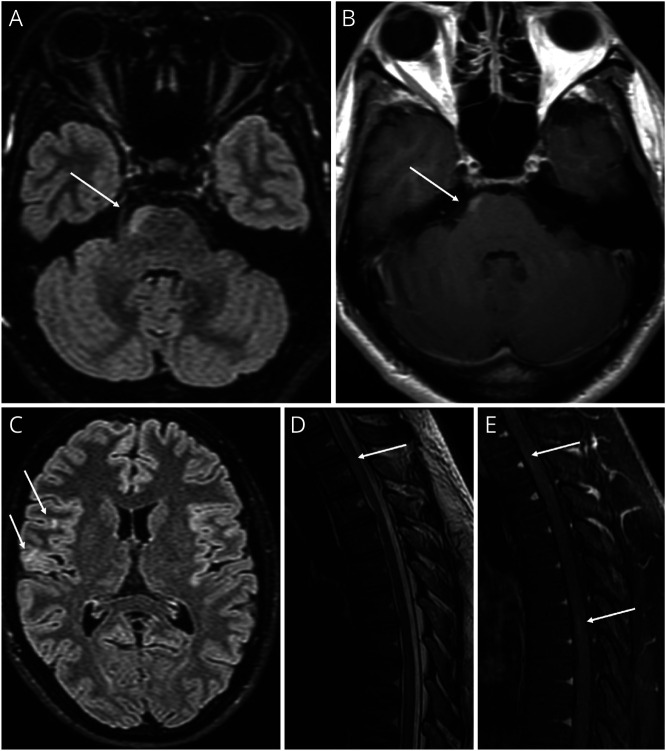
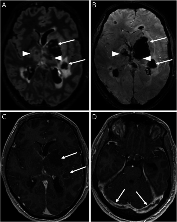
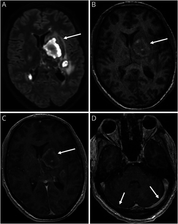
Acute disseminated encephalomyelitis (ADEM) is one characteristic manifestation of myelin oligodendrocyte glycoprotein antibody-associated disease (MOGAD). A previously healthy man presented with retro-orbital headache and urinary retention 14 days after Tdap vaccination. Brain and spine MRI suggested a CNS demyelinating process. Despite treatment with IV steroids, he deteriorated, manifesting hemiparesis and later impaired consciousness, requiring intubation. A repeat brain MRI demonstrated new bilateral supratentorial lesions associated with venous sinus thrombosis, hemorrhage, and midline shift. Anti-MOG antibody was present at a high titer. CSF IL-6 protein was >2,000 times above the upper limits of normal. He improved after plasma exchange, then began monthly treatment alone with anti-IL-6 receptor antibody, tocilizumab, and has remained stable. This case highlights how adult-onset MOGAD, like childhood ADEM, can rapidly become life-threatening. The markedly elevated CSF IL-6 observed here supports consideration for evaluating CSF cytokines more broadly in patients with acute MOGAD.


Conflict of interest statement

A. Virupakshaiah is supported by fellowship support from Biogen, EMD Serono and Novartis. C.E. Moseley is supported by a National Multiple Sclerosis Society Clinician Scientist Development Award # FAN-2107-38301. S.S. Zamvil is supported by NIH grants 1 R01 AI131624-01A1 and 1 RO1 AI170863-01A1. All other authors report no disclosures relevant to the manuscript. Go to Neurology.org/NN for full disclosures.

Figures

Figure 1
Figure 1. Brain and Spinal Cord Imaging at the Time of Clinical Presentation
Brain MRI at presentation demonstrates small T2-FLAIR hyperintense lesions of the right frontal subcortical white matter and right lateral pons (A, C, arrows) with mild enhancement (B, arrow). Thoracic spine MRI demonstrates T2 hyperintensity at T2-3 (D, arrow) and enhancement involving the ventral cord at T2-3 and T6-7 (E, arrows).
Figure 2
Figure 2. Brain Imaging at the Time of Clinical Deterioration
Brain MRI at the time of clinical deterioration demonstrates new supratentorial T2-FLAIR hyperintense lesions involving the white matter and left globus pallidus (A, arrows) with extensive hemorrhage (B, arrows) and peripheral enhancement (C, arrows). Bilateral thalamic T2-FLAIR hyperintensity (A, arrowheads) with hemorrhage (B, arrowheads) and bilateral transverse sinus thrombosis (D, arrows). Internal cerebral vein thrombosis not shown.
Figure 3
Figure 3. Brain Imaging After Plasma Exchange (PLEX)
Brain MRI after treatment with 5 PLEX sessions demonstrates decrease in edema associated with supratentorial T2-FLAIR hyperintense lesions (A, arrow) with intrinsic T1 hyperintensity (B, arrow) and minimal peripheral enhancement (C, arrow). Improvement in previously seen transverse sinus thrombosis (D, arrows).

References

    1. Zamvil SS, Slavin AJ. Does MOG Ig-positive AQP4-seronegative opticospinal inflammatory disease justify a diagnosis of NMO spectrum disorder? Neurol Neuroimmunol Neuroinflamm. 2015;2(1):e62. doi:10.1212/NXI.0000000000000062 - DOI - PMC - PubMed
    1. Banwell B, Bennett JL, Marignier R, et al. . Diagnosis of myelin oligodendrocyte glycoprotein antibody-associated disease: international MOGAD Panel proposed criteria. Lancet Neurol. 2023;22(3):268-282. doi:10.1016/S1474-4422(22)00431-8 - DOI - PubMed
    1. Nurieva RI, Chung Y. Understanding the development and function of T follicular helper cells. Cell Mol Immunol. 2010;7(3):190-197. doi:10.1038/CMI.2010.24 - DOI - PMC - PubMed
    1. Hacohen Y, Banwell B. Treatment approaches for MOG-Ab-associated demyelination in children. Curr Treat Options Neurol. 2019;21(1):2. doi:10.1007/S11940-019-0541-X - DOI - PMC - PubMed
    1. McLendon LA, Gambrah-Lyles C, Viaene A, et al. . Dramatic response to anti-IL-6 receptor therapy in children with life-threatening myelin oligodendrocyte glycoprotein-associated disease. Neurol Neuroimmunol Neuroinflamm. 2023;10(6):e200150. doi:10.1212/NXI.0000000000200150 - DOI - PMC - PubMed