. 2024 Jun:110:110134.
doi: 10.1016/j.clinimag.2024.110134. Epub 2024 Mar 22.

NTRK-rearranged spindle cell neoplasm: Initial observation of imaging appearance and clinicopathologic correlation

Affiliations

NTRK-rearranged spindle cell neoplasm: Initial observation of imaging appearance and clinicopathologic correlation

Matthew Spano et al. Clin Imaging. 2024 Jun.

Abstract

Objective: To explore pre-treatment imaging findings of neurotrophic tyrosine receptor kinase (NTRK)-rearranged spindle cell neoplasm, an emerging group of molecularly defined soft tissue tumors and summarize the clinical course, including TRK inhibitor therapy response.

Materials and methods: This retrospective study included 8 women and 4 men with NTRK-rearranged spindle cell neoplasm (median age, 35.5 years, range, 0-66). Available pre-treatment MRI, CT, PET, and US imaging were reviewed. Tumor histology and the patients' clinical course were reviewed.

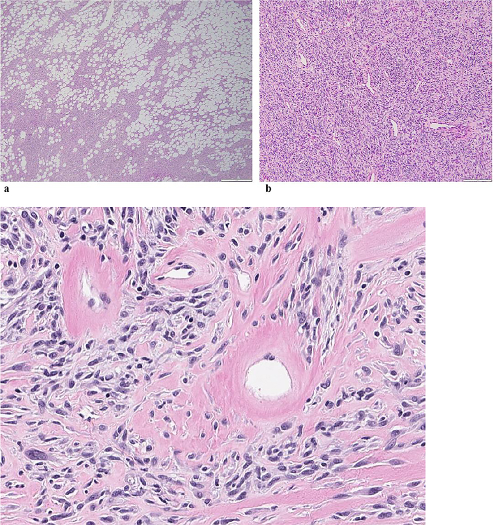
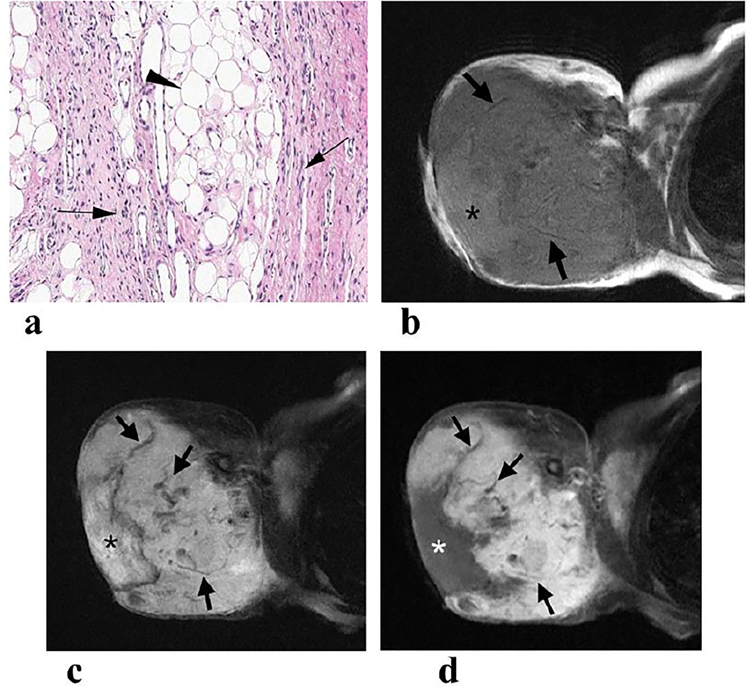
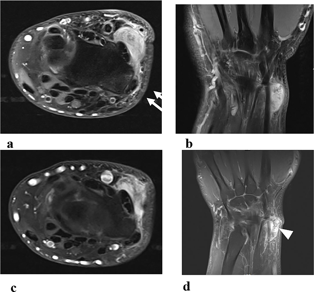
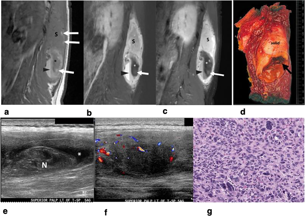
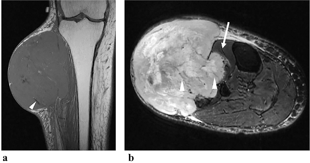
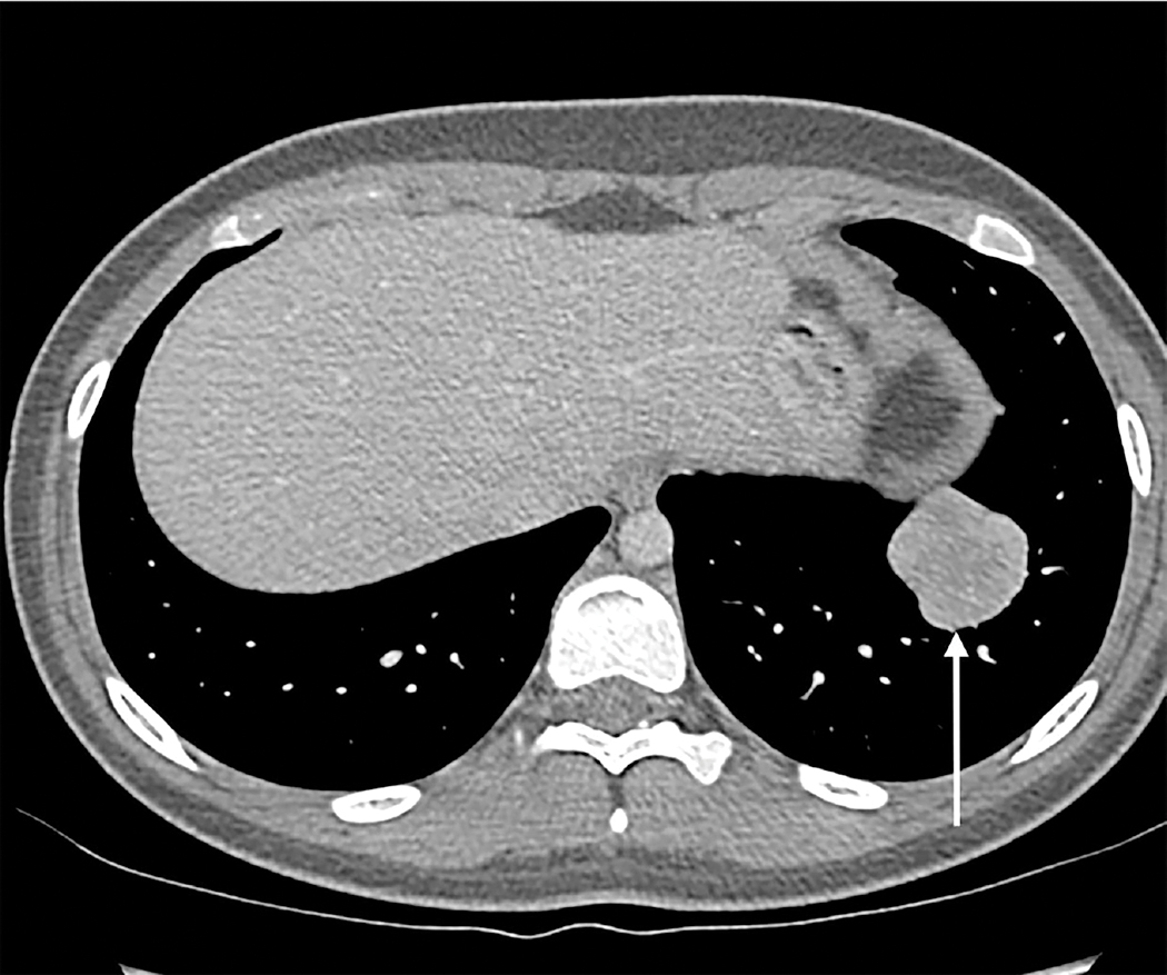
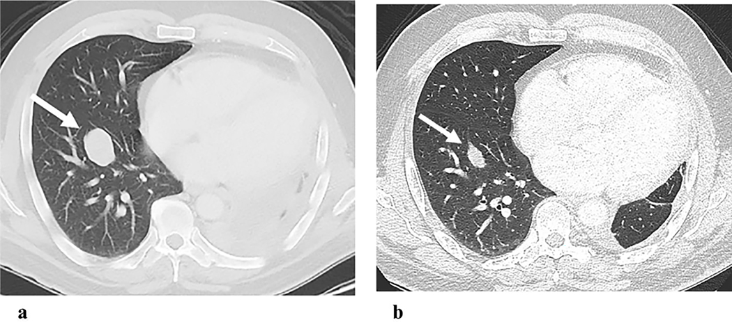
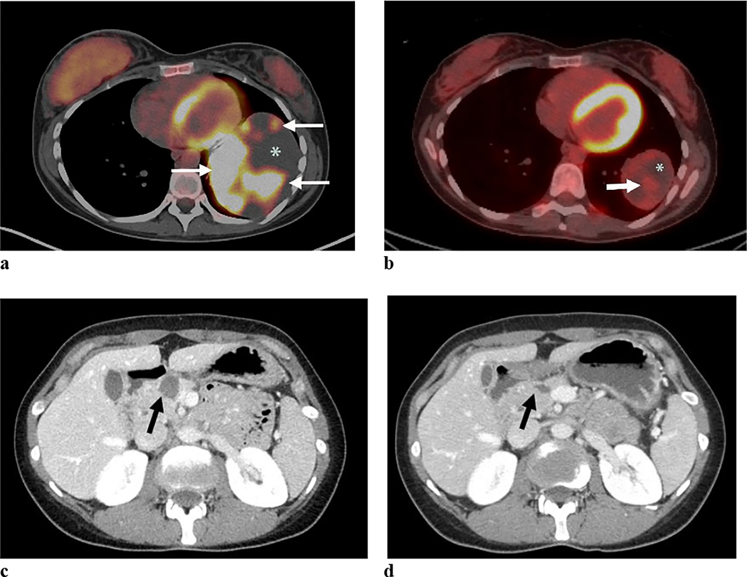
Results: Primary tumors were located within the soft tissue, lungs, kidney, and breast with soft tissue being the most prevalent site (n = 6). Pre-treatment MRI (n = 4) revealed linear hypointense signal foci and contrast enhancement in all patients with hemorrhage in half of the tumors. A tail sign (n = 1) and fluid levels (n = 1) were less frequent. Ultrasound showed well-marginated hypoechoic masses with internal flow. Primary tumors were all non-calcified on CT (4/4). Metastases were FDG-avid (4/4). Among the 8 patients who developed metastasis, 7 developed pulmonary metastases. All four patients who received NTRK inhibitor therapy showed an initial decrease in tumor size or FDG uptake.

Conclusion: NTRK-rearranged neoplasms may occur as enhancing masses with linear hypointense signal foci on MRI and FDG avid metastases on PET. Pulmonary metastases were frequent in our study. Initial treatment response is observed in most patients.

Keywords: CT; Imaging; MR; NTRK; PET; Soft tissue sarcoma; Ultrasound.


Conflict of interest statement

Declaration of competing interest None.

Figures

Fig. 1
Fig. 1
NTRK-rearranged tumors have a wide histologic spectrum. (a) Histology of lipofibromatosis-like neural tumor showing monomorphic spindle cells infiltrating fat (TPM3::NTRK1 fusion). (b) Cellular spindle cell tumor with monomorphic spindle cells and hemangiopericytoma-like vascular pattern (LMNA::NTRK1 Fusion). (c) Histology with prominent stromal collagen bands and perivascular rings of hyalinized collagen.
Fig. 2
Fig. 2
One-day-old newborn with a lipofibromatosis-like neural tumor in her right shoulder. Histological assessment of the biopsied specimen yielded an interpretation of infantile fibrosarcoma at an outside institution. Re-review at a tertiary cancer center yielded a diagnosis of lipomatosis-like neural tumor with NTRK rearrangement. (a) Photomicrograph (medium power) shows bland monomorphic spindle cells (arrows) infiltrating subcutaneous adipose tissue (arrowhead). The tumor involved the dermis, subcutis, and skeletal muscle. (b) Axial T1-weighted and (c) T2-weighted fat-suppressed images show a mass with intermediate T1 signal and heterogeneously high T2 signal. The mass infiltrates the deltoid and triceps muscles, subcutaneous fat, and skin. (d) Axial T1-weighted fat-suppressed image shows diffuse contrast enhancement. Multiple linear hypointense signal foci (arrows) were present in the mass, with a non-enhancing area (*) of slightly high T1 and high T2 signal, suspicious for hemorrhage and necrosis.
Fig. 3.
Fig. 3.
32-year-old man with a lipofibromatosis-like neural tumor in his left wrist. (a) Axial and (b) coronal proton density fat-suppressed images show a mass with hyperintense T2 signal overlying the extensor tendons. Curvilinear tapering soft tissue with hyperintense T2 signal, a “tail sign,” overlies the first extensor compartment tendons. (c) Axial and (d) coronal T1-weighted fat-suppressed post-contrast images show heterogeneous contrast enhancement and linear hypointense signal foci resembling flow voids from vessels (arrowhead) in the mass.
Fig. 4.
Fig. 4.
A 21-year-old woman with high-grade spindle and pleomorphic sarcoma in her left paraspinal muscle. (a) Sagittal T1-weighted image, (b) sagittal T2-weighted fat-suppressed image, and (c) sagittal T1-weighted fat-suppressed post-contrast image demonstrate a mass that contains an enhancing solid cranial component (S) and a cystic caudal component. The cystic component contains a T1 and hypointense T2 signal nodule (*), suggestive of chronic hemorrhage and a fluid level (arrowheads). Foci of hyperintense T1 signal and hypointense T2 signal (arrows) in the nodule suggest subacute hemorrhage. (d) The gross specimen picture shows a cranial solid cranial component (solid) and a caudal cystic component (dotted oval area) containing a clot (arrow). (e) A longitudinal US image demonstrates an internal nodule (N) and fluid (*) in the cystic caudal component of the tumor. (f) Longitudinal Color Doppler US image demonstrates multiple foci of vascular flow in the solid cranial component. (g) Photomicrograph of the tumor in medium power shows a cellular neoplasm composed of spindle cells with foci of marked cytological atypia consistent with high-grade spindle and pleomorphic sarcoma.
Fig. 5.
Fig. 5.
A 60-year-old woman with a lower extremity soft tissue tumor. (a) Coronal T1-weighted image and (b) axial T2-weighted fat-suppressed image show a mass with hypointense T1 and hyperintense T2 signal compared to muscle. There are linear hypointense signal foci (arrowheads) resembling flow voids caused by vessels. The tumor invades the anterior compartment (arrow).
Fig. 6.
Fig. 6.
An 18-year-old man with low-grade spindle cell neoplasm in his lung and multiple recurrent pigmented villonodular synovitis in his ankle. The axial contrast-enhanced CT image shows a left lower lobe mass (arrow) without calcification or fat attenuation.
Fig. 7.
Fig. 7.
Treatment response to NTRK inhibitor targeted therapy in a 47-year-old man with the primary tumor in his thigh and pulmonary metastasis. (a) Pre-treatment axial CT image of the chest shows a 3.5 cm right lower lobe pulmonary metastasis (arrow). (b) The post-treatment axial CT image shows that the metastasis decreased in size to 2.1 cm (arrow).
Fig. 8
Fig. 8
Treatment response to NTRK inhibitor targeted therapy in a 33-year-old woman with a primary hand tumor as well as lung and pancreatic metastases. (a) Axial fused PET/CT image demonstrates a 10.8 cm left lower lobe metastasis which contains multiple FDG-avid components (arrows) and non-avid necrosis (*). (b) Two months later, post-treatment axial fused PET/CT image demonstrates that the metastasis decreased in size to 6.5 cm with decreased FDG avidity (arrow) and persistent necrosis (*). (c) Axial CT image of the abdomen shows a 2.1 cm-cystic pancreatic head metastasis (arrow) in the same patient. (d) Post-treatment axial CT image shows that the metastasis (arrow) decreased in size to 1.4 cm after five months of therapy.

Similar articles

Cited by

References

    1. Chao MV. Neurotrophins and their receptors: a convergence point for many signalling pathways. Nat Rev Neurosci. 2003;4(4):299–309. doi:10.1038/nrn1078 - DOI - PubMed
    1. Eggert A, Grotzer MA, Ikegaki N, Liu XG, Evans AE, Brodeur GM. Expression of the neurotrophin receptor TrkA down-regulates expression and function of angiogenic stimulators in SH-SY5Y neuroblastoma cells. Cancer Res. 2002; 62(6):1802–1808. - PubMed
    1. Lagadec C, Meignan S, Adriaenssens E, Foveau B, Vanhecke E, Romon R, et al. TrkA overexpression enhances growth and metastasis of breast cancer cells. Oncogene. 2009; 28(18):1960–1970. - PubMed
    1. Vaishnavi A, Le AT, Doebele RC. TRKing down an old oncogene in a new era of targeted therapy. Cancer Discov. 2015;5(1):25–34. doi:10.1158/2159-8290.CD-14-0765 - DOI - PMC - PubMed
    1. International Agency for Research on Cancer WHO, International Academy of Pathology. WHO classification of tumours of soft tissue and bone tumours: International Agency for Research on Cancer (IARC), 2020: 287.

LinkOut - more resources