

Periarticular Proprioception: Analyzing the Three-Dimensional Structure of Corpuscular Mechanosensors in the Dorsal Part of the Scapholunate Ligament
- PMID: 38631298
- PMCID: PMC11793100
- DOI: 10.1159/000538169
Periarticular Proprioception: Analyzing the Three-Dimensional Structure of Corpuscular Mechanosensors in the Dorsal Part of the Scapholunate Ligament
Abstract
Introduction: Sensory nerve endings transmit mechanical stimuli into afferent neural signals and form the basis of proprioception, giving rise to the self-perception of dynamic stability of joints. We aimed to analyze the three-dimensional structure of periarticular corpuscular sensory nerve endings in a carpal ligament to enhance our understanding of their microstructure.
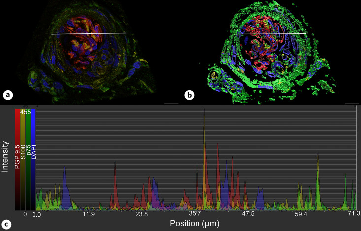
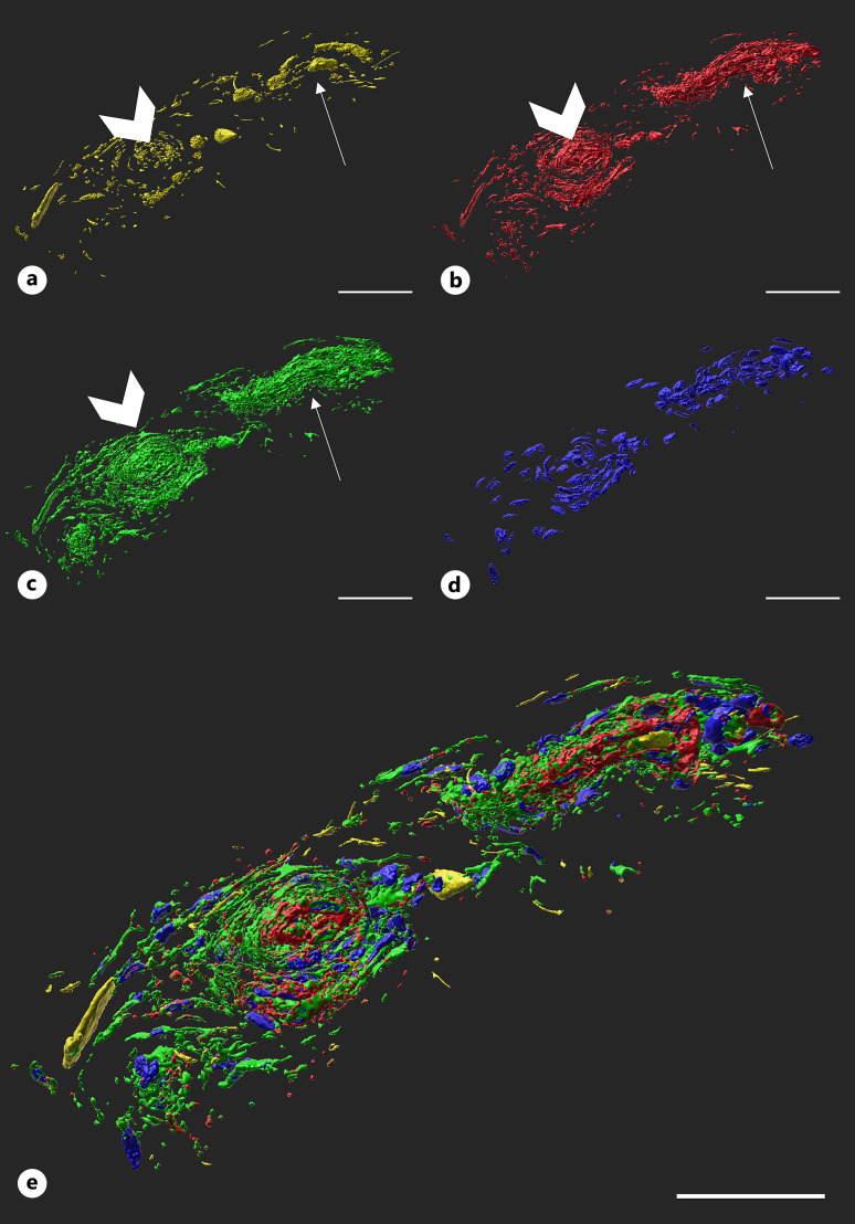
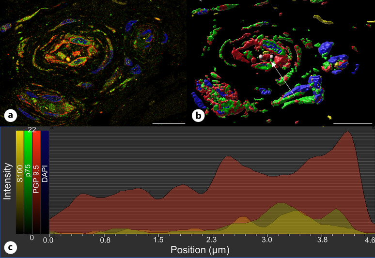
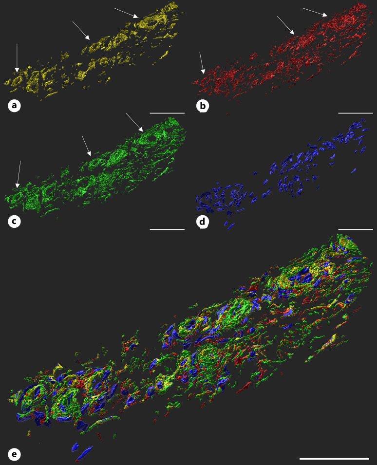
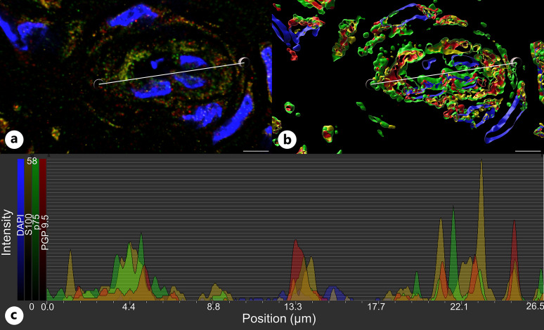
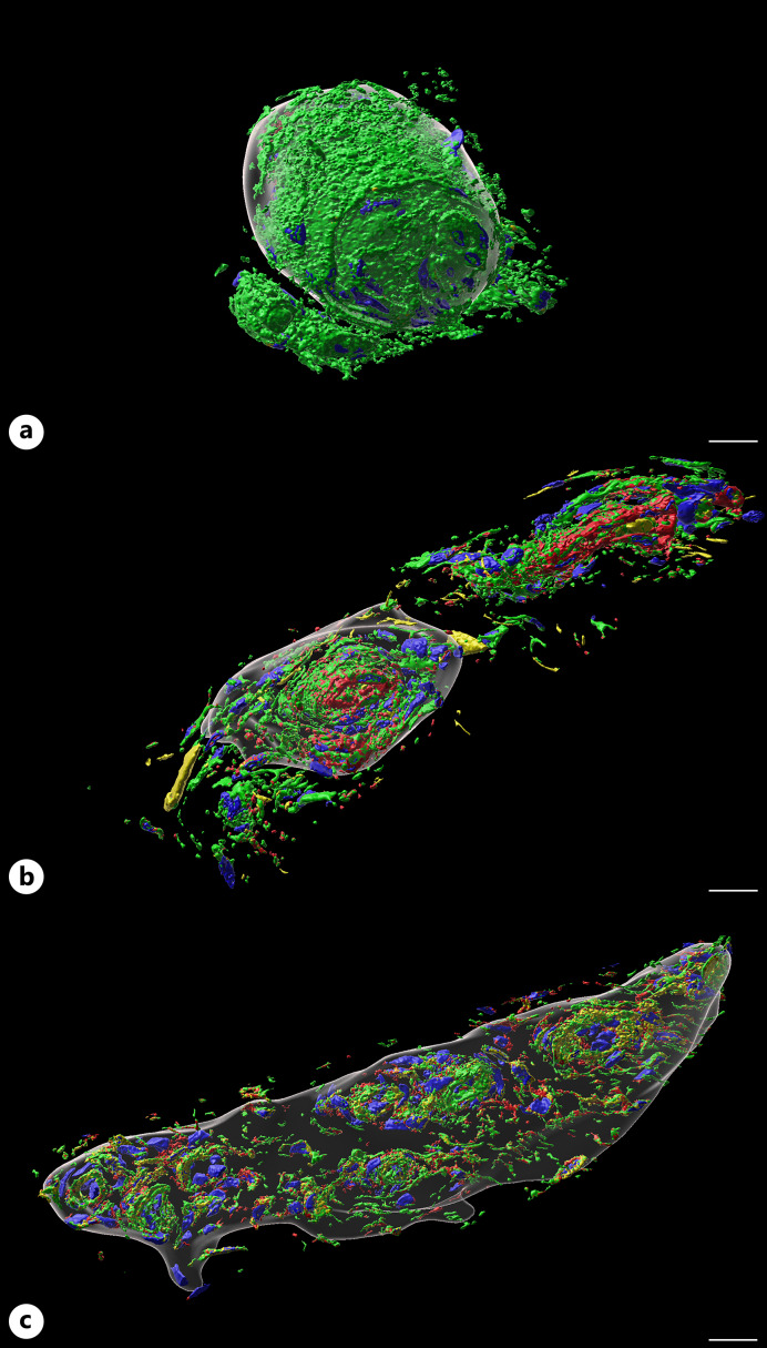
Methods: Two dorsal parts of the scapholunate ligament were excised from two human cadaveric wrist specimens. Consecutive cryosections were stained with immunofluorescence markers protein S100B, neurotrophin receptor p75, protein gene product 9.5 (PGP 9.5), and 4',6-diamidino-2-phenylindole. Three-dimensional images of sensory nerve endings were obtained using confocal laser scanning microscopy, and subsequent analysis was performed using Imaris software.
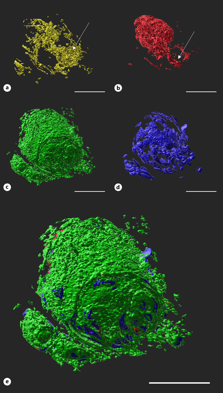
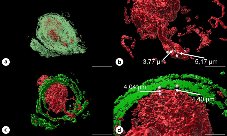
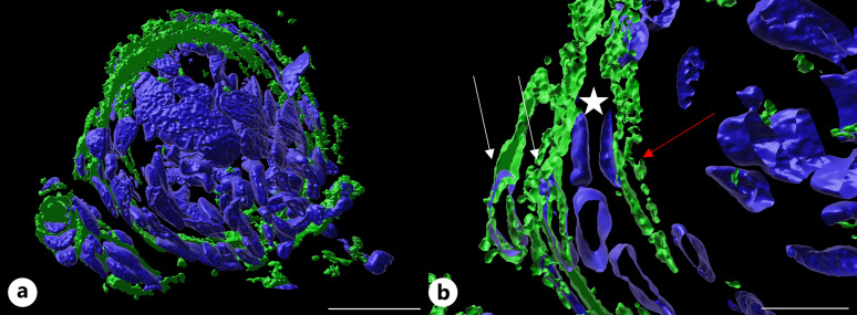
Results: Ruffini endings were characterized by a PGP 9.5-positive central axon, with a median diameter of 4.63 μm and a median of 25 cells. The p75-positive capsule had a range in thickness of 0.94 μm and 15.5 μm, consisting of single to three layers of lamellar cells. Ruffini endings were significantly smaller in volume than Pacini corpuscles or Golgi-like endings. The latter contained a median of three intracorpuscular structures. Ruffini endings and Golgi-like endings presented a similar structural composition of their capsule and subscapular space. The central axon of Pacini corpuscles was surrounded by S100-positive cells forming the inner core which was significantly smaller than the outer core, which was immunoreactive for p75 and PGP 9.5.
Conclusion: This study reports new data regarding the intricate outer and intracorpuscular three-dimensional morphology of periarticular sensory nerve endings, including the volume, number of cells, and structural composition. These results may form a basis to differ between normal and pathological morphological changes in periarticular sensory nerve endings in future studies.
Introduction: Sensory nerve endings transmit mechanical stimuli into afferent neural signals and form the basis of proprioception, giving rise to the self-perception of dynamic stability of joints. We aimed to analyze the three-dimensional structure of periarticular corpuscular sensory nerve endings in a carpal ligament to enhance our understanding of their microstructure.
Methods: Two dorsal parts of the scapholunate ligament were excised from two human cadaveric wrist specimens. Consecutive cryosections were stained with immunofluorescence markers protein S100B, neurotrophin receptor p75, protein gene product 9.5 (PGP 9.5), and 4',6-diamidino-2-phenylindole. Three-dimensional images of sensory nerve endings were obtained using confocal laser scanning microscopy, and subsequent analysis was performed using Imaris software.
Results: Ruffini endings were characterized by a PGP 9.5-positive central axon, with a median diameter of 4.63 μm and a median of 25 cells. The p75-positive capsule had a range in thickness of 0.94 μm and 15.5 μm, consisting of single to three layers of lamellar cells. Ruffini endings were significantly smaller in volume than Pacini corpuscles or Golgi-like endings. The latter contained a median of three intracorpuscular structures. Ruffini endings and Golgi-like endings presented a similar structural composition of their capsule and subscapular space. The central axon of Pacini corpuscles was surrounded by S100-positive cells forming the inner core which was significantly smaller than the outer core, which was immunoreactive for p75 and PGP 9.5.
Conclusion: This study reports new data regarding the intricate outer and intracorpuscular three-dimensional morphology of periarticular sensory nerve endings, including the volume, number of cells, and structural composition. These results may form a basis to differ between normal and pathological morphological changes in periarticular sensory nerve endings in future studies.
Keywords: Histology; Ligament; Morphology; Proprioception; Sensory nerve endings.
© 2024 The Author(s). Published by S. Karger AG, Basel.
Conflict of interest statement
The authors have no conflicts of interest to declare.
Figures
Similar articles
-
Immunofluorescent triple-staining technique to identify sensory nerve endings in human thumb ligaments.Cells Tissues Organs. 2012;195(5):456-64. doi: 10.1159/000327725. Epub 2011 Aug 10. Cells Tissues Organs. 2012. PMID: 21832813
-
Immunofluorescence analysis of sensory nerve endings in the periarticular tissue of the human elbow joint.Arch Orthop Trauma Surg. 2023 Jul;143(7):3779-3794. doi: 10.1007/s00402-022-04604-0. Epub 2022 Sep 7. Arch Orthop Trauma Surg. 2023. PMID: 36070088
-
Development of a 3D-immunofluorescence analysis for sensory nerve endings in human ligaments.J Neurosci Methods. 2022 Dec 1;382:109724. doi: 10.1016/j.jneumeth.2022.109724. Epub 2022 Oct 4. J Neurosci Methods. 2022. PMID: 36207004
-
The Ruffini ending as the primary mechanoreceptor in the periodontal ligament: its morphology, cytochemical features, regeneration, and development.Crit Rev Oral Biol Med. 1999;10(3):307-27. doi: 10.1177/10454411990100030401. Crit Rev Oral Biol Med. 1999. PMID: 10759411 Review.
-
[Morphological basis on periodontal Ruffini endings].Kaibogaku Zasshi. 1998 Apr;73(2):119-34. Kaibogaku Zasshi. 1998. PMID: 9617078 Review. Japanese.
References
-
- Johansson H. Role of knee ligaments in proprioception and regulation of muscle stiffness. J Electromyogr Kinesiol. 1991;1(3):158–79. - PubMed
-
- Hagert E, Persson JKE, Werner M, Ljung BO. Evidence of wrist proprioceptive reflexes elicited after stimulation of the scapholunate interosseous ligament. J Hand Surg Am. 2009;34(4):642–51. - PubMed
-
- García-Piqueras J, García-Suárez O, García-Mesa Y, García-Fernandez B, Quirós LM, Cobo R, et al. . Heparan sulfate in human cutaneous Meissner’s and Pacinian corpuscles. Anat Rec. 2020;303(8):2262–73. - PubMed
MeSH terms
Substances
LinkOut - more resources
Full Text Sources
Medical
Research Materials
Miscellaneous
