. 2024 Apr 16;12(4):e007963.
doi: 10.1136/jitc-2023-007963.

Preclinical evaluation and first-in-dog clinical trials of PBMC-expanded natural killer cells for adoptive immunotherapy in dogs with cancer

Affiliations

Preclinical evaluation and first-in-dog clinical trials of PBMC-expanded natural killer cells for adoptive immunotherapy in dogs with cancer

Aryana M Razmara et al. J Immunother Cancer. .

Abstract

Background: Natural killer (NK) cells are cytotoxic cells capable of recognizing heterogeneous cancer targets without prior sensitization, making them promising prospects for use in cellular immunotherapy. Companion dogs develop spontaneous cancers in the context of an intact immune system, representing a valid cancer immunotherapy model. Previously, CD5 depletion of peripheral blood mononuclear cells (PBMCs) was used in dogs to isolate a CD5dim-expressing NK subset prior to co-culture with an irradiated feeder line, but this can limit the yield of the final NK product. This study aimed to assess NK activation, expansion, and preliminary clinical activity in first-in-dog clinical trials using a novel system with unmanipulated PBMCs to generate our NK cell product.

Methods: Starting populations of CD5-depleted cells and PBMCs from healthy beagle donors were co-cultured for 14 days, phenotype, cytotoxicity, and cytokine secretion were measured, and samples were sequenced using the 3'-Tag-RNA-Seq protocol. Co-cultured human PBMCs and NK-isolated cells were also sequenced for comparative analysis. In addition, two first-in-dog clinical trials were performed in dogs with melanoma and osteosarcoma using autologous and allogeneic NK cells, respectively, to establish safety and proof-of-concept of this manufacturing approach.

Results: Calculated cell counts, viability, killing, and cytokine secretion were equivalent or higher in expanded NK cells from canine PBMCs versus CD5-depleted cells, and immune phenotyping confirmed a CD3-NKp46+ product from PBMC-expanded cells at day 14. Transcriptomic analysis of expanded cell populations confirmed upregulation of NK activation genes and related pathways, and human NK cells using well-characterized NK markers closely mirrored canine gene expression patterns. Autologous and allogeneic PBMC-derived NK cells were successfully expanded for use in first-in-dog clinical trials, resulting in no serious adverse events and preliminary efficacy data. RNA sequencing of PBMCs from dogs receiving allogeneic NK transfer showed patient-unique gene signatures with NK gene expression trends in response to treatment.

Conclusions: Overall, the use of unmanipulated PBMCs appears safe and potentially effective for canine NK immunotherapy with equivalent to superior results to CD5 depletion in NK expansion, activation, and cytotoxicity. Our preclinical and clinical data support further evaluation of this technique as a novel platform for optimizing NK immunotherapy in dogs.

Keywords: adoptive cell therapy (ACT); clinical trials as topic; computational biology; immunotherapy, adoptive; killer cells, natural.


Conflict of interest statement

Competing interests: None declared.

Figures
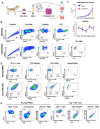
Figure 1
Figure 1
Expansion of canine NK cells in vitro. (A) Schema of experimental strategy expanding NK cells from PBMC and CD5-depleted starting populations. After processing, respective cell populations were co-cultured with irradiated K562 clone 9 feeder cells and 100 mIU/mL rhIL-2 for 14 days. (B) Representative flow cytometry gating of cells prior to co-culture and following magnetic bead separation for CD5 depletion. Depleted cells showed virtually no CD5 expression in contrast to positively selected CD5+ cells, confirming the efficacy of magnetic separation and phenotype of CD5-depleted cells as a starting population. (C) Cell counts and viability were calculated on days 0, 7, 10, 12, and 14 of the 14-day co-culture using bulk PBMCs (blue) and CD5-depleted cells (red) as starting populations. Mean and SEM for 11 healthy donor dogs are plotted against time. (D) Representative flow cytometry gating of bulk PBMCs before and after 14-day co-culture, corroborating the expansion of NKp46+ NK cells from PBMCs without preceding NK-isolation. NK cells were identified as CD3-NKp46+, reaching a majority at day 14 with minimal CD3+ T cell infiltrate. (E) Representative flow cytometry gating of PBMCs at rest and following 14-day co-culture confirming minimal contamination of CD3+γδTCR+ T cells. NK, natural killer; PBMC, peripheral blood mononuclear cell.
Figure 2
Figure 2
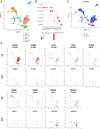
Genomic analysis of expanded NK cells by scRNA-seq. Cells from PBMC-expanded NK cells at D14 were collected and scRNA-seq was completed. The resulting dataset was integrated with a dataset for resting PBMCs. (A) uMAP plot of clusters present in resting PBMCs color-coded by cell type. Cell identities were determined by analysis of differentially expressed genes that distinguished each cluster from all other clusters. (B) Dot plot visualizing a selection of gene markers used to annotate the cells present. Dot size represents the percent of cells expressing the gene while color correlates with average expression within a cell. (C) uMAP of overlapping datasets included within the integration, showing the differences in resting PBMCs (D0, blue) and PBMC-expanded NK cells (D14, red). (D) uMAPs showing distribution and percent of cells expressing various genes associated with cell types in the integrated conditions, D0 (above) and D14 (below). Percentages are calculated as the percent of cells with expression of the specified gene out of the total cells present. The threshold for gene expression is set at 0. D, day; NK, natural killer; PBMC, peripheral blood mononuclear cell; scRNA-seq, single-cell RNA sequencing; uMAP, Uniform Manifold Approximation and Projection.
Figure 3
Figure 3
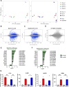
Genomic analysis of expanded NK cells by bulk RNA-seq. Cells from bulk PBMC and CD5-depleted cell starting populations were collected at days 0, 7, and 14 of the 14-day co-culture from four separate healthy beagle donors and 3’-Tag-RNA-seq was performed. (A) Principal component analysis (PCA) of cells color-coded by donor dog (left) or starting population (right) demonstrated variability at days 0 (squares) and 7 (triangles) of co-culture with convergence of cell signatures at day 14 (circles). Certain samples from donor three did not meet RNA quantity standards and were exluded, including day 0 depleted, day 14 depleted, and day 14 bulk. MA plots, using a p<0.05 significance threshold, corroborate PCA plot patterns with (B) 3961 differentially expressed genes (DGEs, blue) between PBMCs at day 14 vs day 0 of co-culture, (C) 3107 DGEs between CD5-depleted cells at day 14 vs day 0 of co-culture and (D) zero DGEs between bulk PBMCs and CD5-depleted cells at day 14 co-culture. The log fold change of NK cell-related gene expression was assessed between day 14 and day 0 co-culture in (E) the bulk PBMC group and (F) the CD5-depleted cell group. Several key genes were significantly different following co-culture compared with day 0 (bold, green). P values were determined using the DESeq2 package in R. (G) Absolute normalized counts for CD16, KLRB1, NKG2D, and GZMB were visualized for CD5-depleted cells at day 0 co-culture (Dep 0) and day 14 co-culture (Dep 14) as well as PBMCs at day 0 co-culture (bulk 0) and day 14 co-culture (bulk 14). Bars show median of normalized counts for donor dogs and p values were determined using the DESeq2 package in R. *P<0.05, **p<0.01, ***p<0.001. NK, natural killer; PBMC, peripheral blood mononuclear cell; RNA-seq, RNA sequencing.
Figure 4
Figure 4
Functional assessment of expanded NK cells. (A) Representative flow cytometry showing gating strategy for NK killing assays using PBMC and CD5-depleted expanded NK cells against osteosarcoma cell line targets (OSCA) at a 1:1 effector-to-target (E:T) ratio. Target cells were identified from effector cells by carboxyfluorescein succinimidyl ester (CFSE)+ labeling with separate viability dye staining to identify dead cells. (B) Mean cytotoxicity (±SEM) of NK cells at day 14 from four donor dogs at increasing E:T ratios from bulk PBMC (blue) and CD5-depleted (red) starting populations against melanoma (M5, left) and osteosarcoma (OSCA, right) targets. (C) At the 1:1 E:T ratio, mean cytotoxicity of expanded NK cell effectors varied against osteosarcoma and melanoma targets, with increased PBMC-expanded NK cell killing against melanoma targets compared with osteosarcoma (p=ns). (D) Supernatant cytokine levels were measured by canine Luminex assay. Bars depict mean values of eight or nine healthy donor dogs for PBMC (blue) and CD5-depleted cells (red) following 14-day co-culture. GM-CSF and IFN-y concentrations were significantly greater in the PBMC group, while MCP-1 was significantly greater in the CD5-depleted group. P values were determined using the Mann-Whitney U test. *P<0.05, **p<0.01. GM-CSF, granulocyte macrophage colony-stimulating factor; IFN, interferon; IL, interleukin; MCP, monocyte chemoattractant protein; NK, natural killer; ns, not significant; PBMC, peripheral blood mononuclear cell; TNF, tumor necrosis factor.
Figure 5
Figure 5
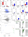
Genomic analysis of human bulk PBMCs versus purified NK cells. Human cells from bulk PBMC and purified NK cell starting populations were collected at days 0, 7, and 14 of 14-day co-culture from three separate human donors and 3’Tag-RNA-seq was performed. Absolute normalized counts for (A) NCAM1/CD56 and NCR1/NKp46 and (B) T and B cell-related genes were visualized for purified NK cells (NK 0) and PBMCs (bulk 0) at day 0 co-culture. Certain samples did not meet RNA quantity standards and were excluded, including day 7 NK isolated and day 14 NK isolated. Floating bars show minimum to maximum of normalized counts for human cells and p values were determined using the DESeq2 package in R. *P<0.05, **p<0.01, ***p<0.001. MA plots, using a p<0.05 significance threshold, reveal starting populations that are distinct at rest, with (C) 1739 DGEs between bulk PBMCs and purified NK cells at day 0, but converge when activated, with (D) only four DGEs between bulk PBMCs and purified NK cells at day 14 co-culture. (E) Principal component analysis (PCA) of cells aligns with MA plot patterns with high variability at day 0 (blue) and concentration of cell signatures at day 7 (green) and furthermore at day 14 (peach) of co-culture. (F) The log fold change of NK cell-related gene expression was assessed between day 14 and day 0 co-culture from purified NK cells (top) and bulk PBMCs (bottom). Several key genes were significantly different following co-culture compared with day 0 (bold, green). P values were determined using the DESeq2 package in R. NK, natural killer; PBMC, peripheral blood mononuclear cell.
Figure 6
Figure 6
First-in-dog clinical trial of adoptive transfer of autologous canine NK cells. (A) Schema of trial design combining adoptive transfer of PBMC-expanded autologous NK cells with inhaled IL-15. PBMCs were isolated from whole blood drawn from patient dogs 14 and 7 days before the start of treatment for the 14-day expansion of autologous NK cells. Dogs received two intravenous injections of autologous NK cells at a dose of 7.5 million cells/kg. Additionally, on day 0, dogs began twice daily treatments with inhaled rhIL-15 continuing for 14 days total. (B) Characteristics of the nine total dogs with pulmonary metastatic melanoma (MEL) or osteosarcoma (OSA) that met entry criteria and were enrolled in the trial. Response was determined by RECIST criteria defining partial response (PR), stable disease (SD), progressive disease (PD), and not evaluable (NE). Survival was calculated from initiation of treatment to death or humane euthanasia. (C) Survival plotted as bars color-coded by RECIST criteria defining responders (PR and SD, blue), non-responders (PD, red), and not evaluable (NE, gray). (D) Cell counts, fold change, and viability were calculated on days 0, 7, 10, 12, and 14 of the 14-day co-culture for autologous PBMCs isolated from patient blood (green) compared with PBMCs isolated from healthy beagle blood (purple). Mean (±SEM) for expansions of both injections of PBMC-expanded autologous NK cells were compared with healthy donor PBMC expansions and plotted over time. P values were determined by mixed effects model. **p<0.01. NK, natural killer; PBMC, peripheral blood mononuclear cell; RECIST, Response Evaluation Criteria for Solid Tumors.
Figure 7
Figure 7
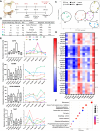
First-in-dog clinical trial of adoptive transfer of allogeneic canine NK cells. (A) Schema of trial design combining adoptive transfer of PBMC-expanded allogeneic NK cells with palliative radiotherapy (RT). PBMCs were isolated from healthy donor beagle blood 14 days before the scheduled NK cell infusion for each patient. Donor PBMC-expanded NK cells at a dose of 7.5 million cells/kg were injected intravenous in patients on completion of RT. (B) Characteristics of the five total dogs with malignant melanoma that met entry criteria and were enrolled in the trial. Dogs ID 1, 4, and 5 had known lymph node metastasis on enrollment and dog ID 4 had pulmonary metastasis on enrollment. Survival was calculated from initiation of RT to death or humane euthanasia. PBMCs were isolated from patients’ whole blood at six timepoint and submitted for 3’-Tag-RNA-seq. (C) Principal component analysis (PCA) of cells color-coded by patient and symbols distinguishing timepoint, demonstrated PBMCs clustered based on the dog they originated from rather than timepoint of treatment. (D) A heatmap depicting log counts per million (logCPM) transformed expression of key NK-related genes shows variation across both patients and treatment. (E) Absolute normalized counts for CD16, KLRB1, NKG2D, and TIGIT were visualized as an aggregate of all patients at each timepoint (box and whiskers, left) and individual values by patient (right). While distinct peaks and trends in expression were recognized, changes between timepoints were not significant for the genes assessed. (F) Samples across timepoints were combined based on donor and compared between the dog with the longest (ID5) and the dog with the shortest (ID1) survival. Induced gene pathways in the dog with the highest survival are depicted as a dot plot of gene ontology (GO) analysis. NK, natural killer; PBMC, peripheral blood mononuclear cell.

Similar articles

Cited by

References

    1. Houot R, Schultz LM, Marabelle A, et al. . T-cell-based immunotherapy: adoptive cell transfer and checkpoint inhibition. Cancer Immunol Res 2015;3:1115–22. 10.1158/2326-6066.CIR-15-0190 - DOI - PubMed
    1. Miller JS, Soignier Y, Panoskaltsis-Mortari A, et al. . Successful adoptive transfer and in vivo expansion of human haploidentical NK cells in patients with cancer. Blood 2005;105:3051–7. 10.1182/blood-2004-07-2974 - DOI - PubMed
    1. Gras Navarro A, Björklund AT, Chekenya M. Therapeutic potential and challenges of natural killer cells in treatment of solid tumors. Front Immunol 2015;6:202. 10.3389/fimmu.2015.00202 - DOI - PMC - PubMed
    1. Rezvani K, Rouce RH. The application of natural killer cell Immunotherapy for the treatment of cancer. Front Immunol 2015;6:578. 10.3389/fimmu.2015.00578 - DOI - PMC - PubMed
    1. Lupo KB, Matosevic S. Natural killer cells as allogeneic effectors in adoptive cancer immunotherapy. Cancers (Basel) 2019;11:769. 10.3390/cancers11060769 - DOI - PMC - PubMed