. 2024 Apr 16;12(4):e008287.
doi: 10.1136/jitc-2023-008287.

Efficacy of LCMV-based cancer immunotherapies is unleashed by intratumoral injections of polyI:C

Affiliations

Efficacy of LCMV-based cancer immunotherapies is unleashed by intratumoral injections of polyI:C

Celia Gomar et al. J Immunother Cancer. .

Abstract

Background: Lymphocytic choriomeningitis virus (LCMV) belongs to the Arenavirus family known for inducing strong cytotoxic T-cell responses in both mice and humans. LCMV has been engineered for the development of cancer immunotherapies, currently undergoing evaluation in phase I/II clinical trials. Initial findings have demonstrated safety and an exceptional ability to activate and expand tumor-specific T lymphocytes. Combination strategies to maximize the antitumor effectiveness of LCMV-based immunotherapies are being explored.

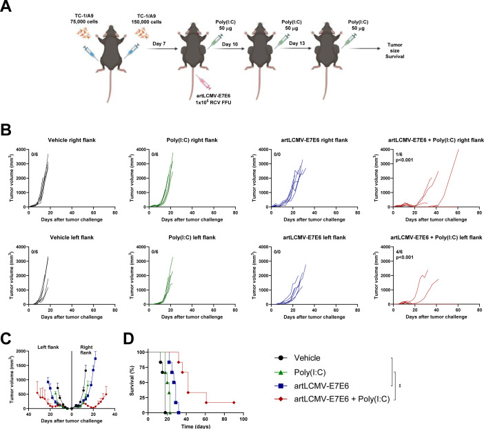
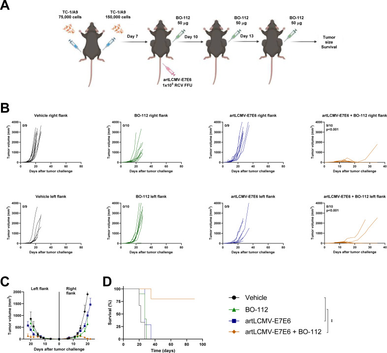
Methods: We assessed the antitumor therapeutic effects of intratumoral administration of polyinosinic:polycytidylic acid (poly(I:C)) and systemic vaccination using an LCMV-vector expressing non-oncogenic versions of the E6 and E7 antigens of human papillomavirus 16 (artLCMV-E7E6) in a bilateral model engrafting TC-1/A9 cells. This cell line, derived from the parental TC-1, exhibits low MHC class I expression and is highly immune-resistant. The mechanisms underlying the combination's efficacy were investigated through bulk RNA-seq, flow cytometry analyses of the tumor microenvironment, selective depletions using antibodies and clodronate liposomes, Batf3 deficient mice, and in vivo bioluminescence experiments. Finally, we assessed the antitumor effectiveness of the combination of artLCMV-E7E6 with BO-112, a GMP-grade poly(I:C) formulated in polyethyleneimine, currently under evaluation in clinical trials.

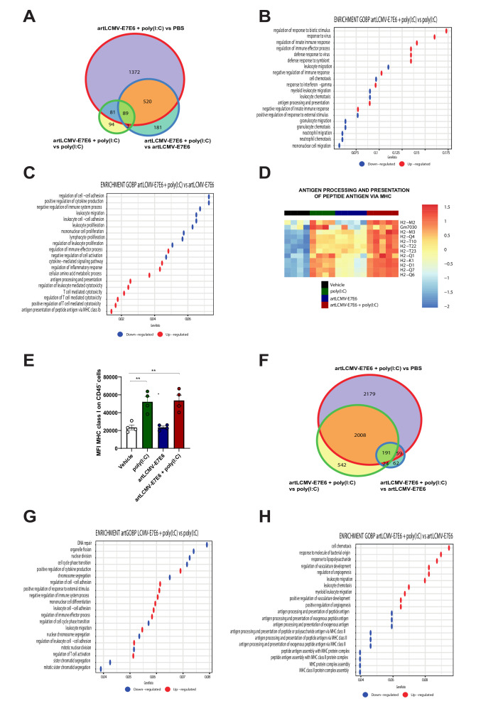
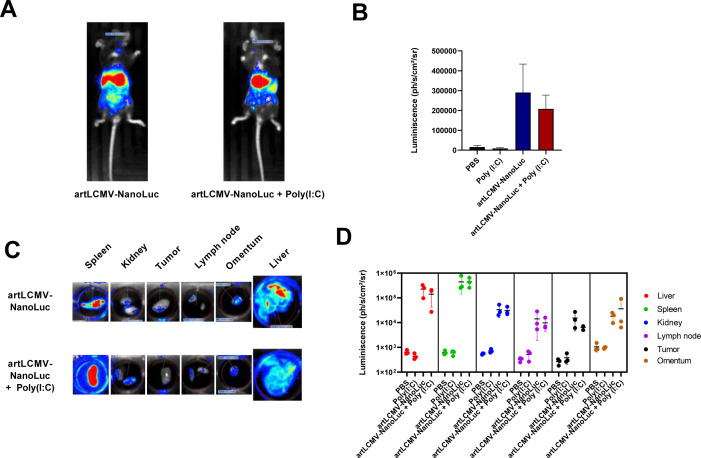
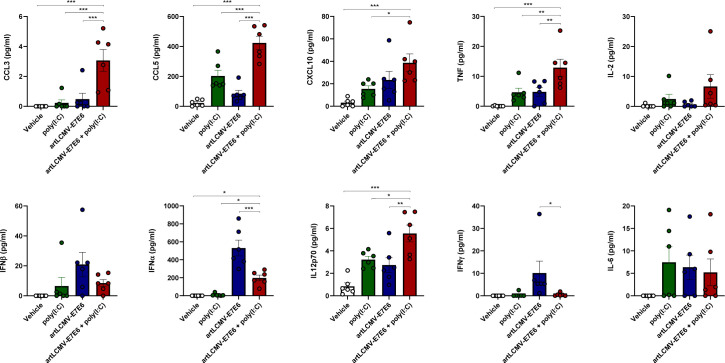
Results: Intratumoral injection of poly(I:C) enhanced the antitumor efficacy of artLCMV-E7E6 in both injected and non-injected tumor lesions. The combined treatment resulted in a significant delay in tumor growth and often complete eradication of several tumor lesions, leading to significantly improved survival compared with monotherapies. While intratumoral administration of poly(I:C) did not impact LCMV vector biodistribution or transgene expression, it significantly modified leucocyte infiltrates within the tumor microenvironment and amplified systemic efficacy through proinflammatory cytokines/chemokines such as CCL3, CCL5, CXCL10, TNF, IFNα, and IL12p70. Upregulation of MHC on tumor cells and a reconfiguration of the gene expression programs related to tumor vasculature, leucocyte migration, and the activation profile of tumor-infiltrating CD8+ T lymphocytes were observed. Indeed, the antitumor effect relied on the functions of CD8+ T lymphocytes and macrophages. The synergistic efficacy of the combination was further confirmed when BO-112 was included.

Conclusion: Intratumoral injection of poly(I:C) sensitizes MHClow tumors to the antitumor effects of artLCMV-E7E6, resulting in a potent therapeutic synergy.

Keywords: Adjuvants, Immunologic; Drug Evaluation, Preclinical; Immunogenicity, Vaccine; Immunotherapy; Vaccination.


Conflict of interest statement

Competing interests: PB received research funding from Hookipa Pharma. IM reports receiving commercial research grants from AstraZeneca, BMS, Highlight Therapeutics, Alligator, Pfizer Genmab and Roche; has received speakers bureau honoraria from MSD; and is a consultant or advisory board member for BMS, Roche, AstraZeneca, Genmab, Pharmamar, F-Star, Bioncotech, Bayer, Numab, Pieris, Gossamer, Alligator and Merck Serono. MA declares receiving a commercial research grant from Highlight Therapeutics. TS, KKO, and HL are employees of Hookipa Pharma. MQ is an employee of Highlight Therapeutics. The rest of the authors have no conflict of interest to declare.

Figures

Figure 1
Figure 1
Intratumoral poly(I:C) enhances the antitumor activity of an LCMV-based vaccine in both injected tumor lesions and distant non-injected engrafted tumors. Mice were subcutaneously injected with 1.5×10⁵ TC-1/A9 cells in the right flank and 7.5×10⁴ TC-1/A9 cells in the left flank. Seven days later, mice were intravenously treated with 1×10⁵ RCV FFU artLCMV-E7E6 and received intratumoral injections of 50 µg of poly(I:C) in the right-sided tumor lesion on days 7, 10, and 13. (A) Schematic representation of the experimental setup. (B) Tumor growth (mm3) is depicted for individual mice in both injected (right flank) and non-injected (left flank) tumors. The numbers in each graph represent the fraction of mice achieving complete tumor regression. (C) The average of in vivo tumor growth is presented for injected (right flank) and non-injected (left flank) tumors. (D) Survival percentage over time is displayed for the experiments in (B). Data are representative of two independent experiments with six mice per group (mean±SEM). Extra sum-of-squares F test (C) or log-rank (D) tests were used to assess significance. Significant differences are indicated for comparisons of each group with the artLCMV-E7E6+poly(I:C) group (**p<0.01). LCMV, lymphocytic choriomeningitis virus; RCV FFU, replication competent virus focus forming units; poly(I:C), polyinosinic:polycytidylic acid.
Figure 2
Figure 2
The combination of systemic artLCMV-E7E6 and intratumoral poly(I:C) induces a distinct gene expression program compared with each monotherapy in the tumor microenvironment. TC1/A9 tumor-bearing mice received intravenous treatment with 1×10⁵ RCV FFU artLCMV-E7E6 on day 7 and intratumoral injections of 50 µg of poly(I:C) in the right tumor lesion on days 7, 10, and 13. On day 8 and day 14, mice were euthanized, and RNA from tumors was subjected to RNA-seq analysis. (A) Venn diagram illustrating the comparison of differentially expressed genes (p<0.05% and FDR<0.05%) in the various experimental groups 24 hours after treatment. (B) Gene Set Enrichment Analysis (GSEA) showing the top upregulated and downregulated Gene Ontology Biological Process (GO:BP) terms comparing artLCMV-E7E6+poly(I:C) with poly(I:C) 24 hours after treatment (p. adj<0.05%). (C) GSEA illustrating the top upregulated and downregulated GO:BP terms comparing artLCMV-E7E6+poly(I:C) with artLCMV-E7E6 24 hours after treatment (p. adj<0.05%). (D) Heatmap displaying the level of gene expression related to the pathway of antigen processing and presentation via MHC. (E) Mean fluorescence intensity (MFI) of MHC class I on tumor cells 24 hour after treatment analyzed by using flow cytometry. Data are presented as mean±SEM, and statistical significance was determined with one-way ANOVA followed by Dunnet’s post-test comparing the combined treatment with the other experimental groups (**p<0.01). (F) Venn diagram demonstrating the comparison of differentially expressed genes (p<0.05% and FDR<0.05%) in the various experimental groups 7 days after treatment. (G) GSEA showing top upregulated and down-regulated GO:BP terms comparing artLCMV-E7E6+poly(I:C) with poly(I:C) 7 days after treatment. (p. adj<0.05%). (H) GSEA showing top upregulated and downregulated GO:BP terms comparing artLCMV-E7E6+poly(I:C) with artLCMV-E7E6 7 days after treatment (p. adj<0.05%). ANOVA, analysis of variance; LCMV, lymphocytic choriomeningitis virus; PBS, phosphate-buffered saline; poly(I:C), polyinosinic:polycytidylic acid; RCV FFU, replication competent virus focus forming units; FDR, false discovery rate; MHC, major histocompatibility complex.
Figure 3
Figure 3
Biodistribution of LCMV vectors remains unaffected by intratumoral administration of poly(I:C). C57BL/6 mice were subcutaneously injected in the right flank with 1.5×10⁵ TC-1/A9 cells. Seven days later, mice received intravenous injections of 1×10⁵ RCV FFU artLCMV-NanoLuc. (A) Representative image of in vivo bioluminescence measured 24 hours post-LCMV injection. (B) Quantification of the in vivo bioluminescence measured in all mice. (C) After in vivo imaging, mice were euthanized, and the bioluminescence in different organs was assessed. Representative images are shown. (D) Quantification of the ex vivo bioluminescence in different organs. Data are presented as mean±SEM, and statistical significance was determined with one-way ANOVA followed by Dunnet’s post-test for panels (B, D). ANOVA, analysis of variance; LCMV, lymphocytic choriomeningitis virus; PBS, phosphate-buffered saline; poly(I:C), polyinosinic:polycytidylic acid; RCV FFU, replication competent virus focus forming units.
Figure 4
Figure 4
The combination of systemic artLCMV-E7E6 and intratumoral poly(I:C) upregulates specific chemokines/cytokines. TC1/A9 tumor-bearing mice received intravenous treatment with 1×10⁵ RCV FFU artLCMV-E7E6 and intratumoral injection of 50 µg of poly(I:C) in the right tumor lesion on day 7. After 24 hours, multiple cytokine/chemokines were assessed in serum samples. Data are presented as mean±SEM, and statistical significance was determined with one-way ANOVA followed by Dunnet’s post-test (*p<0.05; **p<0.01; ***p<0.001). ANOVA, analysis of variance; LCMV, lymphocytic choriomeningitis virus; poly(I:C), polyinosinic:polycytidylic acid; RCV FFU, replication competent virus focus forming unit.
Figure 5
Figure 5
The combination of systemic artLCMV-E7E6 and intratumoral poly(I:C) induces a significant infiltration of highly activated tumor-specific lymphocytes in the tumor microenvironment. TC1/A9 tumor-bearing mice were intravenously treated with 1×10⁵ RCV FFU artLCMV-E7E6 at day 7 and received intratumoral injections of 50 µg of poly(I:C) in the right tumor lesion on days 7, 10, and 13. On day 15, mice were euthanized, and immune cells from tumors were analyzed using multiparametric flow cytometry. (A) Uniform Manifold Approximation and Projection (UMAP) plots illustrating the immune subsets in the different experimental conditions. (B) Quantification of the percentage of each immune subset and comparison between pairs of experimental conditions. Statistical differences were determined using the Wilcoxon test (ns, non-significant; *p<0.05). (C) Heatmap displaying differentially expressed proteins in CD8+ T lymphocytes in artLCMV-E7E6+poly(I:C) vs artLCMV-E7E6. LCMV, lymphocytic choriomeningitis virus; poly(I:C), polyinosinic:polycytidylic acid; RCV FFU, replication competent virus focus forming units.
Figure 6
Figure 6
The antitumor activity of the combination of systemic artLCMV-E7E6 and intratumoral poly(I:C) is preserved in Batf3-deficient mice and requires the presence of macrophages and CD8+ T lymphocytes but not of CD4+ T lymphocytes or NK cells. (A) C57BL/6 mice or Batf3−/− mice were subcutaneously injected with 1.5×10⁵ TC-1/A9 cells in the right flank. Seven days later, mice were intravenously treated with 1×10⁵ RCV FFU artLCMV-E7E6 and received intratumoral injections of 50 µg of poly(I:C) on days 7, 10 and 13. Individual tumor growth (mm3) is shown for each mouse, with numbers in each graph representing the fraction of mice achieving complete tumor regression. (B) The average in vivo tumor growth is shown. (C) The percentage of survival over time. Data are represented as mean±SEM. Extra sum-of-squares F test (A) or log-rank (C) tests were used to assess the significance between the treated mice and control mice in each mouse strain. No significant differences were observed when comparing artLCMV-E7E6+poly(I:C) treatment in wild type versus Batf3−/− mice. (D) Mice were subcutaneously injected with 1.5×10⁵ TC-1/A9 cells in the right flank. Seven days later, mice were intravenously treated with 1×10⁵ RCV FFU artLCMV-E7E6 and received intratumoral injections of 50 µg of poly(I:C) in the right tumor lesion on days 7, 10, and 13. Depleting monoclonal antibodies or clodronate liposomes were administered on days 6, 9, and 13. Individual tumor growth (mm3) is shown for each mouse. (E) The average in vivo tumor growth is presented. (F) The percentage of survival over time. Data are represented as mean±SEM Extra sum-of-squares F test (A, D) or log-rank (C, F) tests were used to assess significance (**p<0.01, ***p<0.001). LCMV, lymphocytic choriomeningitis virus; PBS, phosphate-buffered saline; poly(I:C), polyinosinic:polycytidylic acid; RCV FFU, replication competent virus focus forming units.
Figure 7
Figure 7
Intratumoral BO-112 enhances the antitumor activity of artLCMV-E7E6 in both injected tumor lesions and distant non-injected engrafted tumors. Mice were subcutaneously injected with 1.5×10⁵ TC-1/A9 cells in the right flank and with 7.5×10⁴ TC-1/A9 cells in the left flank cells. Seven days later, mice received intravenous treatment with 1×10⁵ RCV FFU artLCMV-E7E6 and intratumoral injections of 50 µg of BO-112 in the right tumor lesion on days 7, 10, and 13. (A) Schematic representation of the experimental setup. (B) The tumor growth (mm3) is depicted for each individual mouse in both injected (right flank) and non-injected (left flank) tumors. The numbers in each graph represent the fraction of mice achieving complete tumor regression. (C) The average in vivo tumor growth is presented for injected (right flank) and non-injected (left flank) tumors. (D) The percentage of survival over time is shown for experiments in B. n=9–10 (mean±SEM). Extra sum-of-squares F test (C) or log-rank (D) tests were used to assess significance. Significant differences are indicated for comparisons of each group with the artLCMV-E7E6+BO-112 group (**p<0.01). LCMV, lymphocytic choriomeningitis virus; poly(I:C), polyinosinic:polycytidylic acid; RCV FFU, replication competent virus focus forming units.

References

    1. Stachura P, Stencel O, Lu Z, et al. . Arenaviruses: old viruses present new solutions for cancer therapy. Front Immunol 2023;14:1110522. 10.3389/fimmu.2023.1110522 - DOI - PMC - PubMed
    1. Bonthius DJ. Lymphocytic choriomeningitis virus: an underrecognized cause of neurologic disease in the fetus, child, and adult. Semin Pediatr Neurol 2012;19:89–95. 10.1016/j.spen.2012.02.002 - DOI - PMC - PubMed
    1. Lledó L, Giménez-Pardo C, Gegúndez MI. Screening of forestry workers in Guadalajara province (Spain) for antibodies to lymphocytic Choriomeningitis virus, Hantavirus. IJERPH 2019;16:4500. 10.3390/ijerph16224500 - DOI - PMC - PubMed
    1. Kallio-Kokko H, Laakkonen J, Rizzoli A, et al. . Hantavirus and Arenavirus antibody prevalence in rodents and humans in Trentino, northern Italy. Epidemiol Infect 2006;134:830–6. 10.1017/S0950268805005431 - DOI - PMC - PubMed
    1. Wilson EB, Brooks DG. Translating insights from persistent LCMV infection into anti-HIV immunity. Immunol Res 2010;48:3–13. 10.1007/s12026-010-8162-1 - DOI - PMC - PubMed

Publication types