. 2024 Apr 18;15(1):3317.
doi: 10.1038/s41467-024-47605-w.

Modulation of peroxisomal import by the PEX13 SH3 domain and a proximal FxxxF binding motif

Affiliations

Modulation of peroxisomal import by the PEX13 SH3 domain and a proximal FxxxF binding motif

Stefan Gaussmann et al. Nat Commun. .

Abstract

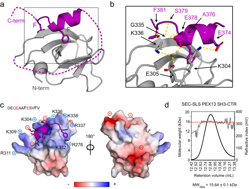
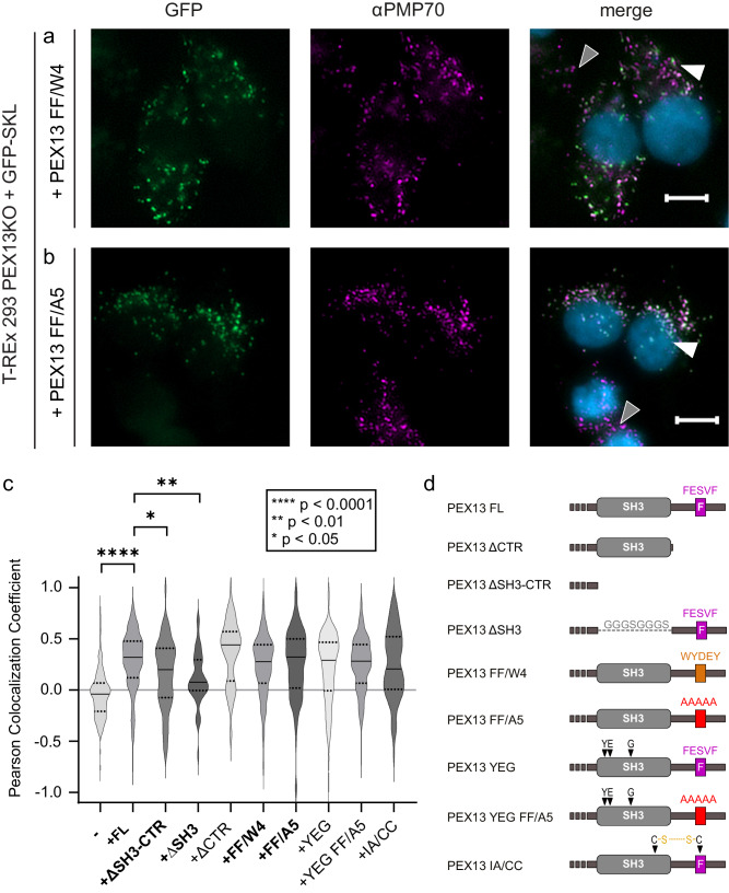
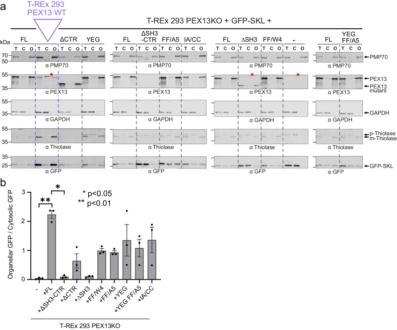
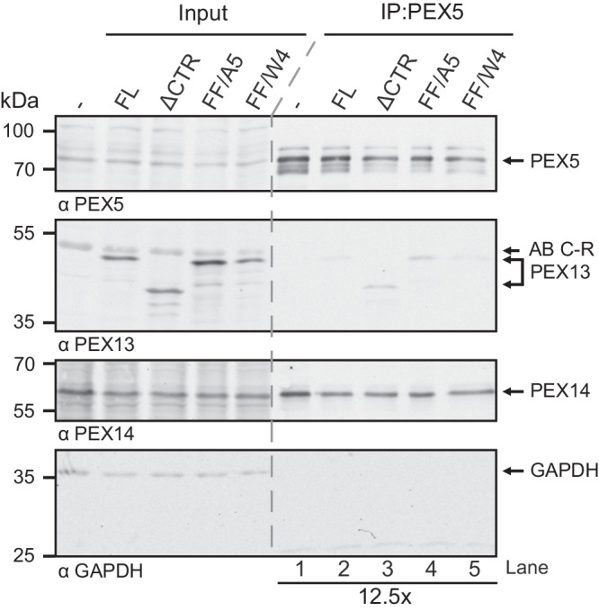
Import of proteins into peroxisomes depends on PEX5, PEX13 and PEX14. By combining biochemical methods and structural biology, we show that the C-terminal SH3 domain of PEX13 mediates intramolecular interactions with a proximal FxxxF motif. The SH3 domain also binds WxxxF peptide motifs in the import receptor PEX5, demonstrating evolutionary conservation of such interactions from yeast to human. Strikingly, intramolecular interaction of the PEX13 FxxxF motif regulates binding of PEX5 WxxxF/Y motifs to the PEX13 SH3 domain. Crystal structures reveal how FxxxF and WxxxF/Y motifs are recognized by a non-canonical surface on the SH3 domain. The PEX13 FxxxF motif also mediates binding to PEX14. Surprisingly, the potential PxxP binding surface of the SH3 domain does not recognize PEX14 PxxP motifs, distinct from its yeast ortholog. Our data show that the dynamic network of PEX13 interactions with PEX5 and PEX14, mediated by diaromatic peptide motifs, modulates peroxisomal matrix import.


Conflict of interest statement

The authors declare no competing interests.

Figures

Fig. 1
Fig. 1. Schematic representation of PTS1 import with interactions of the core components and NMR-based analysis of the conformation and dynamics of PEX13 C-terminal region.
a Schematic overview of cargo recognition, docking and cargo translocation. b Schematic representation of domain architecture and intermolecular interactions of the human peroxins PEX5, PEX14 and PEX13 respectively. Lines 1 and 2 between the peroxins indicate known binding events involving the targeted structure elements or motifs,–,. c Asterisks indicate proline or missing assignment. Top: 13C secondary chemical shifts (Δδ13Cα - Δδ13Cβ) analysed with TALOS + . The propensity for the secondary structure elements random coil, α-helix or β-sheet are represented in gray, blue or green, respectively. Our data support the typical β-sandwich fold of the SH3 domain and the presence of a short α-helical motif comprising the FxxxF motif. Middle: elevated {1H}-15N heteronuclear NOE values indicate an extended SH3 fold (265-345) and a folded FxxxF motif with similar values to the SH3 domain. Bottom: 15N R1*R2 relaxation rates as a function of amino acid sequence. SH3 core residues (266-335) show an average of 16.6. C-terminal residues R337, K341, A375, F377 and F381 show values of 28.4 ± 1.8, 25.2 ± 0.6, 45 ± 2.6, 34,2 ± 0.6 and 42.0 ± 3.0, respectively. Error bars are calculated from fitting errors of R1 and R2 values. Values higher than the average in structured regions indicate the presence of conformational dynamics/and or transient interactions. Secondary structure elements are illustrated at the bottom. d Zoomed view of NMR spectra of PEX13 SH3-CTR (black) and PEX13 SH3 (red). Source data are provided as a Source Data file.
Fig. 2
Fig. 2. Structural analysis of PEX13 SH3 in complex with FxxxF motif.
a Crystallographic structure of PEX13 SH3 2GS FxxxF showing the α-helical FxxxF motif, which clamps β1 and β5 between the two Phe residues. b Zoom view visualizing the hydrogen bond network between the SH3 domain and the FxxxF peptide. Polar backbone contacts are mediated by A376 and G335 as well as S379, F381 and K336 and sidechains E374 and E378 are coordinated by K304, E305, K336 and a water molecule. c Electrostatic surface representation showing the positively charged FxxxF binding site which is caused by seven Arg or Lys residues. The peptide in contrast, is negatively charged which is favored for the binding (Q373, E374 and E387). A 180° rotation on the Y axis reveals a negatively charged backside. d Static light scattering analysis of PEX13 SH3-CTR shows the molecular weight (red dashed line) of 15.64 ± 0.1 kDa versus the calculated mass of 15.56 kDa indicating a monomeric state. Source data are provided as a Source Data file.
Fig. 3
Fig. 3. Interaction of PEX14 NTD with PEX13 SH3-CTR in comparison with PEX19 (66-77).
a 1H,15N correlation spectra of 15N-labeled recombinant PEX14(NTD) free (black), and in complex with PEX13 SH3-CTR (green scale). b Sequence alignment of PEX13 and PEX19 FxxxF motifs. Red and yellow boxes indicate identical and similar residues. c NMR chemical shift perturbations of PEX14 NTD in the presence of PEX13 SH3-CTR (green) or PEX19 peptide (brown) (Neufeld et al.) plotted on the sequence with indicated secondary structure elements above. Asterisk indicate proline or missing assignment. d Chemical shift perturbations (0.08 to 0.3 ppm) of PEX13 SH3-CTR (left) and PEX19 peptide (right) mapped on the PEX14 NTD/PEX19 66-77 structure (2w85). e ITC experiments of PEX14 NTD with PEX13 SH3-CTR (green) and PEX13 FxxxF (pink) showing very different energetics but the same one to one stoichiometry. f Energetic contribution of the PEX14 NTD interaction with PEX13 SH3-CTR (left graph) and PEX13 FxxxF peptide (right graph). Average values and error bars (SD) are calculated from 3 different experiments (n = 3). Source data are provided as a Source Data file.
Fig. 4
Fig. 4. NMR titration of PEX14 NTD onto 15N PEX13 SH3-CTR.
a Spectra overlay from NMR titration of unlabeled PEX14 NTD onto 15N labeled PEX13 SH3-CTR where large chemical shift perturbations of resonances from the FxxxF motif were observed. b Spectra overlay of free PEX13 SH3-CTR (black), PEX13 SH3-CTR + 4x PEX14 NTD (dark red) and apo PEX13 SH3 (blue) showing the transfer from the closed conformation of the PEX13 SH3-CTR back to apo form of PEX13 SH3. c Chemical shift perturbations mapped on the sequence and structural elements (above) of PEX13 SH3-CTR visualizing the effect of the opening on the structural elements β1, β5 and the FxxxF motif. Signals that experience large chemical shift perturbations or line-broadening down to beyond detection are indicated with negative values. d Schematic representation of the opening process. Source data are provided as a Source Data file.
Fig. 5
Fig. 5. Interaction of PEX5 (di)aromatic peptide motifs with PEX13 SH3 or SH3-CTR.
a Overview of PEX5 (di)aromatic peptide motifs. W0 was expressed as PEX5 1-76 while other W motifs were purchased as peptides as listed. b Induced Chemical shifts changes of PEX13 SH3 or SH3-CTR upon addition of 4x or 8x PEX5 (di)aromatic peptide motifs represented as the sum of 10 involved residues. The star indicates W3 which shows less chemical shift perturbation but extensive line-broadening. c Schematic representation of PEX13 SH3 / W peptide binding (top) or PEX13 SH3-CTR / W peptide competition. d Plot of KD values from ITC experiments of PEX13 SH3 or PEX14 NTD with PEX5 W and FxxxF peptide motifs. Average values and error bars (SD) are calculated from 3 different experiments (n = 3). e KD values from ITC experiments in numbers. f Electrostatic surface representation of the PEX13 SH3 GS W4 structure at 2.3 Å. g Zoomed view of the W binding site showing polar interactions marked with yellow circles. h Schematic representation of the conserved hydrogen-bond network of PEX13 SH3 / FxxxF (pink) and PEX5 W4 (orange) interaction. Additional contact sites are marked in gray. Source data are provided as a Source Data file.
Fig. 6
Fig. 6. The PEX13 SH3 domain is essential for PTS1 import.
T-REx 293 cells and PEX13-deficient (PEX13 KO) T-REx 293 cells were transfected with bicistronic expression plasmids encoding GFP-PTS1 and different PEX13 truncation and mutation variants as indicated on the left side. Rescue of the PTS1 import defect of PEX13 KO cells was monitored by fluorescence microscopy with GFP-SKL as reporter (left panels). Detection of peroxisomal membranes was achieved via PMP70 specific antibody indicated by magenta fluorescence punctate pattern (middle panels). Overlap of green and magenta dots in the merge (right panels) represent functional matrix import. Nuclei were stained with DAPI (blue, right panels). For each experiment, one cell showing no peroxisomal protein import is marked with a gray triangle and one cell showing import is marked by a white triangle, if present. Scale bar: 5 μm. In one representative analysis a total of 1216 cells were analysed. a Expression of GFP-SKL in PEX13 KO cells did not rescue PTS1 import indicated by a diffuse cytosolic green GFP signal (left and right panels) and served as negative control. b Expression of full length PEX13 led to an almost complete restored import (p < 0.0001). The introduction of a variant lacking the full C-terminal region (ΔSH3-CTR) (c) or the SH3 domain (ΔSH3) (d) did not sufficiently restore PTS1 import. Source data are provided as a Source Data file.
Fig. 7
Fig. 7. The PEX13 SH3-proximal FxxxF motif modulates PTS1 import.
PEX13-deficient (PEX13 KO) T-REx 293 cells were transfected with bicistronic expression plasmids encoding GFP-PTS1 and PEX13 truncation or mutation variants as indicated. Rescue of the PTS1 import defect of PEX13 KO cells was monitored by fluorescence microscopy with GFP-SKL as reporter (left panels). Detection of peroxisomal membranes was achieved via PMP70 specific antibody indicated by magenta fluorescence punctate pattern (middle panels). Overlap of green and magenta dots in the merge (right panels) represent functional matrix import. Nuclei were stained with DAPI (blue, right panels). For each experiment, a cell showing no peroxisomal protein import is marked with a gray triangle and a cell showing import is marked by a white triangle. Scale bar: 5 μm. Expression of PEX13 with enhanced (a) or reduced (b) structural autoinhibition of the SH3 domain showed reduced PTS1 import in comparison the expression of full length PEX13. c The import defect from different PEX13 mutants was automated analyzed with the Pearson Colocalization Coefficient using the GFP-SKL and PMP70 signals. The Coefficient ranges from −1 (no colocalization) to +1 (perfect colocalization). For each experiment, a minimum of 45 transfected cells was analyzed. A Kruskal-Wallis test was performed for statistical analysis, and the mean rank of each cell line was compared to the mean rank of the control cell line PEX13KO complemented with PEX13FL. Correction for multiple comparisons was done with a Dunn’s test. Significance is indicated as ****p < 0.0001, **p = 0.0018, *p = 0.0281. Dotted lines indicate the quartiles, while the black horizontal line indicates the median. Bold mutants are shown in main figures. d Schematic overview of PEX13 mutants. Source data are provided as a Source Data file.
Fig. 8
Fig. 8. The efficacy of PTS1- and PTS2 import is reduced in cells expressing PEX13 variants.
a, b T-REx 293 cells transfected with GFP-SKL and T-REx 293 PEX13KO cells transfected with a bicistronic vector encoding different PEX13 constructs and GFP-SKL were used for the preparation of total lysates (T) and for fractionation of the cell into cytosol and organelles (C and O, respectively). The localization of the GFP-SKL in cytosolic and organellar fractions served as readout for the PTS1 import into peroxisomes. Additionally, PTS2 import can be monitored by detecting Thiolase, which exists in a cytosolic pre-form (p-Thiolase) and in a mature, processed form (m-Thiolase) when imported into peroxisomes. a All immunoblots shown are from different gels of the same biological replicate. Other replicates can be found in Supplementary Fig. 10. PMP70 and GAPDH served as loading controls for the organellar and cytosolic fraction, respectively. Expression of PEX13 variants and GFP-SKL was verified with corresponding antibodies, as indicated. PEX13 ΔSH3-CTR is not recognized by the PEX13 antibodies used as these were generated against the SH3 domain. A cross-reaction of the antibody with an unknown protein below 55 kDa is indicated by a red asterisk. b Total amounts of GAPDH and PMP70 in cell lysates were used to calculate the relative GFP signal in the cytosolic and organellar fractions, respectively. Average values and error bars (SEM) are calculated from 3 different experiments (n = 3). Black dots indicate the exact data points for each replicate. For statistical analysis a Kruskal-Wallis test was performed and the mean rank of each cell line was compared to the mean rank of the control cell line PEX13KO complemented with PEX13FL. Correction for multiple comparisons was done with a Dunn’s test. Significance is indicated as **p = 0.0085 and *p = 0.0409. Source data are provided as a Source Data file.
Fig. 9
Fig. 9. The PEX13 FxxxF motif modulates PEX5 binding in cells.
PEX13 FL, PEX13 ΔCTR and the two mutations FxxxF to A5 and FxxxF to W4 were expressed in T-REx 293 PEX13 KO cells. The cell lysates were subjected to immunoprecipitation with PEX5 antibody and analyzed by immunoblotting. One of three independent experiments is shown, the other two experiments are shown in Supplementary Fig. 11. Source data are provided as a Source Data file.
Fig. 10
Fig. 10. Copartitioning of PEX14 NTD and PEX5 NTD with PEX13 YG condensates and model for the role of PEX13 in peroxisomal matrix import.
Colocalization experiments were performed in 50 mM Tris pH 7.5, 100 mM NaCl and 10% PEG8000 at RT. Scale bars indicate 25 μm. Experiments were conducted as three technical replicates. a At 100 μM concentration, large condensates (brightfield) are formed by PEX13 YG (His-SUMOPEX13 40–120) but not by His-SUMO (negative control) alone (b). Under the same conditions, PEX14 NTD (1–104) GFP (c) and PEX5 NTD (1–330) mCherry (d) do not form condensates by themselves. e Condensates formed by 50 μM PEX13 YG allow simultaneous partition of PEX14 NTD (GFP) and PEX5 NTD (mCherry). f After a docking event, condensate formation of PEX13 PEX5 and PEX14 mediates translocation of the cargo bound PEX5 TPR domain and the PEX5 associated PEX14 NTD. g After translocation, a PEX13 homo-oligomer replaces the PEX5/PEX14 interaction by avidity effects handing PEX5 over to PEX13 to form a transient complex. (Boxes) Schematic representation of the interactions between PEX5, PEX14 and PEX13 in both states.

References

    1. Fujiki Y, Lazarow PB. Post-translational import of fatty acyl-CoA oxidase and catalase into peroxisomes of rat liver in vitro. J. Biol. Chem. 1985;260:5603–5609. - PubMed
    1. Erdmann R, Veenhuis M, Kunau WH. Peroxisomes: Organelles at the crossroads. Trends Cell Biol. 1997;7:400–407. - PubMed
    1. Wanders RJ, Waterham HR. Biochemistry of mammalian peroxisomes revisited. Ann. Rev. Biochem. 2006;75:295–332. - PubMed
    1. Klouwer FCC, et al. Zellweger spectrum disorders: clinical overview and management approach. Orphanet J. Rare Dis. 2015;10:151. - PMC - PubMed
    1. Distel B, et al. A unified nomenclature for peroxisome biogenesis factors. J. Cell Biol. 1996;135:1–3. - PMC - PubMed

MeSH terms

LinkOut - more resources