. 2024 Apr 17;24(1):467.
doi: 10.1186/s12903-024-04219-4.

Comparison of temporomandibular joints in relation to ages and vertical facial types in skeletal class II female patients: a multiple-cross-sectional study

Affiliations

Comparison of temporomandibular joints in relation to ages and vertical facial types in skeletal class II female patients: a multiple-cross-sectional study

Jing Zhou et al. BMC Oral Health. .

Abstract

Background: The temporomandibular joint (TMJ) is closely related to the dynamic balance and stability of mandibular function and orthodontic treatment. Skeletal class II female patients are thought to be at high risk for TMJ disease. The relationship between the TMJ and craniofacial structures is still controversial. This study compared the morphology and position of the TMJ in skeletal class II adolescents and adults with various vertical facial types using cone-beam computed tomography (CBCT).

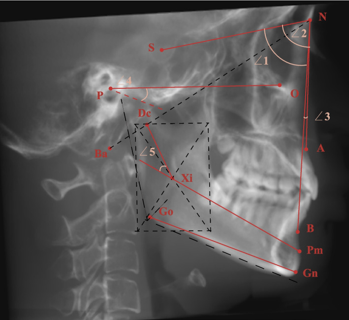
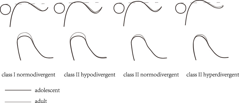
Materials and methods: A total of 117 skeletal class II patients were divided into three groups according to the FH-GoGn angle (hypodivergent, normodivergent and hyperdivergent), with 40 class I normodivergent patients serving as controls. Each group contained two age subgroups (adolescents: 11-14 years old, adults: 18-35 years old). The size (condylar length, height, long and short axis diameter, glenoid fossa width and depth) and shape (condylar neck inclination, condylar head angle and long axis angle, articular eminence inclination) of the condyle and fossa, joint space (anterior, superior, posterior, mesial and lateral), and position of the fossa (vertical, transverse, and anteroposterior distance) and condyle were measured and compared using CBCT.

Results: Class II hypodivergent patients exhibited the greatest condylar length, height, and long- and short-axis diameter; steepest articular eminence; deepest fossa depth; largest superior, mesial and lateral joint spaces; and highest fossa position in both age groups. The manifestations of class II hyperdivergent patients were mostly the opposite. In adults, except for the condylar long axis angle, the measurements of the condyle increased differently among skeletal patterns, while the measurements of the fossa decreased, as the joint spaces and fossa position remained approximately stable compared with those in adolescents.

Conclusion: The vertical skeletal pattern, rather than the class II sagittal skeletal pattern, may be the main factor affecting the morphology and position of the TMJ. Attention should be given to the TMJ area in hyperdivergent patients with a relatively poor-fit condyle-fossa relationship. The changes in the TMJ with age were mainly morphological rather than positional and varied with skeletal pattern.

Keywords: Adolescent; Adult; CBCT; Skeletal pattern; Temporomandibular joint.


Conflict of interest statement

The authors declare that they have no competing interests.

Figures

Fig. 1
Fig. 1
a FH plane parallel to the horizontal plane. b Midsagittal plane passing through the ANS point and Ba point
Fig. 2
Fig. 2
Construction of landmarks used in the cephalometric analysis and angular measurements. (1: SNB ∠2: SNA ∠3: ANB ∠4: FH-GoGn ∠5: mandibular arc)
Fig. 3
Fig. 3
Landmarks and measurements of the sagittal projection of the mandibular ramus (Co: superior point of the condyle; Cc: centre of the largest circle that fits the condylar head arc; Sg: inferior point of the mandibular sigmoid incisure; Cm: intersection of the horizontal line passing through Sg and line 2, which passes through Cc parallel to the tangent of the condylar posterior border; ∠1 Cni: condylar neck inclination, which is the posterior superior angle between the tangent line of the condyle posterior border and a horizontal line; ∠2 Condylar head angle: angle between the condylar head and neck, anterior angle between line 1 and line 2; Line 1: distance between Co and Cc; Line 2: distance between Cc and Cm; Condylar length: line 1 plus line 2; Line 3: condylar height: vertical distance between Co and Sg)
Fig. 4
Fig. 4
Measurements on the largest axial view of the condyle. (∠1 Condylar long axis angle: angle between the condylar mediolateral axis and the midsagittal plane; 1: Condylar long axis diameter: largest mediolateral diameter of the condyle; 2: Condylar short axis diameter: largest anteroposterior diameter of the condyle, perpendicular to line 1)
Fig. 5
Fig. 5
a Midsagittal plane of the condyle. (Ei: inferior point of the articular eminence; Fs: superior point of the glenoid fossa; Fp: intersection of the posterior slope of the glenoid fossa and a line parallel to the FH plane and passing through the Ei. When the posterior slope was shorter than the anterior slope, the inferior point was used. ∠1 Aei: articular eminence inclination, the angle between the best fit line of the posterior slope of the articular eminence and the horizontal plane. 1: Glenoid fossa depth; 2: Glenoid fossa width. A: Anterior joint space: shortest distance from the most prominent anterior point of the condyle to the corresponding glenoid fossa bone. S: Superior joint space: shortest distance from the most superior point of the condyle to the Fs. P: Posterior joint space: shortest distance from the most prominent posterior point of the condyle to the corresponding glenoid fossa bone.) b Midcoronal plane of the condyle (Mes: the medial joint space, the shortest distance from the midpoint between the most medial and superior points of the condyle to the corresponding glenoid fossa bone. Las: lateral joint space, the shortest distance from the midpoint of the most lateral and superior points of the condyle to the corresponding glenoid fossa bone
Fig. 6
Fig. 6
a, b Location of the glenoid fossa centre point and its vertical distance measurement (Fc: glenoid fossa centre point, the most superior point of the glenoid fossa on the sagittal and coronal planes. 1: Glenoid fossa vertical distance, the vertical distance between the Fc point and the FH plane; positive when Fc is higher); c Sella point located on the midsagittal plane, with the coronal plane orientated through it; Fig. 6d Axial view (2: Glenoid fossa sagittal distance, sagittal projection distance from the Fc to S. 3: Glenoid fossa coronal distance, distance from the Fc to the midsagittal plane)
Fig. 7
Fig. 7
Changes in different skeletal patterns of the TMJ with age

Similar articles

Cited by

References

    1. Chang CL, Wang DH, Yang MC, Hsu WE, Hsu ML, et al. Functional disorders of the temporomandibular joints: internal derangement of the temporomandibular joint. Kaohsiung J Med Sci. 2018;34(4):223–230. - PMC - PubMed
    1. Cevidanes LH, Hajati AK, Paniagua B, Lim PF, Walker DG, Palconet G, Nackley AG, Styner M, Ludlow JB, Zhu H, Phillips C, et al. Quantification of condylar resorption in temporomandibular joint osteoarthritis. Oral Surg Oral Med Oral Pathol Oral Radiol Endod. 2010;110(1):110–117. - PMC - PubMed
    1. Noh KJ, Baik HS, Han SS, Jang W, Choi YJ, et al. Differences in mandibular condyle and glenoid fossa morphology in relation to vertical and sagittal skeletal patterns: a cone-beam computed tomography study. Korean J Orthod. 2021;51(2):126–134. - PMC - PubMed
    1. Saccucci M, Polimeni A, Festa F, Tecco S, et al. Do skeletal cephalometric characteristics correlate with condylar volume, surface and shape? A 3D analysis. Head Face Med. 2012;8:15. - PMC - PubMed
    1. Tecco S, Saccucci M, Nucera R, Polimeni A, Pagnoni M, Cordasco G, Festa F, Iannetti G, et al. Condylar volume and surface in Caucasian young adult subjects. BMC Med Imaging. 2010;10:28. - PMC - PubMed

LinkOut - more resources