. 2024 Apr 17;22(1):362.
doi: 10.1186/s12967-024-05133-7.

DB-1310, an ADC comprised of a novel anti-HER3 antibody conjugated to a DNA topoisomerase I inhibitor, is highly effective for the treatment of HER3-positive solid tumors

Affiliations

DB-1310, an ADC comprised of a novel anti-HER3 antibody conjugated to a DNA topoisomerase I inhibitor, is highly effective for the treatment of HER3-positive solid tumors

Xi Li et al. J Transl Med. .

Abstract

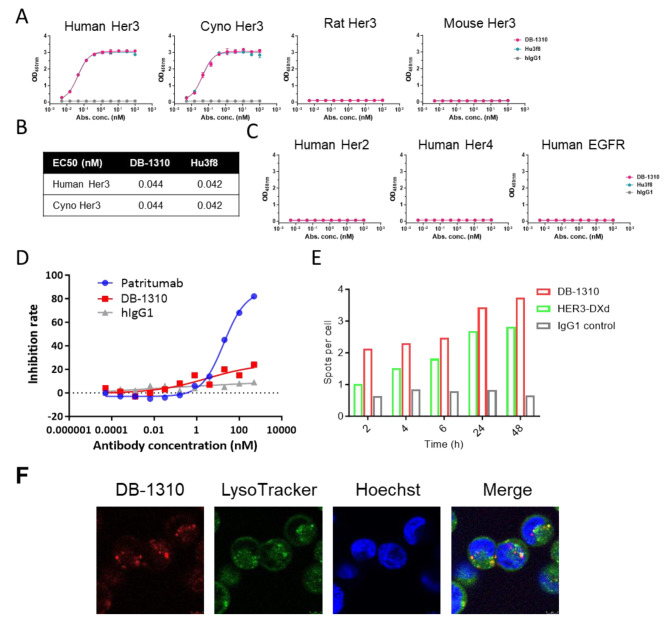
Background: HER3 (ErbB3), a member of the human epidermal growth factor receptor family, is frequently overexpressed in various cancers. Multiple HER3-targeting antibodies and antibody-drug conjugates (ADCs) were developed for the solid tumor treatment, however none of HER3-targeting agent has been approved for tumor therapy yet. We developed DB-1310, a HER3 ADC composed of a novel humanized anti-HER3 monoclonal antibody covalently linked to a proprietary DNA topoisomerase I inhibitor payload (P1021), and evaluate the efficacy and safety of DB-1310 in preclinical models.

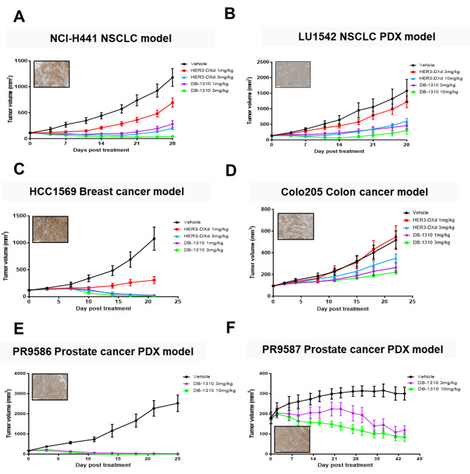
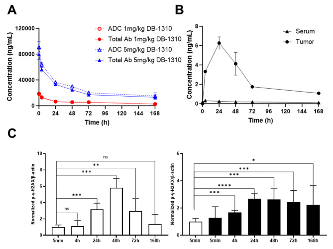
Methods: The binding of DB-1310 to Her3 and other HER families were measured by ELISA and SPR. The competition of binding epitope for DB-1310 and patritumab was tested by FACS. The sensitivity of breast, lung, prostate and colon cancer cell lines to DB-1310 was evaluated by in vitro cell killing assay. In vivo growth inhibition study evaluated the sensitivity of DB-1310 to Her3 + breast, lung, colon and prostate cancer xenograft models. The safety profile was also measured in cynomolgus monkey.

Results: DB-1310 binds HER3 via a novel epitope with high affinity and internalization capacity. In vitro, DB-1310 exhibited cytotoxicity in numerous HER3 + breast, lung, prostate and colon cancer cell lines. In vivo studies in HER3 + HCC1569 breast cancer, NCI-H441 lung cancer and Colo205 colon cancer xenograft models showed DB-1310 to have dose-dependent tumoricidal activity. Tumor suppression was also observed in HER3 + non-small cell lung cancer (NSCLC) and prostate cancer patient-derived xenograft (PDX) models. Moreover, DB-1310 showed stronger tumor growth-inhibitory activity than patritumab deruxtecan (HER3-DXd), which is another HER3 ADC in clinical development at the same dose. The tumor-suppressive activity of DB-1310 synergized with that of EGFR tyrosine kinase inhibitor, osimertinib, and exerted efficacy also in osimertinib-resistant PDX model. The preclinical assessment of safety in cynomolgus monkeys further revealed DB-1310 to have a good safety profile with a highest non severely toxic dose (HNSTD) of 45 mg/kg.

Conclusions: These finding demonstrated that DB-1310 exerted potent antitumor activities against HER3 + tumors in in vitro and in vivo models, and showed acceptable safety profiles in nonclinical species. Therefore, DB-1310 may be effective for the clinical treatment of HER3 + solid tumors.

Keywords: Antibody-drug conjugate; HER3; Preclinical; Solid tumor therapy.


Conflict of interest statement

The authors declare that they have no competing interests.

Figures

Fig. 1
Fig. 1
DB-1310 binds HER3 via a novel epitope with high affinity and internalization capability. (a-c) The binding of DB-1310 and its parental antibody to human HER3, cynomolgus HER3, human HER2, human HER4 and human EGFR was measured by ELISA. (d) MDA-MB-453 cells were stained with 10 nM biotin-labeled patritumab in the presence of DB-1310 or naked patritumab at serial dilutions of up to 500 nM. The binding of biotin-patritumab to cell surface HER3 was detected by streptavidin-PE staining. The Mean fluorescence intensity (MFI) of PE was measured for each condition, and the inhibition rate was calculated as (MFI biotin−patritumab only – MFI test condition)/ MFI biotin−patritumab only x 100%. (e) Internalization of pHrodo-labeled DB-1310 or HER3-DXd was measured by an Operetta CLS high-throughput microplate imager. (f) MDA-MB-453 cells were cultured with pHrodo-labeled DB-1310 for 12 h and images were obtained by confocal microscopy. Lysosomes and nuclei were labeled with LysoTracker (green) and Hoechst (blue), respectively
Fig. 2
Fig. 2
DB-1310 suppresses the growth of tumor cells in vitro. (a) Surface HER3 was detected by FACS in tumor cell lines. HER3 expression was determined as the MFI. (b) Breast cancer, prostate cancer and lung cancer cell lines were cultured with DB-1310. Cell proliferation was measured by a CTG assay, and IC50 values were calculated using linear regression with GraphPad Prism. (c) SK-BR-3 and NCI-H441 cells were treated with DB-1310 or Hu3f8 at 30 nM or 90 nM, and P1021 at 3nM or 30nM for 48 h. The cell cycle was analyzed by FACS. (d) HEK293 cells were stained with CellTrace™ Far Red Dye to trace in analysis and cocultured with HER3-negative HEK293 or HER3-expressing HEK293-ERBB3 cells at a 1:1 ratio for 4 days in the presence of different treatments. Cells were stained with PI, and labeled cell killing was analyzed by FACS. (e) HER3-expressing HEK293-ERBB3 cells were labeled with CellTrace™ Far Red Dye and cocultured with PBMCs at a 1:20 ratio in the presence of DB-1310 for 6 h to access ADCC effect. Cells were stained with PI and subjected to flow cytometric analysis to determine the lysis rate of HEK293-ERBB3 cells. (f) Human serum complement and HEK293-ERBB3 cells were mixed and incubated with DB-1310 for 1 h. Complement-mediated lysis of target cells was measured by CytoToxi96® Non-Radioactive Cytotoxicity Assay kit
Fig. 3
Fig. 3
DB-1310 inhibits tumor growth in vivo. Immunodeficient mice were inoculated with NCI-H441 NSCLC cells (a), LU1542 NSCLC patient-derived tumor xenografts (b), HCC1569 breast cancer cells (c), Colo25 colon cancer cells (d), PR9586 prostate cancer patient-derived tumor xenografts (e) or PR9587 prostate cancer patient-derived tumor xenografts (f) and were then treated with DB-1310 or HER3-DXd. The tumor volume in each animal was measured twice a week
Fig. 4
Fig. 4
Tumor-specific release of P1021 payload contributes to DB-1310 in vivo efficacy in the NCI-H441 model. NU/NU mice were subcutaneously inoculated with 5 × 106 NCI-H441 NSCLC cells and intravenously injected with either 1 or 5 mg/kg DB-1310. Serum and tumor samples were collected at different time points. Total antibody and ADC concentrations in serum were measured by ELISA (a). Free payload concentrations in serum and tumor samples were measured by LC-MS/MS (b). (c) The level of P-H2AX in tumors was measured by western blotting and normalized to β-actin
Fig. 5
Fig. 5
DB-1310 suppresses EGFRm NSCLC tumor growth. (a) Surface HER3 expression on NCI-H1975 NSCLC cells was measured by FACS after osimertinib stimulation for 24 h. (b) NCI-H1975 cells were treated with vehicle or 25 nM osimertinib for 24 h. HER3 internalization was measured by treatment with pHrodo-labeled DB-1310. (c) NCI-H1975 cells were treated with 25 nM osimertinib with or without DB-1310 at 200nM or 1000nM for 5 days. Cell viability was measured by a CTG assay. (d) NCI-H1975 tumor cells were inoculated into BALB/c nude mice, and the mice were treated with DB-1310 (i.v. one treatment) and/or osimertinib (p.o. daily). Tumor growth and body weight were measured. (e) An osimertinib resistant PDX model (LD1-0025-200717) was established with samples from osimertinib-refractory NSCLC patients, and the mice were treated with DB-1310.

References

    1. Sierke SL, Cheng K, Kim HH, Koland JG. Biochemical characterization of the protein tyrosine kinase homology domain of the ErbB3 (HER3) receptor protein. Biochem J. 1997;322(Pt 3):757–63. doi: 10.1042/bj3220757. - DOI - PMC - PubMed
    1. Inaki K, Shibutani T, Maeda N, Eppenberger-Castori S, Nicolet S, Kaneda Y, et al. Pan-cancer gene expression analysis of tissue microarray using EdgeSeq oncology biomarker panel and a cross-comparison with HER2 and HER3 immunohistochemical analysis. PLoS ONE. 2022;17:e0274140. doi: 10.1371/journal.pone.0274140. - DOI - PMC - PubMed
    1. Ocana A, Vera-Badillo F, Seruga B, Templeton A, Pandiella A, Amir E. HER3 overexpression and survival in solid tumors: a meta-analysis. J Natl Cancer Inst. 2013;105:266–73. doi: 10.1093/jnci/djs501. - DOI - PubMed
    1. Haikala HM, Jänne PA. Thirty years of HER3: from basic biology to therapeutic interventions. Clin Cancer Res. 2021;27:3528–39. doi: 10.1158/1078-0432.CCR-20-4465. - DOI - PMC - PubMed
    1. Jänne PA, Baik C, Su WC, Johnson ML, Hayashi H, Nishio M, et al. Efficacy and safety of patritumab deruxtecan (HER3-DXd) in EGFR inhibitor-resistant, EGFR-mutated non-small cell lung cancer. Cancer Discov. 2022;12:74–89. doi: 10.1158/2159-8290.CD-21-0715. - DOI - PMC - PubMed

Publication types

MeSH terms