Review
. 2024 Apr 17;13(1):23.
doi: 10.1186/s40035-024-00409-w.

Focusing on mitochondria in the brain: from biology to therapeutics

Affiliations
Review

Focusing on mitochondria in the brain: from biology to therapeutics

Nanshan Song et al. Transl Neurodegener. .

Abstract

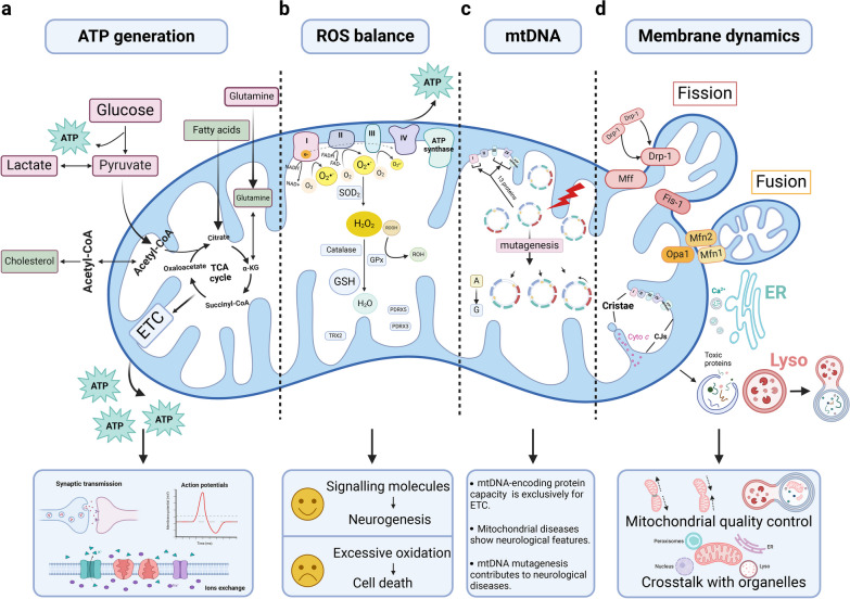
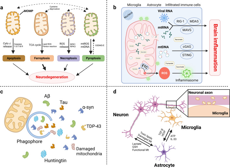
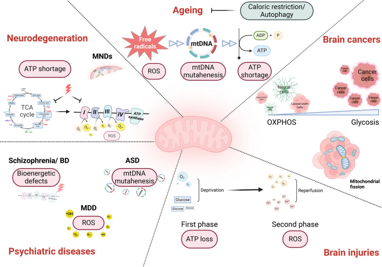
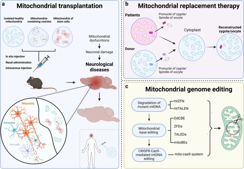
Mitochondria have multiple functions such as supplying energy, regulating the redox status, and producing proteins encoded by an independent genome. They are closely related to the physiology and pathology of many organs and tissues, among which the brain is particularly prominent. The brain demands 20% of the resting metabolic rate and holds highly active mitochondrial activities. Considerable research shows that mitochondria are closely related to brain function, while mitochondrial defects induce or exacerbate pathology in the brain. In this review, we provide comprehensive research advances of mitochondrial biology involved in brain functions, as well as the mitochondria-dependent cellular events in brain physiology and pathology. Furthermore, various perspectives are explored to better identify the mitochondrial roles in neurological diseases and the neurophenotypes of mitochondrial diseases. Finally, mitochondrial therapies are discussed. Mitochondrial-targeting therapeutics are showing great potentials in the treatment of brain diseases.

Keywords: Brain; Mitochondria; Mitochondrial transfer; Neurological disorders.


Conflict of interest statement

The authors declare no competing interests.

Figures

Fig. 1
Fig. 1
Mitochondrial biology maintains brain physiology. a Mitochondria are the power house and generate ATP through relevant processes of glucose, FA and amino acid metabolism. They tightly support normal brain functions dominated by neuronal activity including synaptic transmission, neuroelectrical activity, and ion exchange. b The mitochondrial ETC is the site of mitochondrial ROS generation. During oxidative metabolism, electrons combine prematurely with oxygen to form O2•−, which is dismutated to H2O2 by SOD2 and then converted to H2O by catalase and GPx. There are also mitochondria-targeted antioxidants essential for controlling ROS homeostasis in the brain, such as PDRX3, PDRX5 and TRX2. c The entire protein-coding capacity of mtDNA is devoted to the synthesis of mitochondrial complexes except complex II. Mutagenesis in mitochondrial genome occurs at a much higher rate than that in the nuclear genome, leading to the collapse of mitochondrial functions, which is closely related to neurological diseases. d Mitochondrial membrane dynamics including mitochondrial fission/fusion, membrane interactions with other organelles and ultra-structural membrane remodeling, renders the multifaceted involvement of mitochondria in cell biology. ATP, adenosine triphosphate; cyto c, cytochrome c; ER, endoplasmic reticulum; ETC, electron transport chain; FAs: fatty acids; GPx, glutathione peroxidases; GSH, glutathione; H2O2, hydrogen peroxide; lyso, lysosome; O2•−, superoxide; PDRX, peroxiredoxin; ROH, organic alcohol; ROS, reactive oxygen species; SOD2, manganese-dependent superoxide dismutase; TCA, tricarboxylic acid; TRX, thioredoxin
Fig. 2
Fig. 2
Mitochondria as a multifaceted hub of the brain pathophysiology. a Under cellular stresses, mitochondrial outer membrane permeabilization leads to release of cyto c and ROS, activating the downstream pathways of apoptosis and necroptosis. Ferroptosis is also induced by mitochondrial ETC-promotive lipid peroxide. Pyroptosis is the downstream signal of mitochondrial dysfunctions, and is controlled by mitochondria to initiate apoptosis/necrosis. b Mitochondria contain endogenous inflammatory inducers, including mtDNA, mtRNA, metabolic products and ROS. Mitochondria outer membrane acts as a platform for immune signaling through inflammasome and MAVS activation. MAVS also endows cells with antiviral immunity. c Mitochondria participate in multiple steps of autophagy including autophagy initiation, phagophore elongation, autophagic flux formation and autophagy gene induction. d Mitochondria participate in cellular communication in the brain through membrane contact and cellular organelle transfer. α-syn, α-synuclein; Aβ, β-amyloid; ATP, adenosine triphosphate; cyto c, cytochrome c; ETC, electron transport chain; GSH, glutathione; MAVS, mitochondrial antiviral signaling; ROS, reactive oxygen species; TDP-43: TAR DNA-binding protein 43; TGF-β, transforming growth factor β; VEGF, vascular endothelial growth factor
Fig. 3
Fig. 3
Mitochondria in neurological diseases: commonality and specificity. Mitochondrial dysfunctions are commonly seen in neurological diseases, with both commonality and disease specificity from the perspective of mechanism. ASD, autism spectrum disorder; ATP, adenosine triphosphate; BD, bipolar disorder; MDD: major depressive disorder; MNDs, motor neuron diseases; OXPHOS, oxidative phosphorylation; ROS, reactive oxygen species; TCA, tricarboxylic acid
Fig. 4
Fig. 4
Advanced mitochondrial therapies for neurological diseases and mitochondrial diseases. a Mitochondrial transplantation via injection of isolated mitochondria, mitochondria-containing vesicles and mitochondria-loaded stem cells is promising for the treatment of brain diseases. b Mitochondrial replacement therapy is conducted by pronuclear transfer or spindle transfer. For pronuclear transfer, a zygote is generated by fertilization and then pronuclei of the zygote containing mutated mtDNA are transferred to the donor’s enucleated zygote. For spindle transfer, the spindle of the oocyte with mtDNA mutation is transferred to the donor’s enucleated oocyte, followed by fertilization. c Mitochondrial genome editing is conducted by editing the nuclease systems using the ZFNs, the TALENs and the CRISPR/Cas9 systems. mtTALENs and mtZFNs are mitochondria-targeted DNA nucleases and promote the degradation of mutant mtDNA for heteroplasmic shifting of mutant mtDNA. Mitochondrial base editing is achievable by DdCBEs, TALED, ZFD and mitoBEs to effectively correct the homoplasmic mtDNA mutation. The mito-Cas9 system enables successful knockin of exogenous DNA into mtDNA, which is promising for manipulating more types of mtDNA base editing. CRISPR-Cas9: clustered regularly interspaced short palindromic repeats-associated Cas9; DdCBE: bacterial cytidine deaminase fused with mitochondrial TALE-linked deaminases; mitoBEs: mtDNA base editors; MRT, mitochondrial replacement therapy; TALEN: transcription activator-like effector nuclease; ZFDs: zinc-finger deaminases; ZFNs: zinc finger nucleases

Similar articles

Cited by

References

    1. Pereda AE. Electrical synapses and their functional interactions with chemical synapses. Nat Rev Neurosci. 2014;15:250–263. doi: 10.1038/nrn3708. - DOI - PMC - PubMed
    1. Buzsaki G, Kaila K, Raichle M. Inhibition and brain work. Neuron. 2007;56:771–783. doi: 10.1016/j.neuron.2007.11.008. - DOI - PMC - PubMed
    1. Briscoe J, Marin O. Looking at neurodevelopment through a big data lens. Science. 2020;369(6510):eaaz8627. doi: 10.1126/science.aaz8627. - DOI - PubMed
    1. Pulido C, Ryan TA. Synaptic vesicle pools are a major hidden resting metabolic burden of nerve terminals. Sci Adv. 2021;7:eabi9027. doi: 10.1126/sciadv.abi9027. - DOI - PMC - PubMed
    1. Du F, Zhu XH, Zhang Y, Friedman M, Zhang N, Ugurbil K, et al. Tightly coupled brain activity and cerebral ATP metabolic rate. ProcNatl Acad Sci U S A. 2008;105:6409–6414. doi: 10.1073/pnas.0710766105. - DOI - PMC - PubMed